Review
. 2022 Mar 18;15(3):371.
doi: 10.3390/ph15030371.

Current and Future Prospective of Injectable Hydrogels-Design Challenges and Limitations

Affiliations
Review

Current and Future Prospective of Injectable Hydrogels-Design Challenges and Limitations

Saud Almawash et al. Pharmaceuticals (Basel). .

Abstract

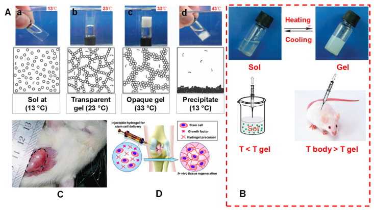
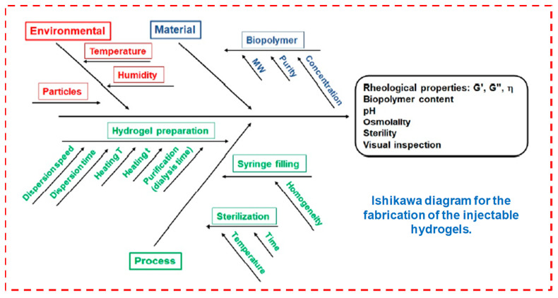
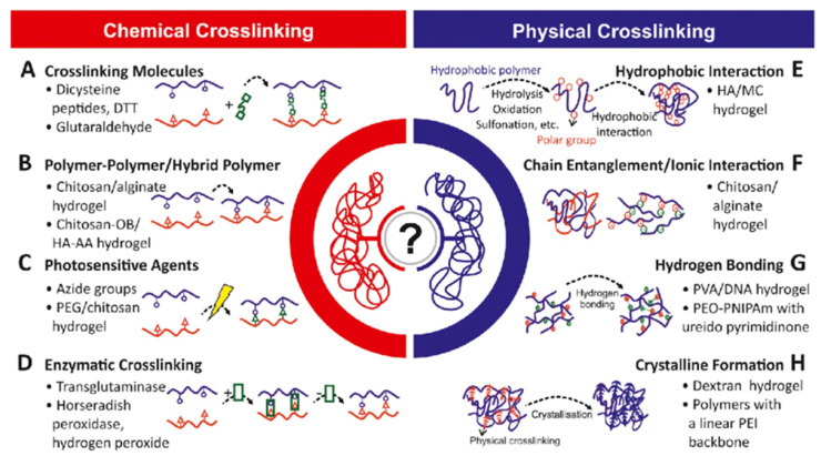
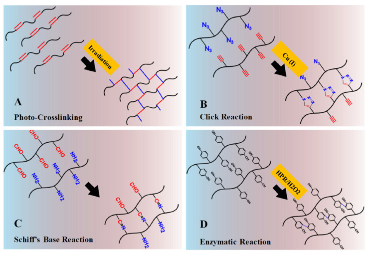
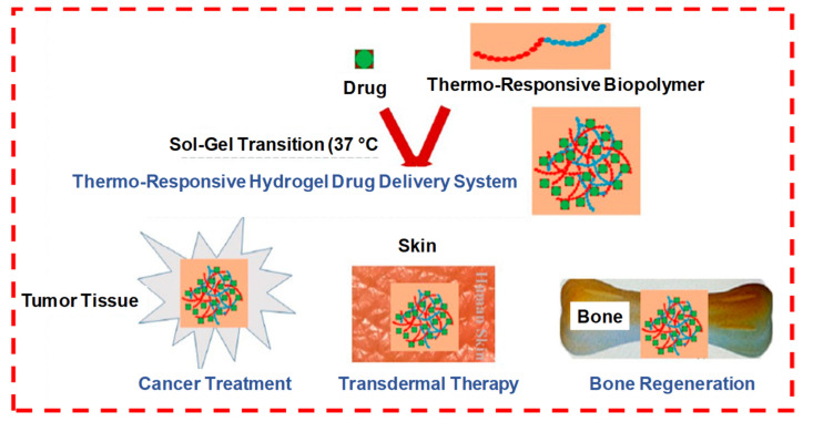
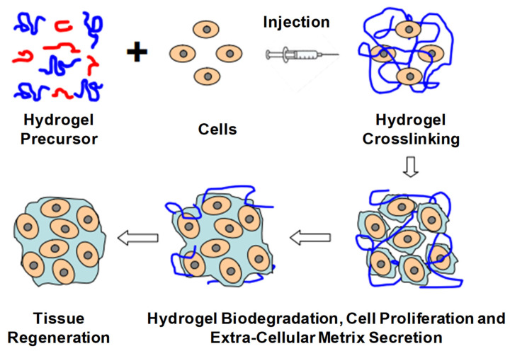
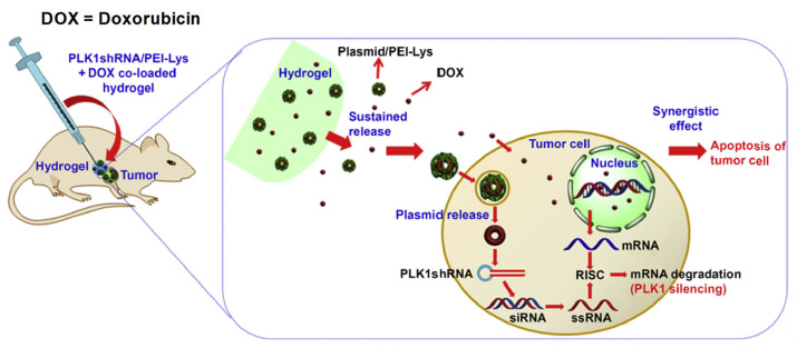
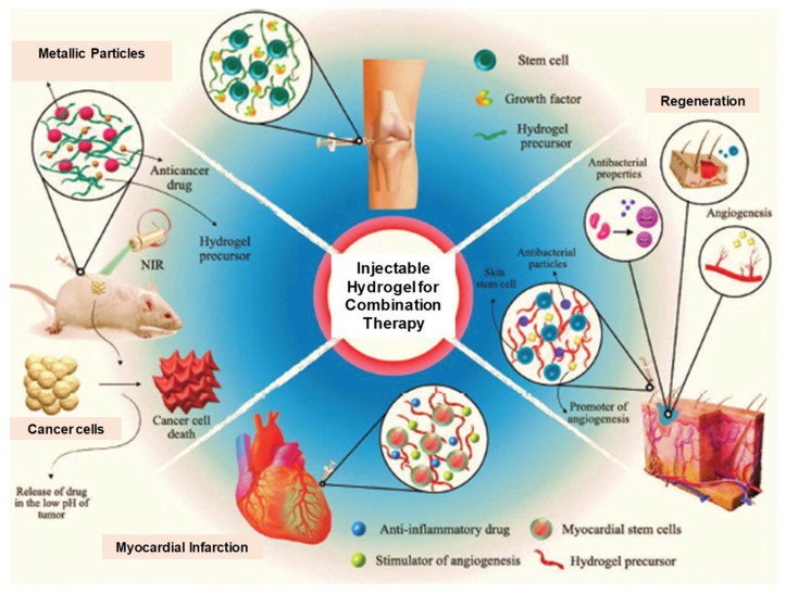
Injectable hydrogels (IHs) are smart biomaterials and are the most widely investigated and versatile technologies, which can be either implanted or inserted into living bodies with minimal invasion. Their unique features, tunable structure and stimuli-responsive biodegradation properties make these IHs promising in many biomedical applications, including tissue engineering, regenerative medicines, implants, drug/protein/gene delivery, cancer treatment, aesthetic corrections and spinal fusions. In this review, we comprehensively analyze the current development of several important types of IHs, including all those that have received FDA approval, are under clinical trials or are available commercially on the market. We also analyze the structural chemistry, synthesis, bonding, chemical/physical crosslinking and responsive release in association with current prospective research. Finally, we also review IHs' associated future prospects, hurdles, limitations and challenges in their development, fabrication, synthesis, in situ applications and regulatory affairs.

Keywords: biodegradable polymers; chemical and physical crosslinking; injectable hydrogels; tissue engineering.


Conflict of interest statement

The authors declare no conflict of interest.

Figures

Figure 1
Figure 1
(A) Swelling ability of hydrogels from lyophilized state. (B) Example of a gelation time measurement by inverted vial method. (C) Cross-section SEM images of freeze-dried injectable hyaluronic acid hydrogel. (D) Physical interactions and chemical linkages of the chemical structure of an injectable hydrogel. Adapted with permission from ref. [9]. Copyright © 2021 Polymer MDPI.
Figure 2
Figure 2
Graphical illustration depicting the design and fabrication of injectable hydrogel for photothermal antitumor therapy and following wound repair and regeneration. Adapted with permission from ref. [10]. Copyright © 2020 Theranostics.
Figure 3
Figure 3
(A) Mechanism of a spontaneous thermos gelling of the appropriate block copolymers in water via the formation of a ‘‘micelle-network’’. For simplicity, a micelle is denoted by a circle, although a micelle owns the core-corona structure and is deformable. (a) The aqueous system takes on a sol-like suspension at a low temperature; (b) the micelles are aggregated into a percolated micelle network in which each micelle is still intact, but micelle aggregation occurs due to the hydrophobic interaction between micelles, and the solvent loses flowability, leading to the so-called sol–gel transition; (c) the micelle network is coarsened until the mesh size is in the order of the visible light wavelength, and the gel is thus opaque; (d) the micelle structure is destroyed due to over-hydrophobicity of the sample at higher temperatures, eventually leading to macroscopic precipitation. (B) A schematic illustration of an injectable hydrogel system exemplified by a physical thermos-gelling material. T gel is the sol–gel transition temperature. The polymers could be dissolved in water to form a sol at low temperatures. Bioactive molecules or cells indicated by the dots in the lower-left image can be incorporated by simple mixing with sols. The sols are injectable, and in situ gelling takes place after injection if the gelling temperature is lower than the body temperature T body. As a result, the encapsulation of drugs or cells and the implantation of biomaterial are carried out with minimal surgical invasiveness. (C) A global observation of a physical gel formed underneath the skin of a rat. The image was taken 21 days after subcutaneous injection of an aqueous solution of PLGA–PEG–PLGA copolymer. The gel region is emphasized by the dashed line. Adapted with permission from ref. [11]. Copyright © 2008 Chemical Society Review. (D) Injectable hydrogel for stem cell therapy. Adapted with permission from ref. [15]. Copyright © 2019 The Royal Society of Chemistry.
Figure 4
Figure 4
Fabrication of injectable hydrogels. Reprinted with permission from ref. [9]. Copyright © 2021 Polymer MDPI.
Figure 5
Figure 5
Polymers and crosslinking physico-chemistry. Reprinted with permission from ref. [22]. Copyright © 2019 Biotechnology advances Elsevier.
Figure 6
Figure 6
Schematic illustrations of chemical crosslinking mechanism: (A) Gelation by photo-crosslinking reaction of vinyl groups bearing polymers. (B) Gelation by alkyne-azide click reaction with Cu (I) as catalyst. (C) Gelation by Schiff’s base reaction between aldehyde and amine groups. (D) Gelation by enzymatic reaction with horseradish peroxidase and hydrogen peroxide as catalyst system. Adapted with permission from ref. [8]. Copyright © 2015 European Polymer Journal.
Figure 7
Figure 7
Schematic thiol-based Michael reaction between vinyl and thiol groups mechanism. Adapted with permission from ref. [8]. Copyright © 2015 European Polymer Journal.
Figure 8
Figure 8
Physically crosslinking stimuli sensitive to hydrogels. Reprinted with permission from ref. [30]. Copyright © 2019 European polymer journal, MDPI.
Figure 9
Figure 9
Schematic physical crosslinking mechanism. (A) Gelation driven by shifting of hydrophobic interaction under the change of temperature. (B) Gelation driven by pH-inducing protonation–deprotonation transition of cationic hydrogel. (C) Gelation by ionic interaction. (D) Gelation by guest–host inclusion complexation between cyclodextrin groups and adamantine groups or PEG chains. Reprinted with permission from ref. [8]. Copyright © 2015 European Polymer Journal.
Figure 10
Figure 10
Mechanism of gelation by stereo-complexation between D-lactide- and L-lactide-bearing polymers. Adapted with permission from ref. [8]. Copyright © 2015 European Polymer Journal.
Figure 11
Figure 11
Crosslinking strategies for forming injectable double network hydrogels. (A) Supramolecular and covalently crosslinked hyaluronic acid hydrogels: by increasing the guest−host concentration or the chemical cross-linker concentration, gel modulus is enhanced. (B) Ionically and covalently crosslinked PVA−CPBA hydrogels: by increasing the concentration of Ca2+ ions, gel modulus is enhanced. n.d. means no significant difference. Adapted with permission from ref. [34]. Copyright © 2020 ACS Applied Polymer material.
Figure 12
Figure 12
Crosslinking strategies for forming injectable double network hydrogels. (A Hydrophobically associated and covalently crosslinked POEGMA hydrogels: by increasing the concentration of the hydrophobic co-monomer, gel modulus is increased. (B) Dual covalently crosslinked hydrogels composed of aldehyde/hydrazide functionalized PNIPAM and thiol/maleimide functionalized PVP: the presence of both kinetically orthogonal crosslinking groups results in enhanced mechanics compared with either single network alone. Adapted with permission from ref. [34]. Copyright © 2020 ACS Applied Polymer material.
Figure 13
Figure 13
Mechanism of action of injectable hydrogel systems. Adapted with permission from ref. [36]. Copyright © 2017 International Journal of Biological Macromolecules, Elsevier B.V.
Figure 14
Figure 14
The formation of a drug-loaded biopolymer-based thermo-responsive hydrogel system via in situ gel formation and its biomedical applications, including cancer treatment, transdermal therapy and bone regeneration. Adapted with permission from ref. [5]. Copyright © 2020 Polymers MDPI.
Figure 15
Figure 15
The sol–gel transition of LCST-type thermo-responsive polymer-based drug delivery system. Formulation over LCST changes to hydrogel from the solution state. LCST-type thermo-responsive polymer in solution forms micelles at low concentration, which further aggregates at high polymer concentration to form gel at a temperature ≥LCST. Adapted with permission from ref. [5]. Copyright © 2021 Polymer MDPI.
Figure 16
Figure 16
Schematic illustration of biodegradable injectable hydrogel for tissue regeneration approaches. Cells are isolated from a small biopsy, expanded in vitro and encapsulated in hydrogel precursors, which are subsequently transplanted into the patient by injection using a needle. Hydrogels provide initial structural support and retain cells in the defective area for cell growth, metabolism and new extracellular matrix (ECM) synthesis. The hydrogel is readily degradable when the cells secrete ECM. This strategy enables the clinician to transplant the cell, growth factor and hydrogel combination in a minimally invasive manner. Adapted with permission from ref. [6]. Copyright © 2010 Material by MDPI.
Figure 17
Figure 17
Schematic illustration of Uox-loaded PEG-PAEU/HSA hydrogels and their controlled release of Uox for degradation of uric acid crystals in hyperuricemia mice models. Reprinted with permission from ref. [57]. Copyright © 2017 from Journal of Control Release, Elsevier.
Figure 18
Figure 18
Schematic illustration for the synergistic effect of PLK1shRNA/PEI-Lys and DOX co-loaded hydrogel on a tumor in nude mice. Reprinted with permission from ref. [58]. Copyright © 2014 from Biomaterial, Elsevier.
Figure 19
Figure 19
Schematic diagram showing injectable and hGH-loaded PNLG-co-PBLG-b-PEG-b-PBLG-co-PNLG hydrogels (a polypeptide copolymer) and their application for protein delivery. The polypeptide copolymers exist as a sol in the syringe and are transformed to a gel after subcutaneous administration. Reprinted with permission from ref. [7]. Copyright © 2021 Journal of Control Release, Elsevier.
Figure 20
Figure 20
Injectable thermosensitive hydrogels for cancer vaccines. Step 1: Sustainable release of GM-CSF from the injectable thermosensitive mPEG- PLGA hydrogels recruits host dendritic cells (DCs) to the site of administration. Step 2: Viral or nonviral vectors carrying immunogens can be delivered in situ to the resident DCs in hydrogels to enhance antigen uptake efficiency, thereby improving anticancer immunity. Reprinted with permission from ref. [63]. Copyright © 2014 Biomacromolecules, American Chemical Society.
Figure 21
Figure 21
Depiction of in situ gel formation process and its sustained release of drugs from the hydrogel into tumor cells. Reprinted with permission from ref. [67]. Copyright © 2020 from Drug delivery, Taylor and Francis.
Figure 22
Figure 22
Schematic diagram showing hydrogel formation. (a) Photograph of excised tumors after mice were euthanized on the 21st day. (b) Changes in relative tumor volume. Reprinted with permission from ref. [36]. Copyright © 2020 Copyright © 2017, International Journal of Biological Macromolecules, Elsevier.
Figure 23
Figure 23
Schematic concept of sol-to-gel phase transition of polyplex-loaded PEGPSMEU copolymers sols, subcutaneous administration, controlled released via diffusion, effective absorption onto skin and wound healing. Reprinted with permission from ref. [7]. Copyright © 2020 from Journal of Controlled Release, Elsevier.
Figure 24
Figure 24
Illustration of the potential application of multifunctional IHs for cancer multi-therapy as well as regeneration after tissue damage. Reprinted with permission from ref. [42]. Copyright © 2020 Advance Health Care Materials, Wiley-VCH GmbH.
Figure 25
Figure 25
Schematic illustration of recruitment of host cells into PCLA-b-PEG-b-PCLA/BSA hydrogels and subsequent activation in response to DNA vaccine-bearing polyplexes released from these hydrogels. Reprinted with permission from ref. [7]. Copyright © 2020 from Journal of Controlled Release, Elsevier.

References

    1. Lee J.H. Injectable hydrogels delivering therapeutic agents for disease treatment and tissue engineering. Biomater. Res. 2018;22:27. doi: 10.1186/s40824-018-0138-6. - DOI - PMC - PubMed
    1. Chao Y., Chen Q., Liu Z. Smart Injectable Hydrogels for Cancer Immunotherapy. Adv. Funct. Mater. 2020;30:1902785. doi: 10.1002/adfm.201902785. - DOI
    1. Wang S., Zheng H., Zhou L., Cheng F., Liu Z., Zhang H., Wang L., Zhang Q. Nanoenzyme-Reinforced Injectable Hydrogel for Healing Diabetic Wounds Infected with Multi-Drug Resistant Bacteria. Nano Lett. 2020;20:5149–5158. doi: 10.1021/acs.nanolett.0c01371. - DOI - PubMed
    1. Liu S., Guo R., Li C., Lu C., Yang G., Wang F., Nie J., Ma C., Gao M. POSS hybrid hydrogels: A brief review of synthesis, properties and applications. Eur. Polym. J. 2021;143:110180. doi: 10.1016/j.eurpolymj.2020.110180. - DOI
    1. Chatterjee S., Hui P.C.L. Review of applications and future prospects of stimuli-responsive hydrogel based on thermo-responsive biopolymers in drug delivery systems. Polymers. 2021;13:2086. doi: 10.3390/polym13132086. - DOI - PMC - PubMed

LinkOut - more resources