Review
. 2022 Aug:43:101739.
doi: 10.1016/j.suronc.2022.101739. Epub 2022 Mar 18.

The importance of MRI for rectal cancer evaluation

Affiliations
Review

The importance of MRI for rectal cancer evaluation

Maria Clara Fernandes et al. Surg Oncol. 2022 Aug.

Abstract

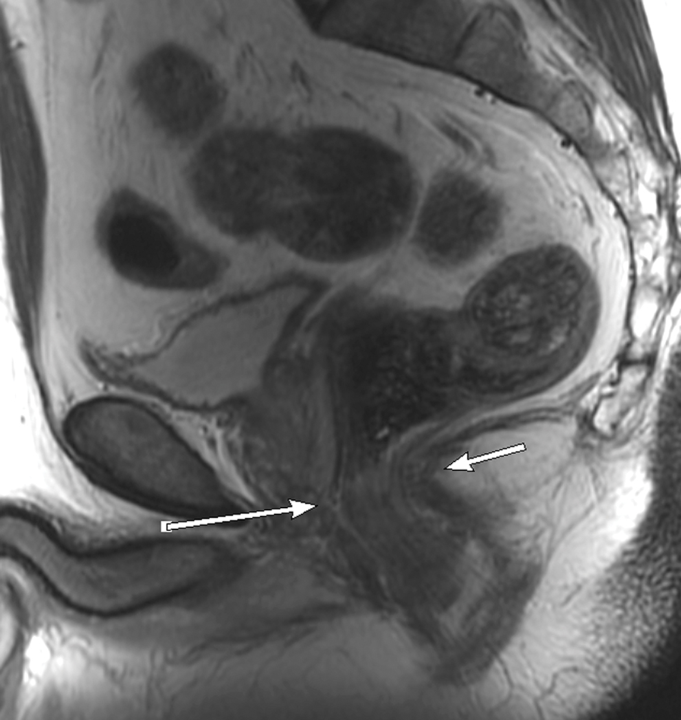
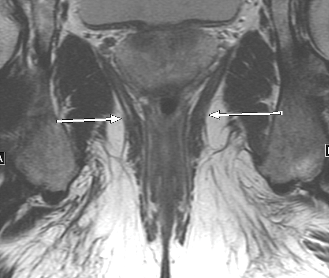
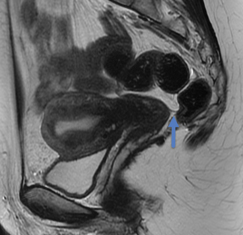
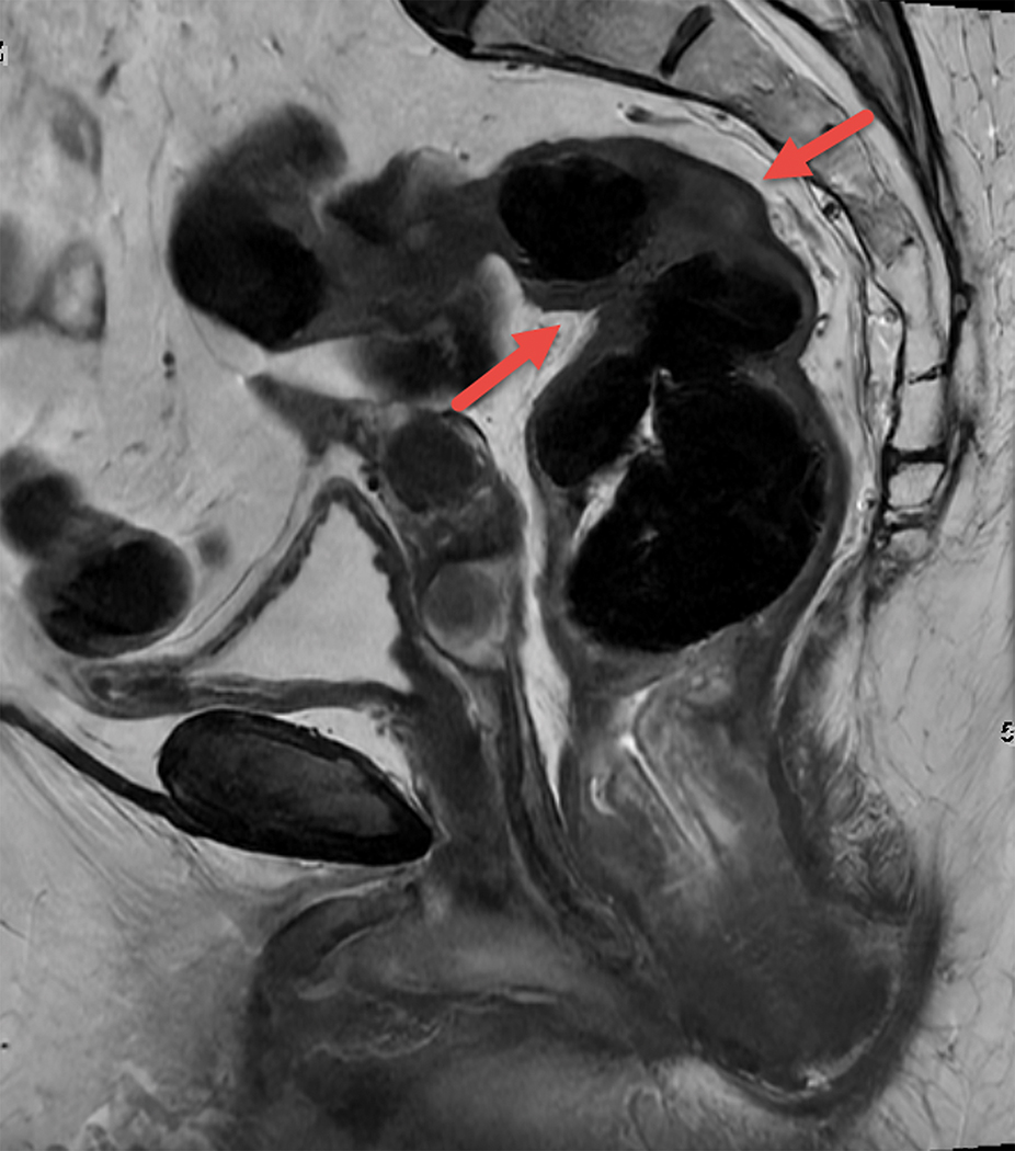
Magnetic resonance imaging (MRI) has gained increasing importance in the management of rectal cancer over the last two decades. The role of MRI in patients with rectal cancer has expanded beyond the tumor-node-metastasis (TNM) system in both staging and restaging scenarios and has contributed to identifying "high" and "low" risk features that can be used to tailor and personalize patient treatment; for instance, selecting the patients for neoadjuvant chemoradiation (NCRT) before the total mesorectal excision (TME) surgery based on risk of recurrence. Among those features, the status of the circumferential resection margin (CRM), extramural vascular invasion (EMVI), and tumor deposits (TD) have stood out. Moreover, MRI also has played a role in surgical planning, especially when the tumor is located in the low rectum, when the relationship between tumor and the anal canal is important to choose the best surgical approach, and in cases of locally advanced or recurrent tumors invading adjacent pelvic organs that may require more complex surgeries such as pelvic exenteration. As approaches using organ preservation emerge, including transanal local excision and "watch-and-wait", MRI may help in the patient selection for those treatments, follow up, and detection of tumor regrowth. Additionally, potential MRI-based prognostic and predictive biomarkers, such as quantitative and semi-quantitative metrics derived from functional sequences like diffusion-weighted imaging (DWI) and dynamic contrast-enhanced (DCE), and radiomics, are under investigation. This review provides an overview of the current role of MRI in rectal cancer in staging and restaging and highlights the main areas under investigation and future perspectives.

Keywords: Magnetic resonance imaging; Rectal cancer.


Figures

Figure 1.
Figure 1.
Sagittal and coronal T2W MR pelvis images through the pelvic floor in a 52-year-old male with rectal cancer. Arrows indicate top of puborectalis/pubococcygeus muscles denoting the anorectal junction or “ring”.
Figure 1.
Figure 1.
Sagittal and coronal T2W MR pelvis images through the pelvic floor in a 52-year-old male with rectal cancer. Arrows indicate top of puborectalis/pubococcygeus muscles denoting the anorectal junction or “ring”.
Figure 2.
Figure 2.
Sagittal T2W MR of the pelvis in a 33-year-old female with rectal cancer. Arrow indicates the thin black line, the Anterior peritoneal reflection, inserting onto the rectum at the cul-de-sac.
Figure 3.
Figure 3.
Sagittal T2W MRI of pelvis in a 73-year-old old female with rectal cancer. Arrows indicate the acute forward angulation of the rectum (sigmoid takeoff) to become the horizontally oriented sigmoid colon.
Figure 4.
Figure 4.
Axial (A) and (B) and Coronal (B) T2W pelvic MRI in 59-year-old male with rectal cancer after treatment. The levator ani muscles comprise the puborectalis (inner thin arrow in (A), the pubococcygeus muscle (outer thin arrow in (A), and the iliococcygeus muscle (small arrows in (C). The anal sphincter muscles include external (black arrow (B)) and the internal (white arrow in (B)). Note the scar in the tumor bed at 6 pm in (A).
Figure 4.
Figure 4.
Axial (A) and (B) and Coronal (B) T2W pelvic MRI in 59-year-old male with rectal cancer after treatment. The levator ani muscles comprise the puborectalis (inner thin arrow in (A), the pubococcygeus muscle (outer thin arrow in (A), and the iliococcygeus muscle (small arrows in (C). The anal sphincter muscles include external (black arrow (B)) and the internal (white arrow in (B)). Note the scar in the tumor bed at 6 pm in (A).
Figure 5.
Figure 5.
Axial T2W pelvic MRI in a 79-year-old female who underwent rectal filling. A sessile tumor between 12–2 PM is either T1 or T2 – but careful scrutiny of the muscularis propria shows its full preservation at the epicenter of the lesion indicating greater certainty of mrT1 stage and potentially amenable to local excision by TEM or TAMIS.
Figure 6.
Figure 6.
Axial T2W pelvic MR in an 87-year-old female with T3 rectal cancer deeply penetrating wall at 5 pm position and mostly consisting of mucin.
Figure 7.
Figure 7.
Axial T2W pelvic MR in middle aged male patient with T3 rectal cancer. On the left with < than 5mm of penetration from 6–9 pm and on the right, in a 48-year-old female with > 5mm at 10pm.
Figure 7.
Figure 7.
Axial T2W pelvic MR in middle aged male patient with T3 rectal cancer. On the left with < than 5mm of penetration from 6–9 pm and on the right, in a 48-year-old female with > 5mm at 10pm.
Figure 8.
Figure 8.
Gross pathologic specimen from TME surgery; anterior and lateral. The blue line demarcates the peritoneal attachment which runs obliquely cephalad on the lateral Rectum. The lower, redder rectum is surrounded by the thin shiny intact mesorectal fascia. Superiorly, the fat of the sigmoid mesocolon is noted. Courtesy of Dr Jinru Shia Attending Pathologist Memorial Sloan Kettering Cancer Center.
Figure 9.
Figure 9.
Coronal T2W pelvic MR in 37-year-old female with rectal cancer and left-sided EMVI
Figure 10.
Figure 10.
Sagittal T2W shows a posterior mesorectal irregular nodule arising from and interrupting the superior rectal vessel consistent with tumor deposit.
Figure 11.
Figure 11.
T2W axial imaging. Note the markedly high signal of the anterior rectal tumor.
Figure 12.
Figure 12.
Restaging MRI in a patient with complete response after chemoradiation. (A) Axial T2W, (B) DWI, and (C) ADC map shows a T2W dark scar in the left lateral rectal wall with no focal restriction diffusion.
Figure 12.
Figure 12.
Restaging MRI in a patient with complete response after chemoradiation. (A) Axial T2W, (B) DWI, and (C) ADC map shows a T2W dark scar in the left lateral rectal wall with no focal restriction diffusion.
Figure 12.
Figure 12.
Restaging MRI in a patient with complete response after chemoradiation. (A) Axial T2W, (B) DWI, and (C) ADC map shows a T2W dark scar in the left lateral rectal wall with no focal restriction diffusion.
Figure 13.
Figure 13.
Coronal T2W pelvic MRI (A) pre and (B) post treatment in a 45 yr. old male with rectal cancer. The left sided EMVI is smaller and T2 darker indicating at least partial regression.

References

    1. Sung H, Ferlay J, Siegel RL, Laversanne M, Soerjomataram I, Jemal A, Bray F, Global Cancer Statistics 2020: GLOBOCAN Estimates of Incidence and Mortality Worldwide for 36 Cancers in 185 Countries, CA Cancer J Clin 71(3) (2021) 209–249. - PubMed
    1. Brown G, Davies S, Williams GT, Bourne MW, Newcombe RG, Radcliffe AG, Blethyn J, Dallimore NS, Rees BI, Phillips CJ, Maughan TS, Effectiveness of preoperative staging in rectal cancer: digital rectal examination, endoluminal ultrasound or magnetic resonance imaging?, Br J Cancer 91(1) (2004) 23–9. - PMC - PubMed
    1. Akasu T, Kondo H, Moriya Y, Sugihara K, Gotoda T, Fujita S, Muto T, Kakizoe T, Endorectal ultrasonography and treatment of early stage rectal cancer, World J Surg 24(9) (2000) 1061–8. - PubMed
    1. Vliegen R, Dresen R, Beets G, Daniels-Gooszen A, Kessels A, van Engelshoven J, Beets-Tan R, The accuracy of Multi-detector row CT for the assessment of tumor invasion of the mesorectal fascia in primary rectal cancer, Abdom Imaging 33(5) (2008) 604–10. - PMC - PubMed
    1. Maizlin ZV, Brown JA, So G, Brown C, Phang TP, Walker ML, Kirby JM, Vora P, Tiwari P, Can CT replace MRI in preoperative assessment of the circumferential resection margin in rectal cancer?, Dis Colon Rectum 53(3) (2010) 308–14. - PubMed