. 2022 Jun;101(6):1116-1125.
doi: 10.1016/j.kint.2022.03.011. Epub 2022 Mar 23.

Integrated single-cell sequencing and histopathological analyses reveal diverse injury and repair responses in a participant with acute kidney injury: a clinical-molecular-pathologic correlation

Collaborators, Affiliations

Integrated single-cell sequencing and histopathological analyses reveal diverse injury and repair responses in a participant with acute kidney injury: a clinical-molecular-pathologic correlation

Rajasree Menon et al. Kidney Int. 2022 Jun.
No abstract available

Keywords: AKI; Kidney Precision Medicine Project; NSAID; kidney; single-cell sequencing.


Conflict of interest statement

DISCLOSURE

All the authors declared no competing interests.

Figures

Figure 1 |
Figure 1 |. Clinical course of acute kidney injury (AKI) and diversity in healthy and injury states and markers revealed by single-nucleus (sn) and single-cell (sc) RNA sequencing (RNAseq).
(a) Periodic serum creatinine levels of the acute kidney injury (AKI) study participant from hospital day 1 up to 2 years’ follow-up. (b) Uniform Manifold Approximation and Projection (UMAP) showing cell clusters from the combined analysis of >400,000 nuclei/cells (after quality filtering) from reference and diseased tissues for the Kidney Precision Medicine Project (KPMP) cell atlas study. UMAP is a dimension reduction technique that can be used for visualization. (c) UMAP showing the cellular diversity in the AKI study participant (in red, 11,379 cells) projected on the kidney disease cell atlas space (gray). (d) Proportion of each cell-type cluster in sc and sn datasets from the AKI study participant. (e) Cell states including adaptive-epithelial (adaptive-epi), adaptive-stromal (adaptive-str), degenerative, cycling, and reference, found in the cell clusters in the AKI study participant. (f) Proportion of the different cell states in the 13 AKI biopsy samples from the KPMP cell atlas study. The study participant is marked by a rectangular box. (g) Dot plots showing the expression of selected key injury markers in the different cell states in the AKI study participant. The dot plot enables the visualization of the expression pattern of a gene across different cell clusters. The size of the dot encodes the percentage of cells within a cluster, whereas the color encodes the average expression level across all cells within a cluster. ATL, ascending thin limb of LOH; CNT, connecting tubule; DCT, distal convoluted tubule; DTL, descending thin limb of LOH; EC, endothelial cells; FIB, fibroblasts; IC, intercalated cells; IMM, immune cells; LOH, loop of Henle; NEU, neural cells; PapE, papillary tip epithelial cells; PC, principal cells; PEC, parietal epithelial cells; POD, podocyte; PT, proximal tubule; TAL, thick ascending limb of LOH; VSM/P, vascular smooth muscle cells/pericyte.
Figure 2 |
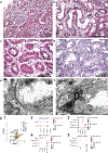
Figure 2 |. Proximal tubule (PT) epithelial cell and podocyte injury and dysfunction.
(a) Low power view showing diffuse interstitial edema and irregular cortical tubular profiles. A glomerulus is unremarkable (hematoxylin and eosin [H&E], original magnification ×200). (b) PTs show focal severe acute injury with coarse clear cytoplasmic vacuolization, reduced cell height, focal simplification of the lining epithelium, and shedding of cytoplasmic fragments into the tubular lumen (arrow; H&E, original magnification ×400). (c) An injured PT at the center shows a mitotic figure in telophase with cytokinesis, giving rise to 2 daughter epithelial cells in situ (arrow; H&E, original magnification ×600). (d) In areas, the pattern of proximal tubular vacuolization forms elongated vertically oriented spaces that evoke widening of basolateral canaliculi (arrows), which represent an extracellular space. There is variable attenuation and loss of brush border (periodic acid-Schiff, original magnification ×600). (e) A PT shows heterogeneous cellular injury. There is focal loss of nuclei with adherence of thin remnants of basal cytoplasm (arrow). Other tubular cells show various stages of detachment and desquamation of brush border or apical cytoplasmic fragments. There is a marked reduction in organelles, and the few residual mitochondria are swollen (electron micrograph, original magnification ×3000). (f) A PT shows focal cytoplasmic condensation appearing “dark” or “mummified” due to loss of cell water with incipient nuclear apoptosis (arrow; electron micrograph, original magnification ×3000). (g) Uniform Manifold Approximation and Projection (UMAP) and dot plots. UMAP showing the different proximal and podocyte (POD) cell types identified from the single-cell/single-nucleus analyses of the acute kidney injury study participant (g1). Dot plots showing podocyte (NPHS1, CLIC5, and PTPRO [g2]), injury (SPP1, HAVCR1, and TMSB10) (g3), cell cycle (TOP2A, MKI67, and PCNA) (g4), and apoptosis (TNF, TNFRSF8) markers (g5). For reference, SLC34A1, a marker of PT cells, is included in injury and cycling dot plots for comparison across different states. To optimize viewing of this image, please see the online version of this article at www.kidney-international.org.
Figure 3 |
Figure 3 |. Molecular and morphologic diversity in the injured distal nephron, collecting tubule, and interstitial and endothelial cells (ECs).
(a) Distal tubules display individual epithelial cell apoptosis and shedding of degenerating cells into the lumen (arrows; hematoxylin and eosin [H&E], original magnification ×600). (b) In the outer medulla, a thick ascending limb of the distal tubule at top left has cytoplasmic vacuolization. Cytoplasmic vacuoles and dilated organelles are also seen involving a collecting duct at right, with injury to both the rounded intercalated cells (ICs; thick arrow) and the thinner principal cells (PCs; thin arrow), (electron micrograph, original magnification ×3000). (c) Distal tubules at the center appear simplified with apical displacement of nuclei. Some proximal tubular epithelial cells are multilayered and multinucleated, suggesting regeneration (arrow). There is prominent interstitial edema with a delicate network of interstitial collagen fibers (periodic acid-Schiff [PAS], original magnification ×600). (d) The interstitium contains plump fibroblasts (FIB) associated with increased loose collagen deposition. There is acute injury of adjacent proximal and distal tubules, including a thick ascending limb with intraluminal Tamm-Horsfall protein (PAS, ×400). (e) Surrounding an injured distal tubule with apical epithelial blebbing, the interstitial capillaries appear dilated with endothelial shedding (arrows; H&E, original magnification ×400). (f) The cortical interstitium contains extravasation of Tamm-Horsfall protein composed of PAS-positive fibrillar material surrounded by a mild mononuclear inflammatory infiltrate and interstitial edema. There is no detectable tubulitis involving the adjacent tubules (PAS, original magnification ×600). (g) Dot plot showing the scaled expression of collagens (COL1A1, COL4A1, and COL4A2), mitochondrial genes (MT-ND1, MT-ND2, and MT-ND3), injury (EGF, LCN2, and S100A6), and repair markers (PROM1 and CD24) in cells of distal nephron, collecting tubule, and interstitium representing different cell states. Rectangular boxes are drawn for degenerative and adaptive cell clusters. (h) Uniform Manifold Approximation and Projection (UMAP) showing the immune cell types identified in the biopsy of the participant. (i) Dot plot showing high expression of endothelial dysfunction markers, MYL9, MYLK2, and TRPM2, in the degenerative ECs compared with other EC types. TRPM2 is shown in a separate dot plot as the scale of this endothelial injury marker is different from MYL9 and MYLK2. (j) Dot plot showing high expression of genes involved in nonsteroidal anti-inflammatory drug metabolism, UGT1A7, UGT1A9, and PTGS1, in the altered cell states compared with reference cells in proximal and distal nephron cells. B, B cell; cDC, classical dendritic cells; CNT, connecting tubule; cycMNP, cycling myeloid cells; cycNKC/T, cycling NKC/T cells; DCT, distal convoluted tubule; MACM2, macrophage M2; MDC, monocyte derived cells; N, neutrophil; ncMON, nonclassical monocyte; NKC, natural killer cells; pDC, plasmacytoid dendritic cells; PL, plasma cells; T, T cell; TAL, thick ascending limb of loop of Henle. To optimize viewing of this image, please see the online version of this article at www.kidney-international.org.
Figure 4 |
Figure 4 |. Biological pathways and genes associated with altered cell states.
(a) Kidney specific functional modules and the associated significantly enriched biological processes that were selected based on supporting pathologic findings. The functional modules (M1–M6) were generated using the HumanBase online resource. Genes that were significantly upregulated in altered cells compared with reference or healthy cells in the combined kidney single-cell/single-nucleus data from the acute kidney injury study participant were used for this analysis. The arrows link the morphology with the functional modules. The pathologic images used in this panel are: (a1) at a focus of tubular basement membrane rupture, lining cuboidal epithelial cells become elongated and appear to migrate out of the tubular confines and integrate into the interstitium (Jones methenamine silver [JMS], original magnification ×600). (a2) A high-power view of a proximal tubule (PT) shows severe mitochondrial injury with partial or complete loss of cristae and focal breaks of the outer mitochondrial membrane (electron micrograph, original magnification ×20,000). (a3) A distal tubule in the center of the field has incipient cellular cast formation by ejected apoptotic cells attached by thin cytoplasmic strands to the originating epithelial lining. Some regions of epithelium are multilayered as possible regenerative response. A pocket of interstitial leukocytes and edema forms an inflammatory niche. There is no associated tubulitis (hematoxylin and eosin, original magnification ×600). (a4) A markedly dilated interstitial capillary containing red blood cells shows focal shedding of its lining endothelial cells into the lumen, leaving denuded segments of interstitial capillary basement membrane (JMS, original magnification ×600). (a5) Tubular basement membrane is focally detached from the tubular epithelium with areas of tubular basement membrane thinning, lamellation, and remodeling (JMS, original magnification ×600). (b) Dot plot showing the expression of mesenchymal markers, COL4A1, VIM, and ZEB1 (transcription factor suggestive of mesenchymal transition), in PT epithelial cell states. (c) Expression of VCAM1, PROM1 in all PT epithelial cells categorized into the indicated cell states. (d) Proportion of cells expressing PROM1 only, VCAM1 only, PROM1 and VCAM1 double positive, and PROM1 and VCAM1 negative in PT epithelial cells. (e) Dot plot showing the expression of myeloid regulated adaptive repair genes, CSF1 and CSF1R, in PT and immune cells. To optimize viewing of this image, please see the online version of this article at www.kidney-international.org.

References

    1. de Boer IH, Alpers CE, Azeloglu EU, et al. Rationale and design of the Kidney Precision Medicine Project. Kidney Int. 2021;99:498–510. - PMC - PubMed
    1. Tuttle KR, Bebiak J, Brown K, et al. Patient perspectives and involvement in precision medicine research. Kidney Int. 2021;99:511–514. - PubMed
    1. Lake BB, Menon R, Winfree S, et al. An atlas of healthy and injured cell states and niches in the human kidney. bioRxiv. Published online July 29, 2021. 10.1101/2021.07.28.454201 - DOI - PMC - PubMed
    1. El-Achkar TM, Eadon MT, Menon R, et al. A multimodal and integrated approach to interrogate human kidney biopsies with rigor and reproducibility: guidelines from the Kidney Precision Medicine Project. Physiol Genomics. 2021;53:1–11. - PMC - PubMed
    1. Sakaguchi K, Green M, Stock N, et al. Glucuronidation of carboxylic acid containing compounds by UDP-glucuronosyltransferase isoforms. Arch Biochem Biophys. 2004;424:219–225. - PubMed

LinkOut - more resources