. 2022 Jun:130:105192.
doi: 10.1016/j.jmbbm.2022.105192. Epub 2022 Mar 23.

Characterization of scar tissue biomechanics during adult murine flexor tendon healing

Affiliations

Characterization of scar tissue biomechanics during adult murine flexor tendon healing

Antonion Korcari et al. J Mech Behav Biomed Mater. 2022 Jun.

Abstract

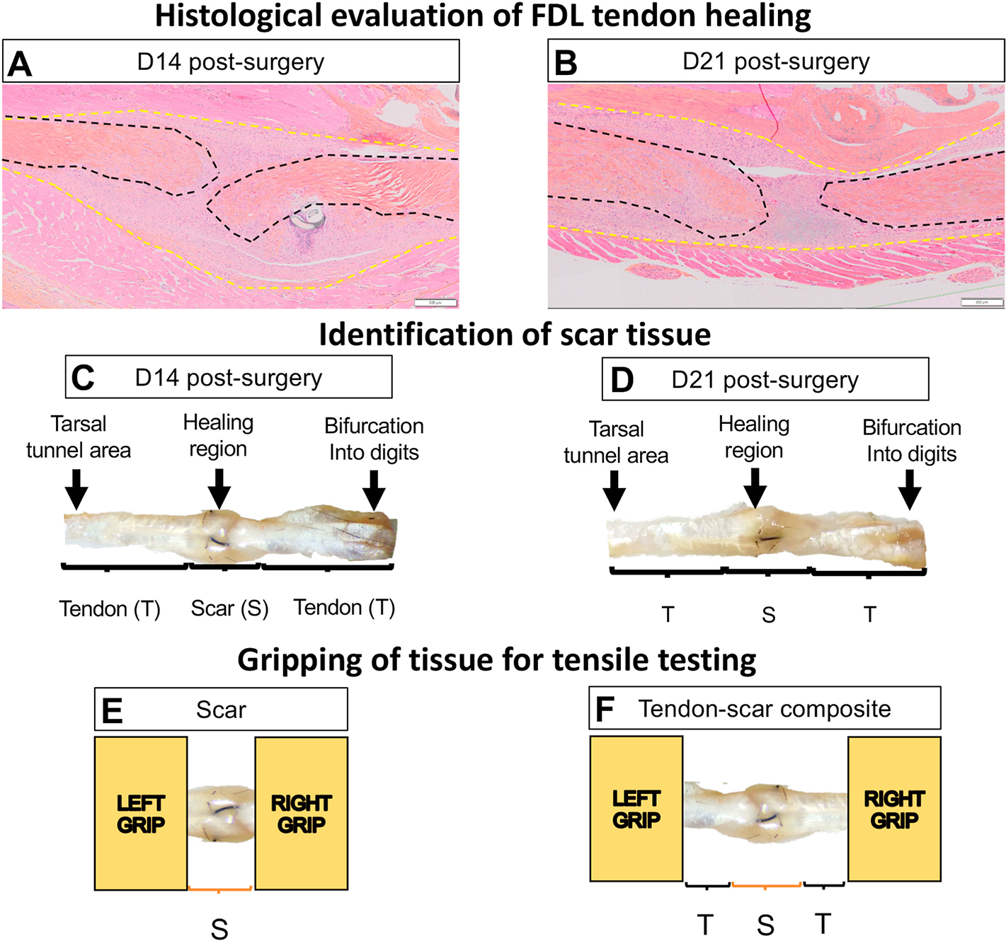
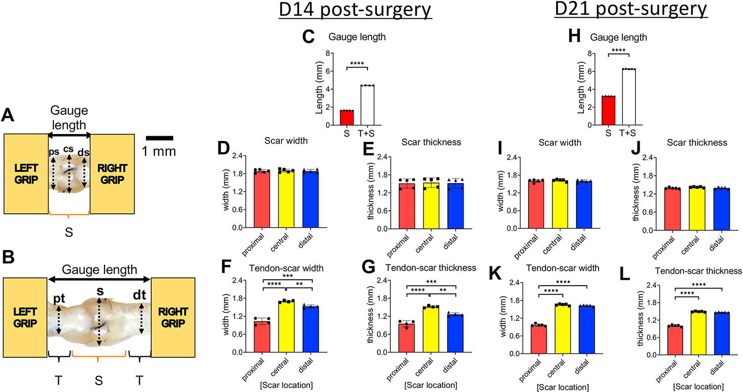
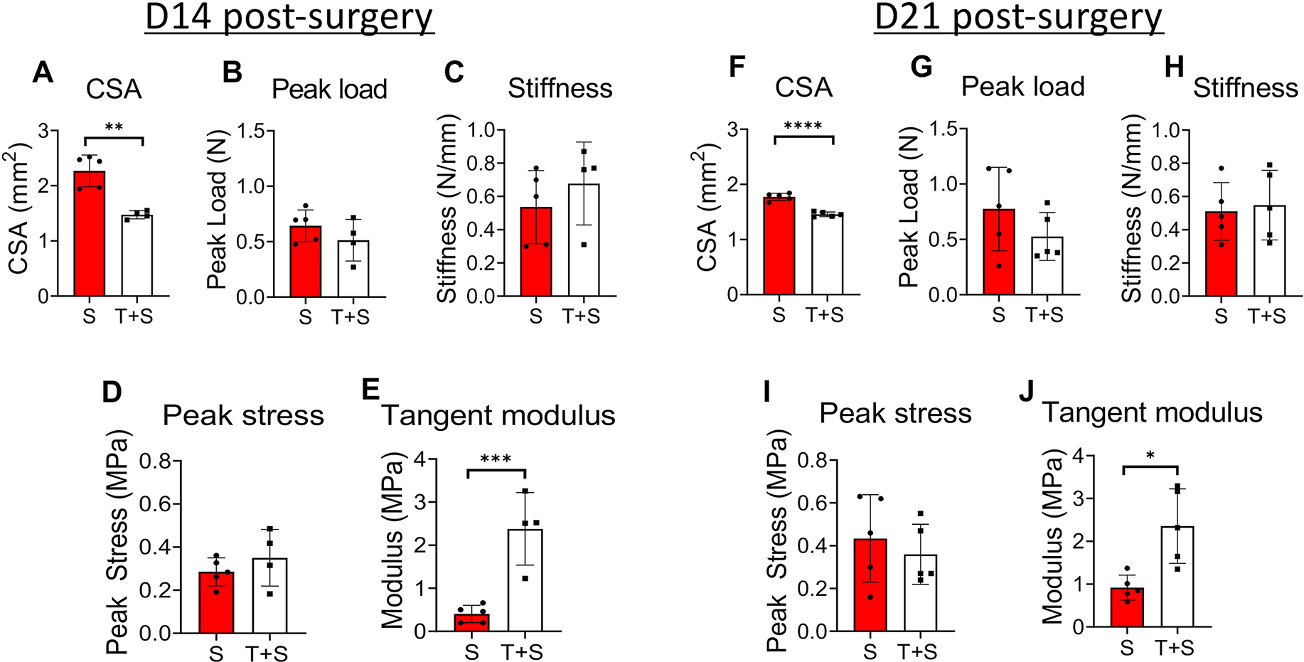
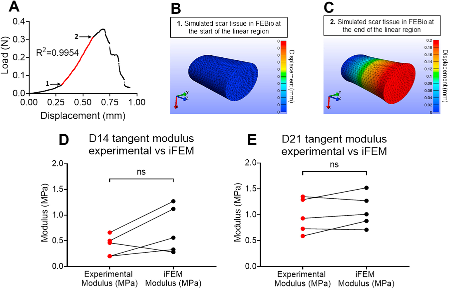
Tendon injuries are very common and result in significant impairments in mobility and quality of life. During healing, tendons produce a scar at the injury site, characterized by abundant and disorganized extracellular matrix and by permanent deficits in mechanical integrity compared to healthy tendon. Although a significant amount of work has been done to understand the healing process of tendons and to develop potential therapeutics for tendon regeneration, there is still a significant gap in terms of assessing the direct effects of therapeutics on the functional and material quality specifically of the scar tissue, and thus, on the overall tendon healing process. In this study, we focused on characterizing the mechanical properties of only the scar tissue in flexor digitorum longus (FDL) tendons during the proliferative and early remodeling healing phases and comparing these properties with the mechanical properties of the composite healing tissue. Our method was sensitive enough to identify significant differences in structural and material properties between the scar and tendon-scar composite tissues. To account for possible inaccuracies due to the small aspect ratio of scar tissue, we also applied inverse finite element analysis (iFEA) to compute mechanical properties based on simulated tests with accurate specimen geometries and boundary conditions. We found that the scar tissue linear tangent moduli calculated from iFEA were not significantly different from those calculated experimentally at all healing timepoints, validating our experimental findings, and suggesting the assumptions in our experimental calculations were accurate. Taken together, this study first demonstrates that due to the presence of uninjured stubs, testing composite healing tendons without isolating the scar tissue overestimates the material properties of the scar itself. Second, our scar isolation method promises to enable more direct assessment of how different treatment regimens (e.g., cellular ablation, biomechanical and/or biochemical stimuli, tissue engineered scaffolds) affect scar tissue function and material quality in multiple different types of tendons.

Keywords: Biomechanics; Function; Regenerative healing; Scar tissue; Tendon injury.


Conflict of interest statement

All authors declare no competing interests in this manuscript

Figures

Figure 1.
Figure 1.. FDL tendon scar and tendon-scar composite tissues can be successfully identified, gripped, and prepared for uniaxial tensile testing.
A. ABHOG staining on FDL tendons at D14 post-surgery. B. ABHOG staining on FDL tendons at D21 post-surgery. C. A representative harvested healing tendon at D14 post-surgery. D. A representative harvested healing tendon at D21 post-surgery. The scar tissue can be easily identified approximately in the middle of the tissue (healing region). The increase of the width and thickness in the healing region due to the scar formation is evident. The anatomical markers used to harvest each tissue are the region where the tendon bifurcates into digits and the region where the tendon goes through the tarsal tunnel area. After dissection under the microscope, (E) only the scar tissue or (F) a composite of tendon-scar-tendon tissue was gripped on each end using sandpaper and superglue and prepared for uniaxial tensile testing. * The black dashed lines indicate each tendon stub, while the yellow dashed lines indicate the total scar tissue.
Figure 2.
Figure 2.. Quantification of dimensions (gauge length, width, thickness) of the scar and tendon-scar composite tissues prior to mechanical testing.
A. The gauge length of the scar tissue and the three sub-equal regions, proximal scar (ps), central scar (cs), and distal scar (ds), used to quantify the width and the thickness of the scar tissue. B. The gauge length of the tendon-scar composite tissue and the three sub-equal regions, proximal tendon (pt), scar (s), distal tendon (dt), used to quantify the width and the thickness of the composite tissue. C. Gauge length between the scar and tendon-scar composite tissue at D14 post-surgery. D. The width of the scar tissue at D14 post-surgery. E. The thickness of the scar tissue at D14 post-surgery. F. The width of the tendon-scar composite tissue at D14 post-surgery. G. The thickness of the tendon-scar composite tissue at D14 post-surgery. H. Gauge length between the scar and tendon-scar composite tissue at D21 post-surgery I. The width of the scar tissue at D21 post-surgery J. The thickness of the scar tissue at D21 post-surgery. K. The width of the tendon-scar composite tissue at D21 post-surgery. L. The thickness of the tendon-scar composite tissue at D21 post-surgery. N=4–5 per group. Statistical significance between regions was determined using a one-way ANOVA followed by a Tukey’s post-hoc analysis. ** indicative of p < 0.01; *** indicative of p < 0.001; **** indicative of p < 0.0001
Figure 3.
Figure 3.. The scar tissue has significantly different cross-sectional area (CSA) and material quality compared to the tendon-scar composite tissue at both 14 and 21 days post-surgery.
A. The CSA of the scar (S) tissue is significantly higher compared to the tendon-scar composite (T+S) tissue at 14 days post-surgery. B. The peak load between the S and T+S tissues is similar at 14 days post-surgery. C. The stiffness between the S and T+S tissues is similar at 14 days post-surgery. D. The peak stress between the S and the T+S tissues is similar at 14 days post-surgery. E. The tangent modulus of the S tissue is significantly lower compared to the T+S tissue at 14 days post-surgery. F. The CSA of the S tissue is significantly higher compared to the T+S tissue at 21 days post-surgery. G. The peak load between the S and T+S tissues is similar at 21 days post-surgery. H. The stiffness between the S and T+S tissues is similar at 21 days post-surgery. N=4–5 per group. Statistical significance between groups was determined using Student’s t-test. I. The peak stress between the scar S and the T+S tissues is similar at 21 days post-surgery. J. The tangent modulus of the S tissue is significantly lower compared to the T+S tissue at 21 days post-surgery. N=4–5 per group. Statistical significance between regions was determined using unpaired Student t-test. * indicative of p < 0.05; *** indicative of p < 0.001.
Figure 4.
Figure 4.. Validation of experimentally calculated scar tissue tangent moduli using a Finite Elements Analysis (FEA) modeling approach.
A. Representative load-displacement graph of a scar tissue where the linear region was identified, values of force and displacement for the start and endpoints of the linear region were recorded and used to simulate the linear region of a displacement-controlled uniaxial tensile stretch in FEBio. B. A representative simulated scar tissue in FEBio right at the beginning of the linear region. C. A representative simulated scar tissue in FEBio after the simulated uniaxial stretching right at the end of the linear region. D. The tangent moduli calculated using the experimental data (Experimental Modulus) versus FEBio (iFEM Modulus) at D14 post-surgery were found not to be significantly different from each other. E. The tangent moduli calculated using the experimental data (Experimental Modulus) versus FEBio (iFEM Modulus) at D21 post-surgery were found not to be significantly different from each other. N=5 per group. Statistical significance between regions was determined using Student t-test. ns indicative of not statistically significant.

Similar articles

Cited by

References

    1. McCormick A, Charlton J & Fleming D Assessing health needs in primary care. Morbidity study from general practice provides another source of information. Bmj 310, 1534, doi:10.1136/bmj.310.6993.1534d (1995). - DOI - PMC - PubMed
    1. Korcari A, Nichols AE, O’Neil M & Loiselle AE Ligament and tendon tissue engineering. Musculoskeletal Tissue Engineering, 81 (2021).
    1. Pennisi E Tending tender tendons. Science 295, 1011, doi:10.1126/science.295.5557.1011 (2002). - DOI - PubMed
    1. Nichols AEC, Best KT & Loiselle AE The cellular basis of fibrotic tendon healing: challenges and opportunities. Transl Res 209, 156–168, doi:10.1016/j.trsl.2019.02.002 (2019). - DOI - PMC - PubMed
    1. Tempfer H & Traweger A Tendon Vasculature in Health and Disease. Front Physiol 6, 330, doi:10.3389/fphys.2015.00330 (2015). - DOI - PMC - PubMed

Publication types