

Challenges and opportunities of integrating imaging and mathematical modelling to interrogate biological processes
- PMID: 35339913
- PMCID: PMC9693675
- DOI: 10.1016/j.biocel.2022.106195
Challenges and opportunities of integrating imaging and mathematical modelling to interrogate biological processes
Abstract
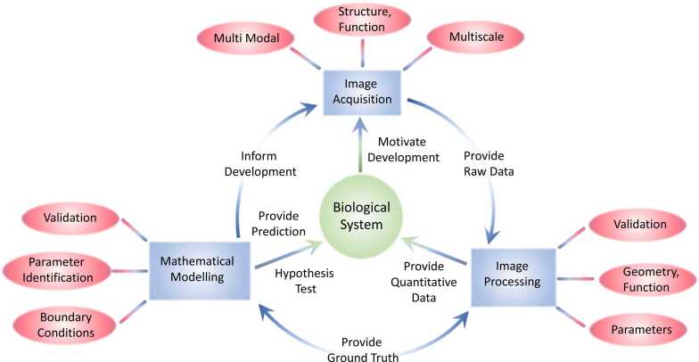
Advances in biological imaging have accelerated our understanding of human physiology in both health and disease. As these advances have developed, the opportunities gained by integrating with cutting-edge mathematical models have become apparent yet remain challenging. Combined imaging-modelling approaches provide unprecedented opportunity to correlate data on tissue architecture and function, across length and time scales, to better understand the mechanisms that underpin fundamental biology and also to inform clinical decisions. Here we discuss the opportunities and challenges of such approaches, providing literature examples across a range of organ systems. Given the breadth of the field we focus on the intersection of continuum modelling and in vivo imaging applied to the vasculature and blood flow, though our rationale and conclusions extend widely. We propose three key research pillars (image acquisition, image processing, mathematical modelling) and present their respective advances as well as future opportunity via better integration. Multidisciplinary efforts that develop imaging and modelling tools concurrently, and share them open-source with the research community, provide exciting opportunity for advancing these fields.
Keywords: Biophysical modelling; Multiscale; Validation.
Copyright © 2022 Elsevier Ltd. All rights reserved.
Conflict of interest statement
The authors have no competing interests to declare.
Figures
References
-
- Abdeladim L., Matho K.S., Clavreul S., Mahou P., Sintes J.-M., Solinas X., Arganda-Carreras I., Turney S.G., Lichtman J.W., Chessel A., Bemelmans A.-P., Loulier K., Supatto W., Livet J., Beaurepaire E. Multicolor multiscale brain imaging with chromatic multiphoton serial microscopy. Nat. Commun. 2019;10(1):1662. - PMC - PubMed
-
- Berg M., Davit Y., Quintard M., Lorthois S. Modelling solute transport in the brain microcirculation: is it really well mixed inside the blood vessels? J. Fluid Mech. 2020;884:A39.
-
- Bernabeu M.O., Köry J., Grogan J.A., Markelc B., Beardo A., d’Avezac M., Enjalbert R., Kaeppler J., Daly N., Hetherington J., Krüger T., Maini P.K., Pitt-Francis J.M., Muschel R.J., Alarcón T., Byrne H.M. Abnormal morphology biases hematocrit distribution in tumor vasculature and contributes to heterogeneity in tissue oxygenation. Proc. Natl. Acad. Sci. U.S.A. 2020;117(45):27811–27819. - PMC - PubMed
-
- Chakraborty T., Driscoll M.K., Jeffery E., Murphy M.M., Roudot P., Chang B.-J., Vora S., Wong W.M., Nielson C.D., Zhang H., Zhemkov V., Hiremath C., De La Cruz E.D., Yi Y., Bezprozvanny I., Zhao H., Tomer R., Heintzmann R., Meeks J.P., Marciano D.K., Morrison S.J., Danuser G., Dean K.M., Fiolka R. Light-sheet microscopy of cleared tissues with isotropic, subcellular resolution. Nat. Methods. 2019;16(11):1109–1113. - PMC - PubMed
Publication types
MeSH terms
Grants and funding
LinkOut - more resources
Full Text Sources
