. 2021 Jun;101(6):408-421.
doi: 10.1124/molpharm.121.000471. Epub 2022 Mar 26.

Novel Small Molecule Fibroblast Growth Factor 23 Inhibitors Increase Serum Phosphate and Improve Skeletal Abnormalities in Hyp Mice

Affiliations

Novel Small Molecule Fibroblast Growth Factor 23 Inhibitors Increase Serum Phosphate and Improve Skeletal Abnormalities in Hyp Mice

Zhousheng Xiao et al. Mol Pharmacol. 2021 Jun.

Abstract

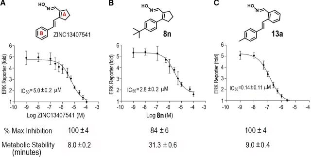
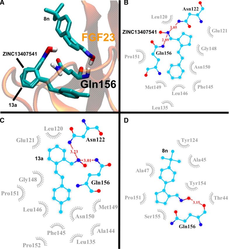
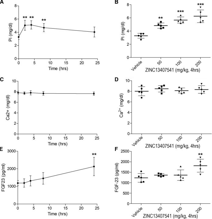
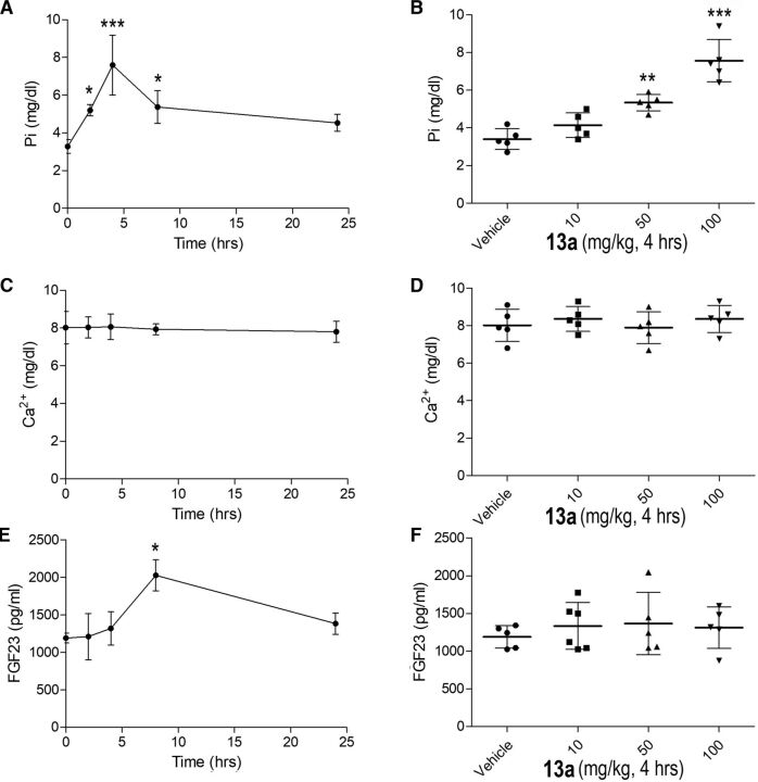
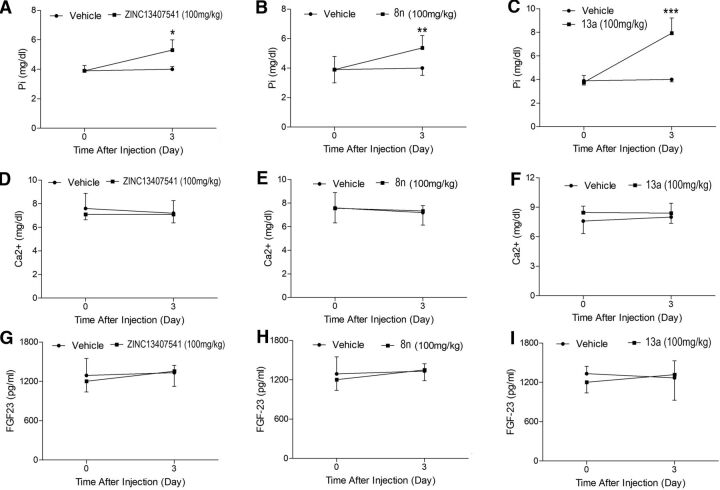
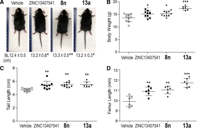
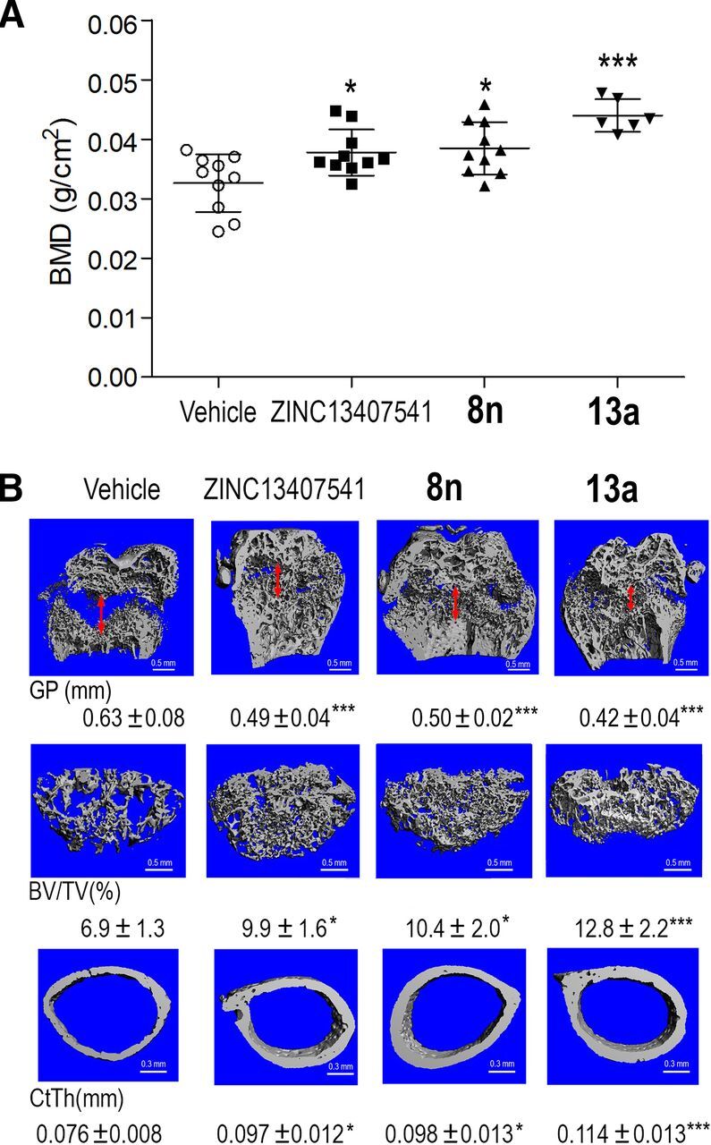
Excess fibroblast growth factor (FGF) 23 causes hereditary hypophosphatemic rickets, such as X-linked hypophosphatemia (XLH) and tumor-induced osteomalacia (TIO). A small molecule that specifically binds to FGF23 to prevent activation of the fibroblast growth factor receptor/α-Klotho complex has potential advantages over the currently approved systemically administered FGF23 blocking antibody. Using structure-based drug design, we previously identified ZINC13407541 (N-[[2-(2-phenylethenyl)cyclopenten-1-yl]methylidene]hydroxylamine) as a small molecule antagonist for FGF23. Additional structure-activity studies developed a series of ZINC13407541 analogs with enhanced drug-like properties. In this study, we tested in a preclinical Hyp mouse homolog of XLH a direct connect analog [(E)-2-(4-(tert-butyl)phenyl)cyclopent-1-ene-1-carbaldehyde oxime] (8n), which exhibited the greatest stability in microsomal assays, and [(E)-2-((E)-4-methylstyryl)benzaldehyde oxime] (13a), which exhibited increased in vitro potency. Using cryo-electron microscopy structure and computational docking, we identified a key binding residue (Q156) of the FGF23 antagonists, ZINC13407541, and its analogs (8n and 13a) in the N-terminal domain of FGF23 protein. Site-directed mutagenesis and bimolecular fluorescence complementation-fluorescence resonance energy transfer assay confirmed the binding site of these three antagonists. We found that pharmacological inhibition of FGF23 with either of these compounds blocked FGF23 signaling and increased serum phosphate and 1,25-dihydroxyvitamin D [1,25(OH)2D] concentrations in Hyp mice. Long-term parenteral treatment with 8n or 13a also enhanced linear bone growth, increased mineralization of bone, and narrowed the growth plate in Hyp mice. The more potent 13a compound had greater therapeutic effects in Hyp mice. Further optimization of these FGF23 inhibitors may lead to versatile drugs to treat excess FGF23-mediated disorders. SIGNIFICANCE STATEMENT: This study used structure-based drug design and medicinal chemistry approaches to identify and optimize small molecules with different stability and potency, which antagonize excessive actions of fibroblast growth factor 23 (FGF23) in hereditary hypophosphatemic rickets. The findings confirmed that these antagonists bind to the N-terminus of FGF23 to inhibit its binding to and activation of the fibroblast growth factor receptors/α-Klotho signaling complex. Administration of these lead compounds improved phosphate homeostasis and abnormal skeletal phenotypes in a preclinical Hyp mouse model.


Figures

Fig. 1.
Fig. 1.
Structure, in vitro efficacy, and metabolic stability of ZINC13407541 (A) and its analogs 8n (B) and 13a (C). IC50 values were extracted from three independent concentration-response experiments examining the compound-dependent inhibition of FGF23-induced ERK reporter activities in HEK293T cells expressing human α-KL after application of ZINC13407541 (n = 3 cells) and its analogs 8n (n = 3 cells) and 13a (n = 3 cells). Maximum percent inhibition values were calculated from the plateaus in inhibition at the highest inhibitor concentration (n = 3 cells for each compound). Metabolic stabilities were expressed as the half-life of the parent compound remaining after an incubation with human liver microsomes (n = 3 liver microsomes for each compound).
Fig. 2.
Fig. 2.
The computationally predicted interaction of ZINC13407541 and its two analogs, 13a and 8n, with Gln156, the potential binding site, in the N-terminal domain of FGF23 (PDB code: 5W21) shown in (A) 3D structure; (B–D) two-dimensional residue-contacting map for ZINC13407541, 13a, and 8n, respectively. Hydrogen bonds are shown in red dash lines with donor-acceptor distances in Å. Hydrophobic interactions are shown in gray. The corresponding estimated free energies of binding (ΔG) for the three poses are shown in Table 2.
Fig. 3.
Fig. 3.
Effects of ZINC13407541 and its analogs on CFP-FGF23WT or CFP-FGF23Q156A induced BiFC-based FRET ratio and ERK reporter activities. (A) Diagrams of fusion constructs for human CFP-FGF23WT, CFP-FGF23Q156A, α-KL-VN155, and FGFR1-VC155. (B) BiFC-based YFP/CFP ratio (n = 3 cells). (C) ERK reporter activities (n = 3 cells). (D) Computational model of FGF23 antagonist that targets FGF23/FGFR1/α-Klotho complex. Data are expressed as the mean ± S.D. from three independent experiments. *, **, and *** indicate statistically significant difference from vehicle control group. P values were determined by one-way ANOVA with Tukey’s multiple-comparisons test.
Fig. 4.
Fig. 4.
Time- and dose-dependent effects of ZINC13407541 on mineral ion homeostasis and FGF23 levels in Hyp mice. In left panels (A), (C), and (E), time-course assessments of serum phosphate, calcium, and FGF23 levels in Hyp mice that were given a single i.p. injection of ZINC13407541 (100mg/kg) during 24 hours. In right panels (B), (D), and (F), dose-response studies of serum phosphate, calcium, and FGF23 levels in Hyp mice that were given a single i.p. injection of ZINC13407541 (50, 100, and 200 mg/kg) after 4 hours. Data are expressed as the mean ± S.D. from serum samples of individual mice (n = 5). *, **, and *** indicate statistically significant difference from vehicle control group. P values were determined by 1-way ANOVA with Dunnett's test.
Fig. 5.
Fig. 5.
Time- and dose-dependent effects of 13a on mineral ion homeostasis and FGF23 levels in Hyp mice. In left panels (A), (C), and (E), time-course assessments of serum phosphate, calcium, and FGF23 levels in Hyp mice that were given a single i.p. injection of 13a (100mg/kg) during 24 hours. In right panels (B), (D), and (F), dose-response studies of serum phosphate, calcium, and FGF23 levels in Hyp mice that were given a single i.p. injection of 13a (10, 50, and 100 mg/kg) after 4 hours. Data are expressed as the mean ± S.D. from serum samples of individual mice (n = 5). *, **, and *** indicate statistically significant difference from vehicle control group. P values were determined by one-way ANOVA with Dunnett's test.
Fig. 6.
Fig. 6.
Short-term effects of ZINC13407541, 8n, and 13a on mineral ion homeostasis and FGF23 levels in Hyp mice. (A–C) Serum phosphate. (D–F) Serum calcium. (G–I) Serum FGF23. Data are expressed as the mean ± S.D. from serum samples of individual mice (n = 5). *, **, and *** indicate statistically significant difference from vehicle control group. P values were determined by two-way ANOVA with Bonferroni post hoc test.
Fig. 7.
Fig. 7.
Long-term effects of ZINC13407541, 8n, and 13a on skeletal phenotypes in Hyp mice. (A) body length (n = 6–10). (B) body weight (n = 6–10). (C) tail length (n = 6–10). (D) femur length (n = 6). Data are expressed as the mean ± S.D. from serum samples of individual mice. *, **, and *** indicate statistically significant difference from vehicle control group. P values were determined by one-way ANOVA with Tukey’s multiple-comparisons test.
Fig. 8.
Fig. 8.
Long-term effects of ZINC13407541, 8n, and 13a on bone mineral density and bone structure in Hyp mice. (A) BMD (n = 6–10). (B) Micro-CT 3D images, including width of the growth plate (GP, double red arrow, n = 6), femoral bone volume (BV/TV, n = 6), and cortical thickness (Ct.Th, n = 6). Data are expressed as the mean ± S.D. from serum samples of individual mice (n = 5). *, **, and *** indicate statistically significant difference from vehicle control group. P values were determined by one-way ANOVA with Tukey’s multiple-comparisons test.

Similar articles

Cited by

References

    1. Andrukhova O, Slavic S, Smorodchenko A, Zeitz U, Shalhoub V, Lanske B, Pohl EE, Erben RG (2014) FGF23 regulates renal sodium handling and blood pressure. EMBO Mol Med 6:744–759 DOI: 10.1002/emmm.201303716. - DOI - PMC - PubMed
    1. Arango Sancho P (2020) Complications of phosphate and vitamin D treatment in X-linked hypophosphataemia. Adv Ther 37 (Suppl 2):105–112 DOI: 10.1007/s12325-019-01170-7. - DOI - PubMed
    1. Ashkenazy H, Abadi S, Martz E, Chay O, Mayrose I, Pupko T, Ben-Tal N (2016) ConSurf 2016: an improved methodology to estimate and visualize evolutionary conservation in macromolecules. Nucleic Acids Res 44:W344–350 DOI: 10.1093/nar/gkw408. - DOI - PMC - PubMed
    1. Carpenter TOImel EARuppe MDWeber TJKlausner MAWooddell MMKawakami TIto TZhang XHumphrey J, et al. (2014) Randomized trial of the anti-FGF23 antibody KRN23 in X-linked hypophosphatemia. J Clin Invest 124:1587–1597 DOI: 10.1172/JCI72829. - DOI - PMC - PubMed
    1. Carpenter TOWhyte MPImel EABoot AMHögler WLinglart APadidela RVan’t Hoff WMao MChen CY, et al. (2018) Burosumab therapy in children with X-linked hypophosphatemia. N Engl J Med 378:1987–1998 DOI: 10.1056/NEJMoa1714641. - DOI - PubMed

MeSH terms

LinkOut - more resources