. 2022 Mar 16:2022:5362115.
doi: 10.1155/2022/5362115. eCollection 2022.

Preserved Left Ventricular Function despite Myocardial Fibrosis and Myopathy in the Dystrophin-Deficient D2.B10-Dmd mdx /J Mouse

Affiliations

Preserved Left Ventricular Function despite Myocardial Fibrosis and Myopathy in the Dystrophin-Deficient D2.B10-Dmd mdx /J Mouse

Holly M Hayes et al. Oxid Med Cell Longev. .

Abstract

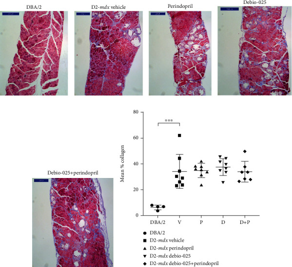
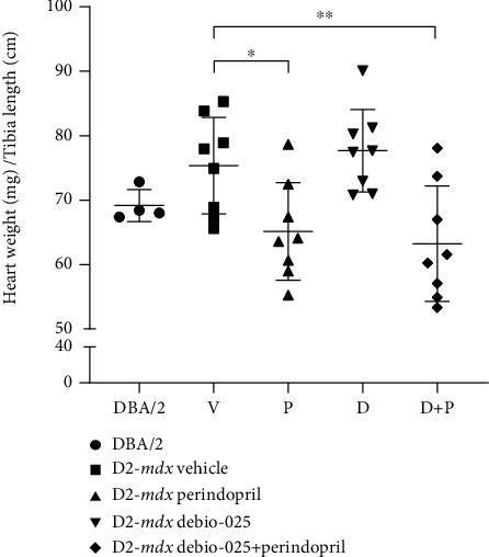
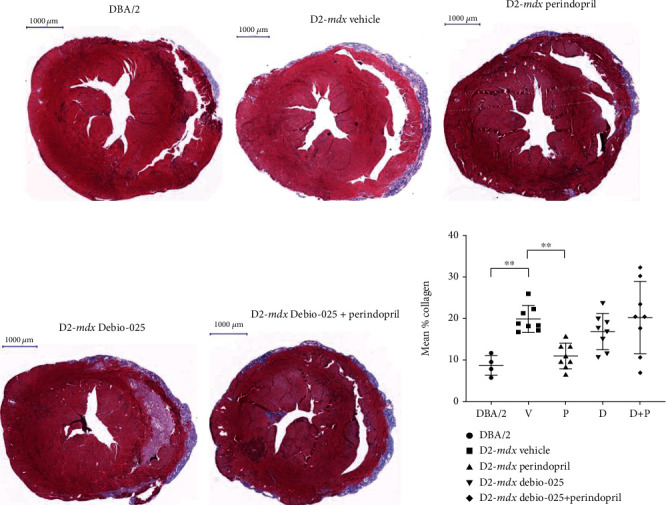
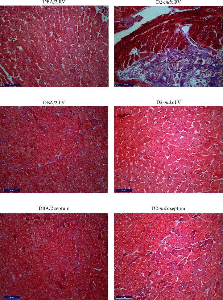
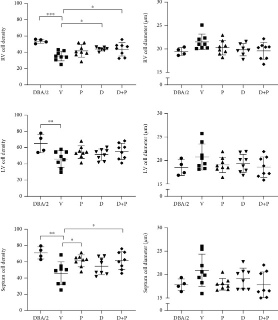
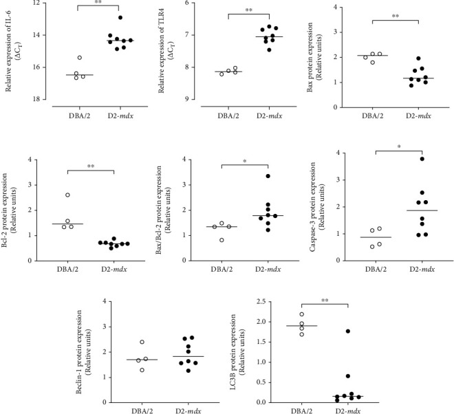
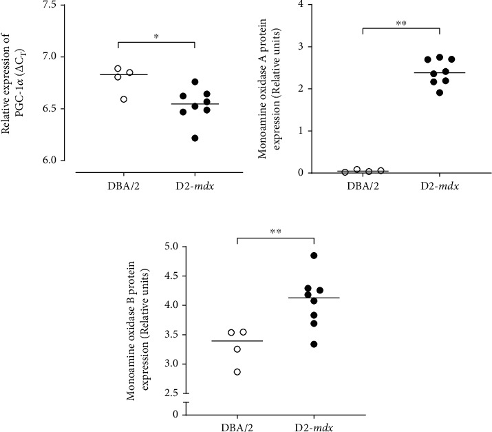
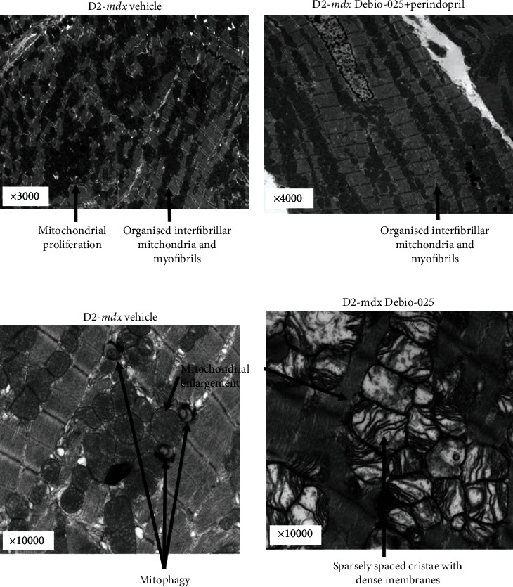
Duchenne muscular dystrophy involves an absence of dystrophin, a cytoskeletal protein which supports cell structural integrity and scaffolding for signalling molecules in myocytes. Affected individuals experience progressive muscle degeneration that leads to irreversible loss of ambulation and respiratory diaphragm function. Although clinical management has greatly advanced, heart failure due to myocardial cell loss and fibrosis remains the major cause of death. We examined cardiac morphology and function in D2.B10-Dmd mdx /J (D2-mdx) mice, a relatively new mouse model of muscular dystrophy, which we compared to their wild-type background DBA/2J mice (DBA/2). We also tested whether drug treatment with a specific blocker of mitochondrial permeability transition pore opening (Debio-025), or ACE inhibition (Perindopril), had any effect on dystrophy-related cardiomyopathy. D2-mdx mice were treated for six weeks with Vehicle control, Debio-025 (20 mg/kg/day), Perindopril (2 mg/kg/day), or a combination (n = 8/group). At 18 weeks, compared to DBA/2, D2-mdx hearts displayed greater ventricular collagen, lower cell density, greater cell diameter, and greater protein expression levels of IL-6, TLR4, BAX/Bcl2, caspase-3, PGC-1α, and notably monoamine oxidases A and B. Remarkably, these adaptations in D2-mdx mice were associated with preserved resting left ventricular function similar to DBA/2 mice. Compared to vehicle, although Perindopril partly attenuated the increase in heart weight and collagen at 18 weeks, the drug treatments had no marked impact on dystrophic cardiomyopathy.


Conflict of interest statement

All authors declare that they have no conflicts of financial interest.

Figures

Figure 1
Figure 1
Fibrosis in diaphragms of DBA/2 and D2-mdx mice at 18 weeks. Representative transverse sections of diaphragms (Masson's trichrome stain, scale bar = 200 μm) from (a) DBA/2 and D2-mdx treated to (b) Vehicle, (c) Perindopril, (d) Debio-025, and (e) Debio-025+Perindopril. Mean % collagen for each group (f). Data analysed by one-way ANOVA and Dunnett's multiple comparisons and presented as mean ± SD and individual values; ∗∗∗p < 0.001.
Figure 2
Figure 2
Heart weights (normalised to tibial length) from DBA/2 and D2-mdx mice at 18 weeks. Tibial bone lengths were similar in all animals and did not differ between groups. Data analysed by one-way ANOVA and Dunnett's multiple comparisons tests and presented as mean ± SD and individual values; p < 0.05 and ∗∗p < 0.01.
Figure 3
Figure 3
Cardiac fibrosis in DBA/2 and D2-mdx mice at 18 weeks. Representative sections of (a) DBA/2, (b) D2-mdx Vehicle, (c) D2-mdx Perindopril, (d) D2-mdx Debio-025, and (e) D2-mdx Debio-025+Perindopril mouse hearts stained with Masson's trichrome stain (scale = 1000 μm). (f) Mean % collagen for DBA/2 and D2-mdx hearts. Data analysed by one-way ANOVA and Dunnett's multiple comparisons tests and presented as mean ± SD and individual values; ∗∗p < 0.01.
Figure 4
Figure 4
Cross-sectional images from the RV, LV, and septum of DBA/2 and D2-mdx hearts at 18 weeks. Representative sections of (a) DBA/2 RV, (b) D2-mdx RV, (c) DBA/2 LV, (d) D2-mdx LV, (e) DBA/2 septum, and (f) D2-mdx septum stained with Masson's trichrome (scale bar = 50 μm).
Figure 5
Figure 5
RV, LV, and septum cell density (number of cells/area) and diameter in DBA/2 and treated D2-mdx mice at 18 weeks. The number of cells within an area of 19,500 μm2 (150 × 130 μm) for (a) RV, (c) LV, and (e) septum and average cell diameter for (b) RV, (d) LV, and (f) septum. Values represent the average of five areas of size 150 × 130 μm assessed for each tissue region. Data were analysed by nested one-way ANOVA and presented as mean ± SD and individual values; p < 0.05, ∗∗p < 0.01, and ∗∗∗p < 0.001.
Figure 6
Figure 6
Relative expression of mRNA and proteins involved in inflammation, apoptosis, and autophagy in DBA/2 and D2-mdx hearts at 18 weeks. Expression of (a) IL-6 mRNA and (b) TLR4 mRNA obtained from RT-qPCR and presented as individual ΔCT values, where a lower ΔCT denotes greater mRNA expression. Relative protein expression of (c) Bax, (d) Bcl-2, (e) Bax/Bcl-2 ratio, (f) caspase-3, (g) Beclin-1, and (h) LC3B obtained from western immunoblots. Intact western blots are shown in Supplement 1 Data analysed by Mann–Whitney U test and presented as median and individual values; p < 0.05 and ∗∗p < 0.01.
Figure 7
Figure 7
Relative expression pattern of PGC-1α gene and protein expression of markers of mitochondrial ROS signalling from monoamine oxidase in DBA/2 and D2-mdx hearts at 18 weeks. (a) Relative expression of PGC-1α obtained from RT-qPCR and presented as individual ΔCT values, where a lower ΔCT denotes greater mRNA expression. (b, c) Relative protein expression of monoamine oxidase A and monoamine oxidase B obtained from western immunoblots. Data analysed by Mann–Whitney U test and presented as median and individual values; p < 0.05 and ∗∗p < 0.01.
Figure 8
Figure 8
Representative electron micrographs of LV myocardial tissue from D2-mdx mice at 18 weeks. (a) An example from the D2-mdx Vehicle group that shows areas of randomly distributed mitochondria between poorly organised mitochondria, mitochondrial proliferation, and also some areas that are preserved (3000x mag). (b) Highly preserved regions with evenly sized and distributed mitochondria in the D2-mdx Debio-025+Perindopril group (4000x mag). (c) Examples of mitophagy and mitochondrial swelling in the D2-mdx Vehicle group (10,000x mag). (d) Examples of enlarged mitochondria with thick membranes and sparse cristae in the D2-mdx Debio-025 group (10,000x mag).

References

    1. Zimmermann J. F., Butler C. R. Diagnosis and management of Duchenne muscular dystrophy, part 1: diagnosis, and neuromuscular, rehabilitation, endocrine, and gastrointestinal and nutritional management. Lancet Neurology . 2018;17(6):p. 251. - PMC - PubMed
    1. Birnkrant D. J., Bushby K., Bann C. M., et al. Diagnosis and management of Duchenne muscular dystrophy, part 2: respiratory, cardiac, bone health, and orthopaedic management. Lancet Neurology . 2018;17(4):347–361. doi: 10.1016/S1474-4422(18)30025-5. - DOI - PMC - PubMed
    1. Fukada S., Morikawa D., Yamamoto Y., et al. Genetic background affects properties of satellite cells and _mdx_ phenotypes. The American Journal of Pathology . 2010;176(5):2414–2424. doi: 10.2353/ajpath.2010.090887. - DOI - PMC - PubMed
    1. Fairclough R. J., Wood M. J., Davies K. E. Therapy for Duchenne muscular dystrophy: renewed optimism from genetic approaches. Nature Reviews Genetics . 2013;14(6):373–378. doi: 10.1038/nrg3460. - DOI - PubMed
    1. Whitehead N. P., Yeung E. W., Allen D. G. Muscle damage in mdx (dystrophic) mice: role of calcium and reactive oxygen species. Clinical and Experimental Pharmacology & Physiology . 2006;33(7):657–662. doi: 10.1111/j.1440-1681.2006.04394.x. - DOI - PubMed