. 2022 Mar 18:2022:2513837.
doi: 10.1155/2022/2513837. eCollection 2022.

Ketone Body β-Hydroxybutyrate Prevents Myocardial Oxidative Stress in Septic Cardiomyopathy

Affiliations

Ketone Body β-Hydroxybutyrate Prevents Myocardial Oxidative Stress in Septic Cardiomyopathy

Liwei Ji et al. Oxid Med Cell Longev. .

Abstract

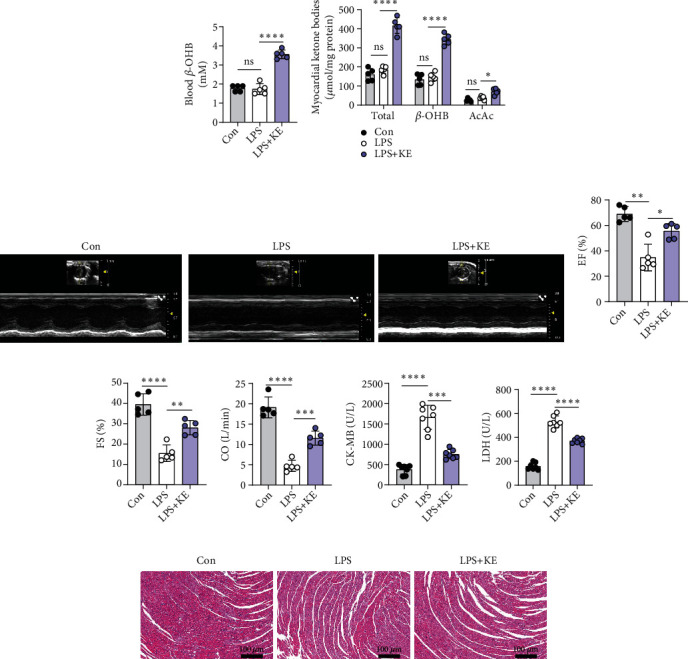
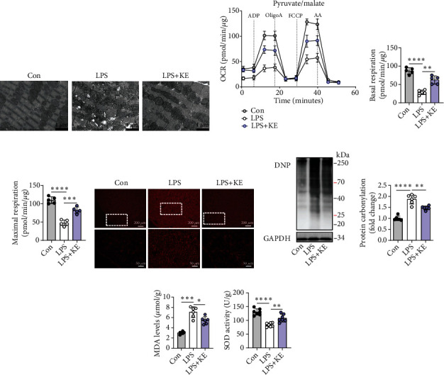
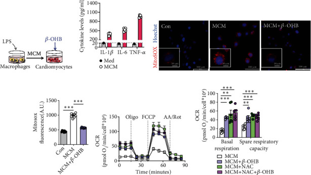
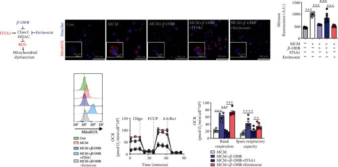
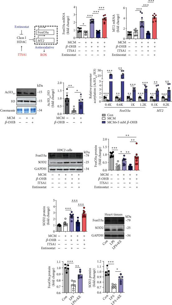
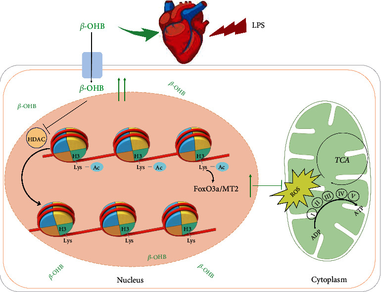
Septic cardiomyopathy is a life-threatening complication of severe sepsis and septic shock. Oxidative stress and mitochondrial dysfunction have been identified as significant abnormalities in septic cardiomyopathy. However, specific treatments are rare. This study aims to investigate the impact of β-hydroxybutyrate (β-OHB) on septic cardiomyopathy and explore the underlying mechanism(s). We found that pretreatment of D-β-hydroxybutyrate-(R)-1,3 butanediol monoester (ketone ester, 3 mg/g body weight, once daily) by gavage for three days elevated the levels of ketone bodies, especially that of β-hydroxybutyrate (β-OHB) in the circulation and mouse hearts, which exerted a protective effect against lipopolysaccharide (LPS, 20 mg/kg)-induced septic cardiomyopathy in mice. In addition, an LPS-stimulated macrophage-conditioned medium (MCM) was used to mimic the pathological process of septic cardiomyopathy. Mechanistically, β-OHB alleviated myocardial oxidative stress and improved mitochondrial respiratory function through the antioxidant FoxO3a/MT2 pathway activated via histone deacetylase (HDAC) inhibition, which ultimately enhanced heart performance in septic cardiomyopathy. Our results, therefore, suggested an unappreciated critical role of β-OHB in septic heart protection as well as highlighted the potential of β-OHB as a simple remedy for the septic cardiomyopathy population.


Conflict of interest statement

The authors declare that they have no conflicts of interest.

Figures

Figure 1
Figure 1
Ketone bodies protected the hearts against LPS-induced acute myocardial injury and cardiac dysfunction. (a). Blood β-OHB levels in Con, LPS, and LPS + KE mice (n = 5 per group). (b). The levels of ketone bodies (β-OHB; AcAc) in the Con, LPS, and LPS + KE hearts (n = 5 per group). (c). Representative echocardiography images of the Con, LPS, and LPS + KE hearts (n = 5 per group). (d–f) Cardiac function was indicated by ejection fractions (EF) (d), shortening fraction (FS) (e), and cardiac output (CO) (f) (n = 5 per group). (g and h) The levels of CK-MB (g) and LDH (h) in the plasma samples (n = 7 per group). (i) Representative images of hematoxylin and eosin- (H&E-) stained left ventricle tissue sections (n = 5 per group). Scale bar = 100 μm. Con, Control; LPS, lipopolysaccharide; and KE, ketone ester. Data are presented as the mean ± SD. Statistical comparisons were conducted by one-way ANOVA, followed by Tukey's multiple comparisons test (a, b, d, e, f, g, and h). The exact P values were reported for the indicated comparisons, and P < 0.05 was considered statistically significant. P < 0.05, ∗∗P < 0.01, ∗∗∗P < 0.001, and ∗∗∗∗P < 0.0001 for the indicated comparisons. ns: no significant difference.
Figure 2
Figure 2
Ketone bodies mitigated cardiac mitochondrial dysfunction and oxidative stress in septic cardiomyopathy. (a) Representative transmission electron microscopy images of the Con, LPS, and LPS + KE heart sections (n = 5 per group). Scale bar = 1 μm. (b–d) The oxygen consumption rate (OCR) curve in the presence of pyruvate and malate of the isolated mitochondria from the mouse hearts (b). The ADP-stimulated OCR (c) and FCCP-stimulated OCR (d) (n = 5 per group). (e) Representative images of dihydroethidium (DHE) staining in Con, LPS, and LPS + KE hearts (n = 50 fields, from 3 hearts per group). Scale bar = 200 μm or 50 μm. (f and g) Western blotting of 2,4-dinitrophenylhydrazone (DNP) in the heart tissues from Con, LPS, and LPS + KE mice (f) (n = 5 per group). The protein levels of DNP were normalized to the GAPDH levels, and the data were expressed as the fold change relative to the control (g) (n =5 per group). (h) Malondialdehyde (MDA) levels in the heart tissues (n = 5 per group). (i) Superoxide dismutase (SOD) activity in the heart tissues (n = 5 per group). Con: Control; LPS: lipopolysaccharide; KE: ketone ester; ADP: adenosine diphosphate; Oligo: oligomycin A; FCCP: trifluorocarbonyl cyanide phenylhydrazone; and AA: antimycin A. Data are presented as the mean ± SD. Statistical comparisons were conducted using one-way ANOVA, followed by Tukey's multiple comparisons test (c, d, g, h, and i). The exact P values were reported for the indicated comparisons, with P < 0.05 considered statistical significance. P < 0.05, ∗∗P < 0.01, ∗∗∗P < 0.001, and ∗∗∗∗P < 0.0001 for the indicated comparisons.
Figure 3
Figure 3
β-OHB alleviated oxidative stress and enhanced aerobic respiration in H9C2 cells exposed to MCM. (a) Schematic representation of the experimental protocol. (b) The levels of inflammatory cytokines in LPS-stimulated macrophage conditioned medium (MCM) (n = 5) and standard cell culture medium (DMEM, Med) with 10% fetal calf serum. (c). Representative fluorescent images of H9C2 cells stained with MitoSOX red after the indicated treatments. Scale bar = 100 μm. The nuclei were visualized with Hoechst staining. Scale bar = 10 μm. (d). The fluorescence intensity of MitoSOX-stained H9C2 cells after the indicated treatments (n = 6 per group). (e and f) The oxygen consumption rate (OCR) curve in the presence of pyruvate and L-glutamine in H9C2 cells after the indicated treatments (e) and the basal respiration and spare respiratory capacity (OCRFCCP–OCRbasal) (f) (n = 5–6 per group). Oligo: Oligomycin A; A A./Rot: antimycin A and rotenone. Data are presented as the mean ± SD. Statistical comparisons were conducted by one-way ANOVA, followed by Tukey's multiple comparisons test (b, d, and f). The exact P values are reported for the indicated comparisons, and P < 0.05 indicates statistical significance. ∗∗P < 0.01 and ∗∗∗P < 0.001 for the indicated comparisons.
Figure 4
Figure 4
β-OHB alleviated oxidative stress and improved mitochondrial function via HDAC inhibition in MCM-treated H9C2 cells. (a) ITSA1 (HDAC activator) and entinostat (HDAC inhibitor) were used in this study. (b) Representative fluorescent images of H9C2 cells stained with MitoSOX red to assess the mitochondrial ROS generation after the indicated treatments (n = 8 per group). Scale bar = 100 μm. The nuclei were visualized with Hoechst staining. Scale bar = 10 μm. (c). The fluorescence intensity of MitoSOX-stained H9C2 cells after the indicated treatments (n = 8 per group). (d). Representative MitoSOX flow cytometry analysis in H9C2 cells after the indicated treatments (n = 6 per group). (e and f) The oxygen consumption rate (OCR) curve in the presence of pyruvate and L-glutamine in H9C2 cells after the indicated treatments (e), and the basal respiration and spare respiratory capacity (OCRFCCP−OCRbasal) (f) (n = 4 per group). Oligo: Oligomycin A; A.A./Rot: antimycin A and rotenone. Data are presented as the mean ± SD. Statistical comparisons were conducted by one-way ANOVA, followed by Tukey's multiple comparisons test (c and f). The exact P values are reported for the indicated comparisons, and P < 0.05 indicates statistical significance. ∗∗P < 0.01 and ∗∗∗P < 0.001 for the indicated comparisons.
Figure 5
Figure 5
The ketone bodies enhanced FoxO3a and MT2 expression by promoting histone H3 lysine 9 acetylation. (a) HDAC exerts oxidative stress resistance through the FoxO3a/MT2 pathway. FoxO3a: Forkhead box O3; MT2: Metallothionein 2A. (b and c) qRT-PCR analysis of FoxOa3 (b) and MT2 (c) in H9C2 cells after the indicated treatments (n = 6–8 per group). (d and e) Western blotting of the acetylation of H3K9 in purified histones from H9C2 cells after the indicated treatments (d). The quantification of the band intensity (e). Acetylation is normalized to the total histone content H3 and reported to be relative to the MCM-exposed cells. Coomassie blue staining was used as the gel loading control (n = 5–6 per group). (f) Chromatin from H9C2 cells after the indicated treatments were immunoprecipitated with anti-histone H3 or anti-AcH3K9, and the purified DNA was analyzed with primer pairs specific for the FoxO3a or MT2 promoters. The results are shown as the ratios of AcH3K9 to total histone H3 (n = 5–6 per group). (g–i) Western blotting of FoxO3a and SOD2 in H9C2 cells after the indicated treatments (g). Quantification of the band intensity (h and i). The protein levels of FoxO3a and SOD2 were normalized to those of GAPDH, and the data are expressed as fold changes relative to the control value (n = 7–9 per group). (j–l) Western blotting of FoxO3a and SOD2 in the heart tissues from control, LPS, and LPS + KE mice (j). Quantification of the band intensity (k and l). The protein levels of FoxO3a and SOD2 were normalized to those of the GAPDH, and the data are expressed as the fold change relative to the control value (n = 5 per group). Data are presented as the mean ± SD. Statistical comparisons were conducted using one-way ANOVA, followed by Tukey's multiple comparisons test (b, c, d, e, f, h, i, k, and l). The exact P values are reported for the indicated comparisons, and P < 0.05 indicates statistical significance. P < 0.05, ∗∗P < 0.01, and ∗∗∗P < 0.001 for the indicated comparisons.
Figure 6
Figure 6
Schematic illustration of the working model for this study. β-OHB is an HDAC inhibitor that enhances histone acetylation to activate the antioxidant FoxO3a/MT2 pathway for preventing ROS production from protecting the mitochondria in septic cardiomyopathy.

Similar articles

Cited by

References

    1. Martin L., Derwall M., al Zoubi S., et al. The septic heart: current understanding of molecular mechanisms and clinical implications. Chest . 2019;155(2):427–437. doi: 10.1016/j.chest.2018.08.1037. - DOI - PubMed
    1. Fleischmann C., Scherag A., Adhikari N. K., et al. Assessment of global incidence and mortality of hospital-treated sepsis. Current estimates and limitations. American Journal of Respiratory and Critical Care Medicine . 2016;193(3):259–272. doi: 10.1164/rccm.201504-0781OC. - DOI - PubMed
    1. Hollenberg S. M., Singer M. Pathophysiology of sepsis-induced cardiomyopathy. Nature Reviews Cardiology . 2021;18(6):424–434. doi: 10.1038/s41569-020-00492-2. - DOI - PubMed
    1. Van Der Poll T., van de Veerdonk F. L., Scicluna B. P., Netea M. G. The immunopathology of sepsis and potential therapeutic targets. Nature Reviews Immunology . 2017;17(7):407–420. doi: 10.1038/nri.2017.36. - DOI - PubMed
    1. Drosatos K., Khan R. S., Trent C. M., et al. Peroxisome proliferator–activated receptor-γ activation prevents sepsis-related cardiac dysfunction and mortality in mice. Circulation Heart Failure . 2013;6(3):550–562. doi: 10.1161/CIRCHEARTFAILURE.112.000177. - DOI - PMC - PubMed