. 2022 Feb 2;5(2):666-682.
doi: 10.1016/j.matt.2021.11.017. Epub 2021 Dec 8.

Immunomodulatory Microneedle Patch for Periodontal Tissue Regeneration

Affiliations

Immunomodulatory Microneedle Patch for Periodontal Tissue Regeneration

Xuexiang Zhang et al. Matter. .

Abstract

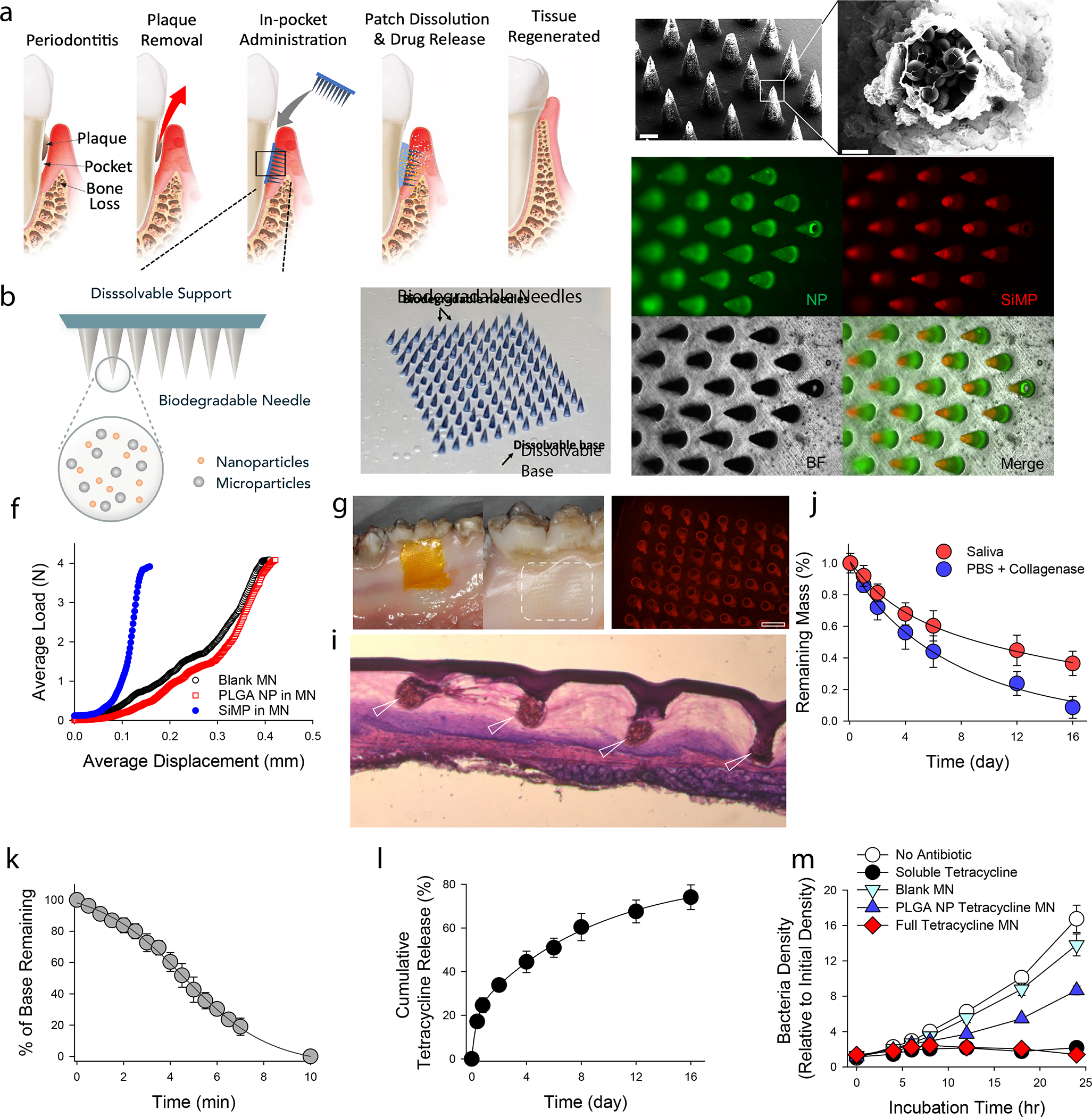
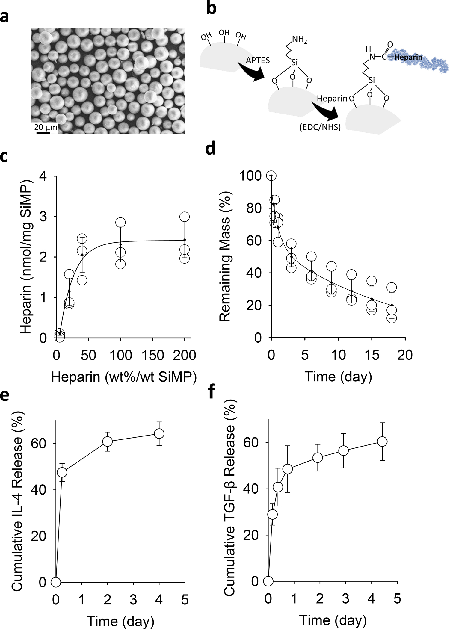
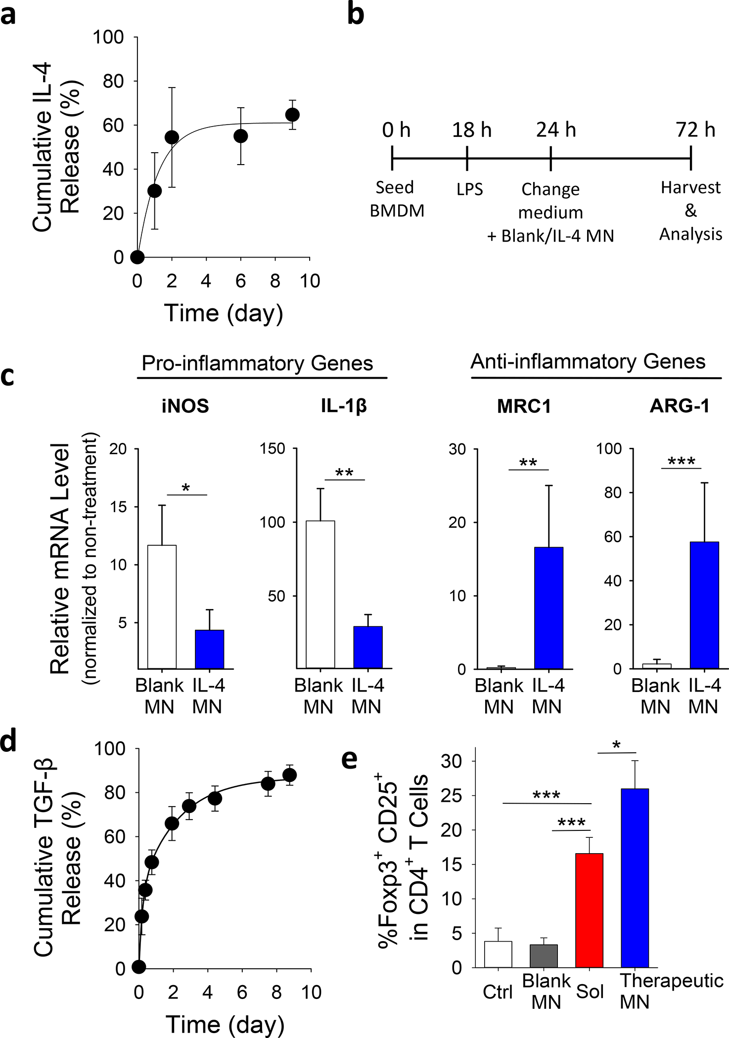
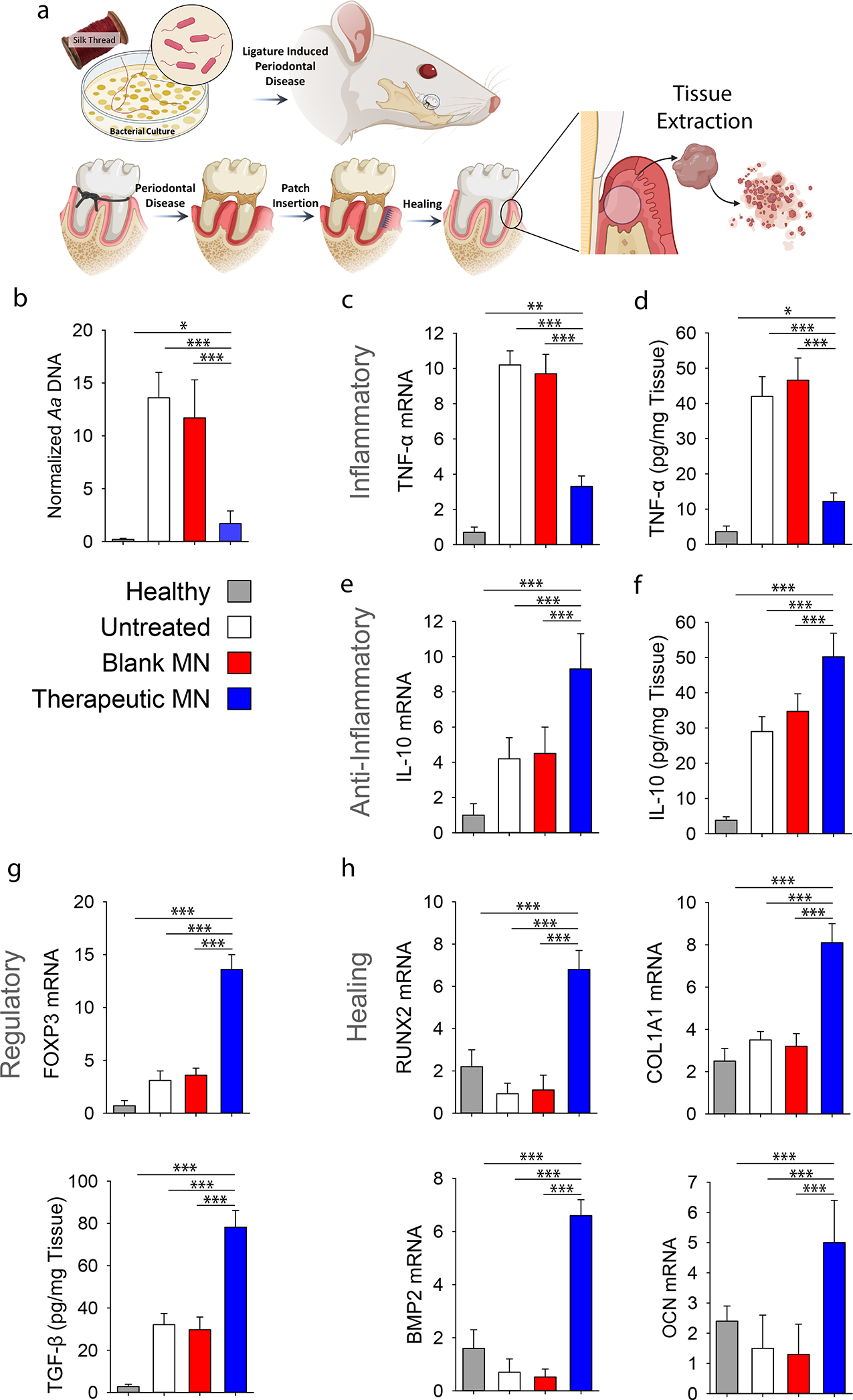
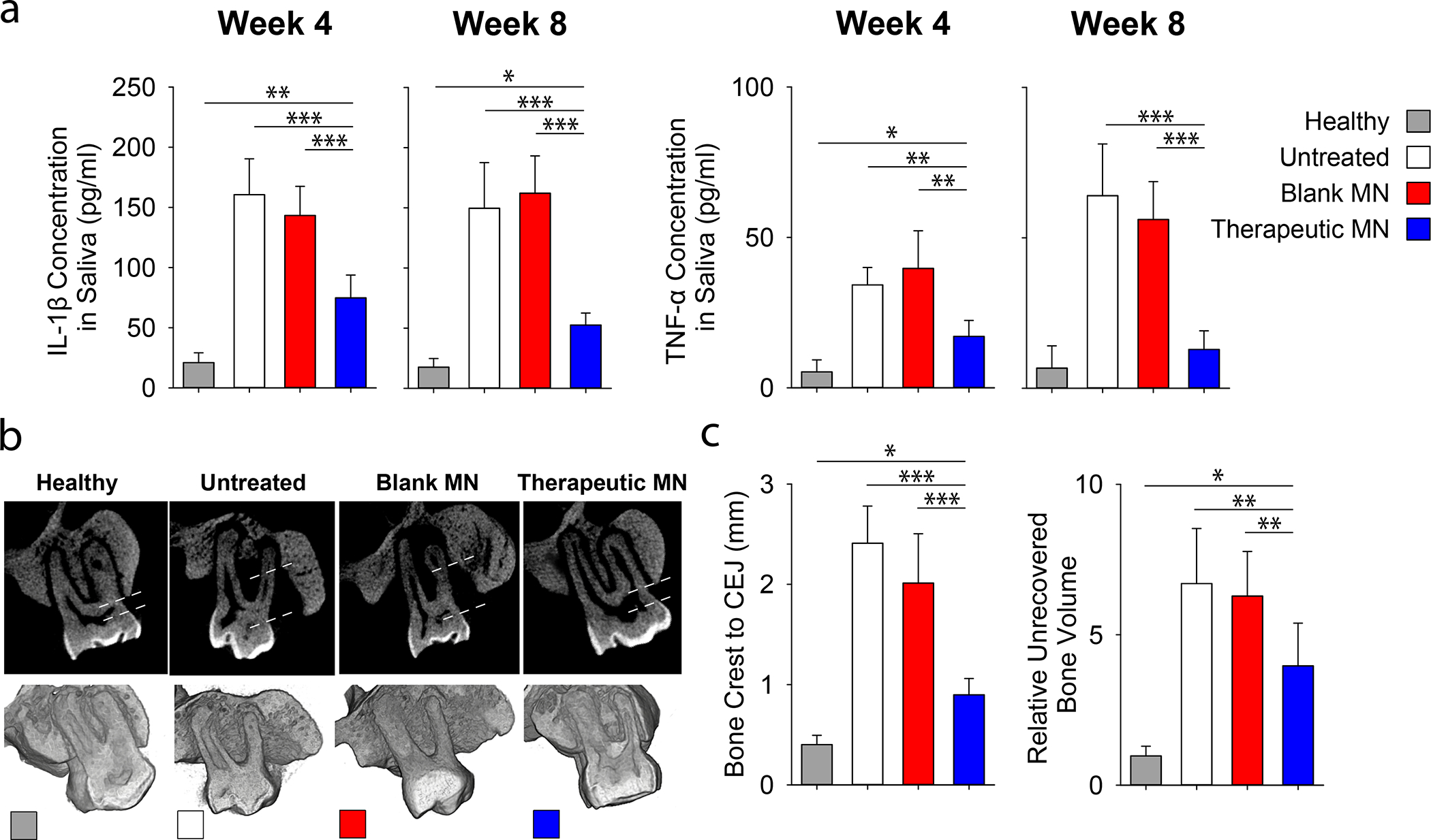
Periodontal diseases are caused by microbial infection and the recruitment of destructive immune cells. Current therapies mainly deal with bacteria elimination, but the regeneration of periodontal tissues remains a challenge. Here we developed a modular microneedle (MN) patch that delivered both antibiotic and cytokines into the local gingival tissue to achieve immunomodulation and tissue regeneration. This MN patch included a quickly dissolvable gelatin membrane for an immediate release of tetracycline and biodegradable GelMA MNs that contained tetracycline-loaded poly(lactic-co-glycolic acid) nanoparticles and cytokine-loaded silica microparticles for a sustained release. Antibiotic release completely inhibited bacteria growth, and the release of IL-4 and TGF-β induced the repolarization of anti-inflammatory macrophages and the formation of regulatory T cells in vitro. In vivo delivery of MN patch into periodontal tissues suppressed proinflammatory factors and promoted pro-regenerative signals and tissue healing, which demonstrated the therapeutic potential of local immunomodulation for tissue regeneration.

Keywords: Microneedles; anti-bacteria; anti-inflammatory; biomaterials; immunoengineering; local immunomodulation.


Conflict of interest statement

Declaration of Interests: S.L., M.M.H.-S., T.A., and A.M. have patent applications (periodontal micropatch and uses thereof, U.S. Provisional Patent Application PCT/US20/58069) related to the current study and, thus, may have related financial interests. The other authors declare no potential conflicts of interest with respect to the authorship and/or publication of this article.

Figures

Figure 1.
Figure 1.. Physical and chemical characterization of MN patches.
a, Proposed working mechanism of the designed immunotherapeutic periodontal MN patch. MN patches are designed to provide transgingival administration of antibiotics and anti-inflammatory cytokines. Following administration, MNs will detach from the supporting membrane upon gelatin dissolution and stay in gingival tissue, release their cargos, and degrade. b, Illustration of the multifunctional MN patch design. The image on the right shows an example of fabricated MN patch. MNs were visualized by encapsulating Trypan Blue dye. c, Scanning electron microscopy (SEM) image of fabricated MNs. d, Encapsulated SiMPs can be seen inside truncated needles. See also Figure S2. e, Fluorescent imaging demonstrates the distribution of fluorophore-conjugated PLGA NPs (in Green) and SiMPs (in Red) inside GelMA-based MNs. f, Mechanical strength of MNs measured by Instron compression test. g, MN patch penetrated on freshly harvested porcine gingiva. Following the administration, the tissue was kept in 37°C incubator for 30 minutes to monitor gelatin dissolution. Pig’s gingival surface is showing insertion of MNs. h, MN-treated pig gingival tissue was imaged to demonstrate the presence of fluorophore-loaded MNs (in Red). Scale bar: 400 μm. i, Penetration of MN into fresh rat gingival tissue was visualized by hematoxylin and eosin (H&E) staining. j, In vitro degradation of MNs over time in PBS containing collagenase enzyme or human saliva at 37°C under gentle shaking. k, FITC-conjugated gelatin was used as the support layer to measure the rate of gelatin base dissolution. l, Cumulative in vitro release of tetracycline was assessed from the dissolution of gelatin base (burst release) and from GelMA MNs loaded with PLGA-tetracycline NPs (sustained release) at 37°C and pH 7.4. m, Antibacterial effect of designed patches against P.g. Full tetracycline MN includes tetracycline-loaded gelatin base and tetracycline-loaded PLGA NPs in MNs. The data in j-m are presented as mean ± standard deviation (SD) (n = 5).
Figure 2.
Figure 2.. Sustained release of IL-4 and TGF-β from silica-heparin microparticles.
a, SEM image of mesoporous SiMPs. b, Surface chemistry to functionalize SiMP surface with heparin. c, The efficiency of heparin-conjugation on SiMPs with various initial amounts of heparin in the reaction mixture. d, In vitro degradation of heparin-functionalized SiMPs over time. e-f, Cumulative release of IL-4 (e) and TGF-β (f) from heparin-functionalized SiMPs at 37°C. The data are presented as mean ± SD (n = 5).
Figure 3.
Figure 3.. Immunomodulatory properties of MN patches in vitro.
a, Cumulative in vitro release of IL-4 from MN patches containing IL-4-loaded SiMPs at 37°C and pH 7.4. b, Experiment procedure of co-culturing LPS-treated BMDMs with MN patches. c, Effects of IL-4 released from MN patches on the gene expression of pro/anti-inflammatory markers in LPS-treated BMDMs as assessed by using qPCR. See also Figure S9 for results from no-contact setup. d, Cumulative in vitro release of TGF-β from MN patches containing TGF-β-loaded SiMPs at 37°C and pH 7.4. e, Effects of MN-released IL-4/TGF-β on the development of induced Treg. Naïve CD4+ T-cells were treated with anti-CD3 and anti-CD28 antibodies for 4 days in the presence of IL-4/TGF-β in solution (Sol), blank MN patches or MN patches with IL-4/TGF-β (Therapeutic MN), followed by flow cytometric analysis of Treg development (co-expression of Foxp3 and CD25). All data are presented as mean ± SD (n = 5). The results were analyzed using one-way ANOVA with post-hoc analysis. Statistical significance is indicated by * p < 0.05, ** p < 0.01 and *** p < 0.001.
Figure 4.
Figure 4.. In vivo evaluation of the immunomodulatory effects in a rat model of periodontitis.
a, Ligature induced periodontal disease model in rats was used. a, Schematic representation of the proposed disease induction strategy patch placement. Mucoperiosteal flaps were elevated, uncovering the alveolar bone adjacent to the lingual aspect of the first maxillary molar to place MN at the defect site. At 8-weeks post MN insertion, buccal and palatal tissues of maxillary molars were isolated and dissociated to assess inflammatory status of tissue microenvironment. b, PCR analysis of A.a. bacterial ribosomal DNA in the periodontal tissue normalized by palatal tissue weight. c, Relative mRNA expression level of pro-inflammatory cytokine TNF-α. See also Figure S10. d, ELISA analysis of TNF-α level in the tissue. e, Relative mRNA expression level of anti-inflammatory cytokine IL-10. f, ELISA analysis of IL-10 in the tissue. g, Relative mRNA expression of Treg marker FOXP3 and ELISA analysis of TGF-β secretion. h, Relative mRNA expression level pro-healing genes RUNX2, COL1A1, BMP2 and OCN in the periodontal tissue. Healthy: healthy rats; Untreated: untreated periodontitis; Blank MN: MN patches without any therapeutic cargo; Therapeutic MN: MN patches containing both tetracycline- and IL-4/TGF-β-loaded SiMPs (Tetracycline: 0.5 mg; IL-2: 100 ng, IL-4: 40 ng, and TGF-β: 40 ng per patch). The presented data are expressed as mean ± SD (n= 5). The results were analyzed by using one-way ANOVA with post-hoc analysis. Statistical significance is indicated by * p < 0.05, ** p < 0.01 and *** p < 0.001.
Figure 5.
Figure 5.. Effects of therapeutic MN patches on the regeneration of periodontal tissue in vivo.
Ligature induced periodontal disease model in rats was used. a, Quantification of inflammatory (TNF-α and IL-1β) cytokines in rat saliva at 4- and 8-weeks post MN implantation. b, The μCT images of the rat maxilla at 8 weeks post MN implantation. All specimens were normalized, and μCT images were calibrated to enable quantitative comparisons. Dotted line indicated the distances between bone crest and CEJ. c, Quantitative analysis of the vertical bone recovery as determined by measuring the distance between the bone crest and CEJ after 8-weeks of treatment. d, Relative volumetric bone recovery was calculated by using 3D reconstructed volume at 8 weeks post MN implantation. All data are presented as mean ± SD (n= 5). Statistical significance is indicated by * p < 0.05, ** p < 0.01 and *** p < 0.001 for differences between samples with different formulations.

References

    1. Pihlstrom BL, Michalowicz BS, and Johnson NW (2005). Periodontal diseases. Lancet 366, 1809–1820. - PubMed
    1. Dye B, Thornton-Evans G, Li X, and Iafolla T (2015). Dental caries and tooth loss in adults in the United States, 2011–2012. NCHS Data Brief, 197. - PubMed
    1. Chen F-M, Zhang J, Zhang M, An Y, Chen F, and Wu Z-F (2010). A review on endogenous regenerative technology in periodontal regenerative medicine. Biomaterials 31, 7892–7927. - PubMed
    1. Patel SK, Greene AC, Desai SM, Rothstein S, Basha IT, MacPherson JS, Wang Y, Zou Y, Shehabeldin M, Sfeir CS, et al. (2021). Biorelevant and screening dissolution methods for minocycline hydrochloride microspheres intended for periodontal administration. Int. J. Pharm. 596, 120261. - PubMed
    1. Khaliq NU, Chobisa D, Richard CA, Swinney MR, and Yeo Y (2021). Engineering microenvironment of biodegradable polyester systems for drug stability and release control. Ther. Deliv. 12, 37–54. - PubMed

LinkOut - more resources