Review
. 2022 Jan 11;14(1):55-66.
doi: 10.1007/s12551-021-00916-4. eCollection 2022 Feb.

Characterizing conformational ensembles of multi-domain proteins using anisotropic paramagnetic NMR restraints

Affiliations
Review

Characterizing conformational ensembles of multi-domain proteins using anisotropic paramagnetic NMR restraints

Xue-Ni Hou et al. Biophys Rev. .

Abstract

It has been over two decades since paramagnetic NMR started to form part of the essential techniques for structural analysis of proteins under physiological conditions. Paramagnetic NMR has significantly expanded our understanding of the inherent flexibility of proteins, in particular, those that are formed by combinations of two or more domains. Here, we present a brief overview of techniques to characterize conformational ensembles of such multi-domain proteins using paramagnetic NMR restraints produced through anisotropic metals, with a focus on the basics of anisotropic paramagnetic effects, the general procedures of conformational ensemble reconstruction, and some representative reweighting approaches.

Keywords: Ensemble reconstruction; Multi-domain proteins; Nuclear magnetic resonance; Pseudocontact shifts; Residual dipolar couplings.


Conflict of interest statement

Conflict of interestThe authors declare no competing interests.

Figures

Fig. 1
Fig. 1
Schematic representation of structural information in a two-domain protein derived from PCSs and RDCs. a PCSs are described by Eq. (1); the observed nucleus (1H, orange circle) of the metal-free domain in three different arrangements (surface, gray) have different PCSs in the fame of the Δχ tensor of the metal (Ln3+, circle, black); PCSs from the metal-bearing domain (surface, limon) can be used to determine the Δχ tensor; b RDCs are described by Eq. (2); the averaged effective tensor (red frame in the center) from the metal-free domain depends on the exchange rate of all conformations (here, three are depicted); c, d illustration of paramagnetic effects PCSs (c) and RDCs (d) in 2D 1H-15 N correlation NMR spectra; for large biomolecules, there might be some overlaid peaks in diamagnetic NMR spectra (concentric circles, blue), while in paramagnetic states, they can be separated due to different metal-nucleus distances (solid circles, green and gray)
Fig. 2
Fig. 2
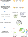
Overview of ensemble reconstruction in a two-domain protein, linear diubiquitin (Ub2), by using PCSs and RDCs: to enable unambiguous chemical shift assignments, the N- or C-terminal Ub was selectively enriched with 15 N-nuclei; the structure of each individual Ub was refined by using paramagnetic data with Xplor-NIH, and the structure model of linear Ub2 was obtained by assembling two Ubs with AIDA; the conformational pool was generated by MESMER; and two approaches were employed for the conformational ensemble reconstruction: MESMER selected a minimal ensemble comprising seven conformations with associated weights that are proportional to their putative population, while MaxOcc depicted the conformational distributions. Almost all conformations selected by MESMER had higher MaxOcc values. Adapt from (Hou et al. 2021)
Fig. 3
Fig. 3
a An example of dramatically changed PCSs by the introduction of mutations in free linear Ub2: PCSs collected from the metal-free Ub of wild-type (cyan bar), E16Rp (orange triangle), and E18Rp (red square) of linear Ub2, with paramagnetic tag (PSPy-6 M-DO3MA-Tm3+) (Yang et al. 2016) at D39C of N-terminal (distal) Ub. b, c MaxOcc of top 1,000 conformers calculated using PCSs from wild-type (purple or green), E16Rp (orange), and E18Rp (red) of linear Ub2 is sorted and plotted in descending order. The rank of MaxOcc of the bound conformer of linear Ub2 in complex with HOIL-1L-NZF (PDB code: 3b0a) (Sato et al. 2011), shown as triangles by corresponding color, dropped (elevated) in E16Rp (E18Rp), implying that the E16Rp (E18Rp) decreased (increased) the probability for free linear Ub2 to adapt the bound state. Correspondingly, dissociation constant (Kd) increased by over 14-fold for E16Rp and decreased by fivefold for E18Rp, implying contribution from the conformational selection mechanism. Adapted from (Hou et al. 2021)

Similar articles

Cited by

References

    1. Andrałojć W, Berlin K, Fushman D, et al. Information content of long-range NMR data for the characterization of conformational heterogeneity. J Biomol NMR. 2015;62:353–371. doi: 10.1007/s10858-015-9951-6. - DOI - PMC - PubMed
    1. Andrałojć W, Luchinat C, Parigi G, Ravera E. Exploring regions of conformational space occupied by two-domain proteins. J Phys Chem B. 2014;118:10576–10587. doi: 10.1021/JP504820W. - DOI - PubMed
    1. Andrałojć W, Ravera E, Salmon L, et al. Inter-helical conformational preferences of HIV-1 TAR-RNA from maximum occurrence analysis of NMR data and molecular dynamics simulations. Phys Chem Chem Phys. 2016;18:5743–5752. doi: 10.1039/c5cp03993b. - DOI - PMC - PubMed
    1. Anthis NJ, Doucleff M, Clore GM. Transient, sparsely populated compact states of apo and calcium-loaded calmodulin probed by paramagnetic relaxation enhancement: interplay of conformational selection and induced fit. J Am Chem Soc. 2011;133:18966–18974. doi: 10.1021/JA2082813. - DOI - PMC - PubMed
    1. Banci L, Bertini I, Bren KL, et al. The use of pseudocontact shifts to refine solution structures of paramagnetic metalloproteins: Met80Ala cyano-cytochrome c as an example. J Biol Inorg Chem. 1996;1:117–126. doi: 10.1007/s007750050030. - DOI