Review
. 2021 Nov 18;14(1):233-255.
doi: 10.1007/s12551-021-00903-9. eCollection 2022 Feb.

Structural biology of cell surface receptors implicated in Alzheimer's disease

Affiliations
Review

Structural biology of cell surface receptors implicated in Alzheimer's disease

Stefan J Hermans et al. Biophys Rev. .

Abstract

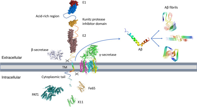
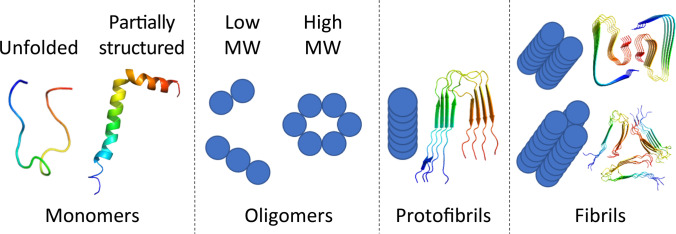
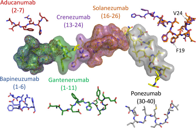
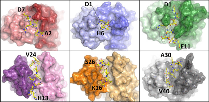
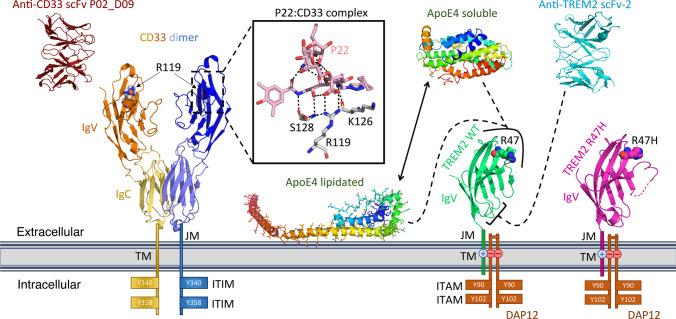
Alzheimer's disease is a common and devastating age-related disease with no effective disease-modifying treatments. Human genetics has implicated a wide range of cell surface receptors as playing a role in the disease, many of which are involved in the production or clearance of neurotoxins in the brain. Amyloid precursor protein, a membrane-bound signaling molecule, is at the very heart of the disease: hereditary mutations in its gene are associated with a greatly increased risk of getting the disease. A proteolytic breakdown product of amyloid precursor protein, the neurotoxic Aβ peptide, has been the target for many drug discovery efforts. Antibodies have been designed to target Aβ production with some success, although they have not proved efficacious in clinical trials with regards to cognitive benefits to date. Many of the recently identified genes associated with late-onset Alzheimer's disease risk are integral to the innate immune system. Some of these genes code for microglial proteins, such as the strongest genetic risk factor for the disease, namely APOE, and the cell surface receptors CD33 and TREM2 which are involved in clearance of the Aβ peptide from the brain. In this review, we show how structural biology has provided key insights into the normal functioning of these cell surface receptors and provided a framework for developing novel treatments to combat Alzheimer's disease.

Keywords: Alzheimer’s disease; Antibodies; Cell surface receptors; Neuroinflammation; Structural biology; X-ray crystallography.


Conflict of interest statement

Conflict of interestWe have a collaboration and license agreement with Janssen Pharmaceuticals on some of our microglia work. Dr Chen Gao is now an employee of Wren Therapeutics Ltd.

Figures

Fig. 1
Fig. 1
Diagrammatic representation of the known amyloid precursor protein (APP) structures, in the context of a cell membrane. Each structure shown is described here with its PDB ID. APP consists of an N-terminal growth factor-like domain (GFD) (red, 1MWP (Rossjohn et al. 1999)) and associated copper-binding domain (CuBD) (blue, 2FMA (Kong et al. 2007a)) which are commonly referred to as the “extracellular domain 1” or E1 (3KTM (Dahms et al. 2010)). This is followed by an anionic acid-rich region containing a Kunitz protease inhibitor domain (orange, 1AAP (Hynes et al. 1990)) that is absent, due to differential mRNA splicing, from the form of APP predominantly found in neurons. A helical bundle region, E2 (brown, 3UMH (Dahms et al. 2012)) is the last extracellular portion of APP, linked to the TM helical region that includes the Aβ peptide (rainbow, 1IYT (Crescenzi et al. 2002)). A short C-terminal intracellular domain is found to be partly helical (blue-white, 3DXC (Radzimanowski et al. 2008)) in complex with binding proteins such as Fe65 (tan, 3DXC (Radzimanowski et al. 2008)) and X11 (green, 1AQC (Zhang et al. 1997)) while other binding proteins are known but lack experimental structures (e.g., PAT1, dark green, AlphaFold2 model AF-Q92624-F1 (Jumper et al. 2021)). Processing of APP by β- and γ-secretases (gray, 5HD0, and colored by chain, 6IYC, respectively (Mandal et al. ; Zhou et al. 2019)) releases the Aβ peptide which is then able to polymerize into a range of different amyloid structures (shown are 5OQV, 6SHS, and 6W0O as examples (Ghosh et al. ; Gremer et al. ; Kollmer et al. 2019))
Fig. 2
Fig. 2
The Aβ peptide folds into a variety of conformations and comes together into multiple oligomeric states. Aβ monomers are found to have a partially helical structure (PDB ID: 1IYT (Crescenzi et al. 2002)), whereas protofibril (PDB ID: 2LMN (Paravastu et al. 2008)) and fibril (PDB IDs: 2LMP and 5OQV (Gremer et al. ; Paravastu et al. 2008)) structures of the peptide reveal a pleated β-sheet fold that can form between multiple peptide chains
Fig. 3
Fig. 3
The Aβ epitopes recognized by the clinical anti-Aβ antibodies listed in Table 1 are depicted as molecular surfaces overlaid onto the helical Aβ42 monomer (PDB ID: 1IYT (Crescenzi et al. 2002)). Residue numbers of the epitope are shown in parentheses. The conformation adopted by each of the Aβ epitopes in the Aβ:antibody complexes are shown alongside in stick fashion, with those for crenezumab and solanezumab superimposed to illustrate their structural similarity. The location of residues F19 and V24 are indicated in the overlay of crenezumab and solanezumab; the linear Aβ conformation is disrupted at F19 and the remainder of the epitope adopts a more helical conformation in both antibodies, while V24 is the last Aβ residue observed in the crenezumab structure and the side-chain adopts a different orientation to that observed in solanezumab (see main text for details)
Fig. 4
Fig. 4
Structures of the clinical anti-Aβ antibodies listed in Table 1; the view is looking down onto the Aβ-binding cleft: (left-right, top-down) aducanumab (PDB ID: 6CO3 (Arndt et al. 2018)), bapineuzumab (PDB ID: 4HIX (Miles et al. 2013)), gantenerumab (PDB ID: 5CSZ (Bohrmann et al. 2012)), crenezumab (PDB ID: 5VZY (Ultsch et al. 2016)), solanezumab (PDB ID: 4XXD (Crespi et al. 2015)), and ponezumab (PDB ID: 3U0T (La Porte et al. 2012)), with the complexed Aβ ligand colored yellow in stick form. The antibodies are depicted as transparent molecular surfaces; the heavy and light chains are identified by dark and light shades, respectively. The N- and C-terminal Aβ residues observed in each crystal complex are labeled
Fig. 5
Fig. 5
Diagrammatic representation of microglial proteins CD33, TREM2, DAP12, and APOE4 structures in the context of a cell membrane. CD33 is thought to signal as a homodimer, or possibly a higher-order oligomer. The extracellular region of CD33 is comprised of an N-terminal IgV domain and a membrane-proximal IgC domain (PDB ID: 5IHB), followed by a flexible JM region connected to a helical TM and an intracellular region containing the signaling ITIM motifs. The insert box shows the polar interactions (black dashed lines) of the sialic acid mimetic P22 with CD33 IgV domain residues (PDB ID: 6D4A (Miles et al. 2019)). In addition to traditional antibodies targeting the extracellular region of CD33, scFv antibodies such as scFv P02_D09 (PDB ID: 6UUP (Park et al. 2021)) have been identified. TREM2 requires the DAP12 receptor to form a signaling complex. The extracellular region of TREM2 is comprised of an IgV domain (PDB ID: 5ELI, wild-type TREM2 (Kober et al. 2016)), followed by a flexible JM region connected to a helical TM domain and a very short non-signaling intracellular region. The TREM2 TM helix interacts with the two TM helices of DAP12 via salt bridge interactions (indicated by the + and −). DAP12 forms a homodimer with a very short N-terminal extracellular region, a disulfide bond connects the two N-terminal regions of the DAP12 monomers. The intracellular tails of each DAP12 monomer contain two signaling ITAM motifs. The extracellular IgV domain of TREM2 is the target for agonistic antibodies, such as scFv-2 (PDB ID: 6YYE (Szykowska et al. 2021)) whose epitope is indicated by the black bracket. Mutation of TREM2 R47 to histidine increases the risk of developing LOAD, the location of this residue is indicated and the structure of the R47H TREM2 mutant is depicted to the right of the wild-type protein (PDB ID: 5UD8 (Sudom et al. 2018)). The soluble (i.e., unlipidated) form and membrane-associated lipidated form of APOE4 are also shown, APOE4 interacts with the extracellular region of TREM2 as well as Aβ oligomers (Figs. 1 and 2). The location of the APOE4 interaction surface is indicated by the black curved line encompassing R47 in wild-type TREM2. The APOE4 structures (residues 19–317) depicted are models constructed using X-ray crystallography structures of APOE4 and APOA-I, for the soluble and lipidated forms respectively (PDB IDs: 6NCO (Petros et al. 2019) and 3R2P (Mei and Atkinson 2011)

References

    1. Adolfsson O, Pihlgren M, Toni N, Varisco Y, Buccarello AL, Antoniello K, Lohmann S, Piorkowska K, Gafner V, Atwal JK, Maloney J, Chen M, Gogineni A, Weimer RM, Mortensen DL, Friesenhahn M, Ho C, Paul R, Pfeifer A, Muhs A, Watts RJ. An effector-reduced anti-beta-amyloid (Abeta) antibody with unique abeta binding properties promotes neuroprotection and glial engulfment of Abeta. J Neurosci. 2012;32:9677–9689. doi: 10.1523/JNEUROSCI.4742-11.2012. - DOI - PMC - PubMed
    1. Alzheimer’s Association Alzheimer’s disease facts and figures. Alzheimer’s Dementia. 2012;8:131–168. doi: 10.1016/j.jalz.2012.02.001. - DOI - PubMed
    1. Anandatheerthavarada HK, Biswas G, Robin MA, Avadhani NG. Mitochondrial targeting and a novel transmembrane arrest of Alzheimer’s amyloid precursor protein impairs mitochondrial function in neuronal cells. J Cell Biol. 2003;161:41–54. doi: 10.1083/jcb.200207030. - DOI - PMC - PubMed
    1. Appelbaum FR, Matthews DC, Eary JF, Badger CC, Kellogg M, Press OW, Martin PJ, Fisher DR, Nelp WB, Thomas ED, et al. The use of radiolabeled anti-CD33 antibody to augment marrow irradiation prior to marrow transplantation for acute myelogenous leukemia. Transplantation. 1992;54:829–833. doi: 10.1097/00007890-199211000-00012. - DOI - PubMed
    1. Argyri L, Dafnis I, Theodossiou TA, Gantz D, Stratikos E, Chroni A. Molecular basis for increased risk for late-onset Alzheimer disease due to the naturally occurring L28P mutation in apolipoprotein E4. J Biol Chem. 2014;289:12931–12945. doi: 10.1074/jbc.M113.538124. - DOI - PMC - PubMed

LinkOut - more resources