

A Pilot Study of Subclinical Non-Capillary Peripapillary Perfusion Changes in Thyroid-Related Orbitopathy Detected Using Optical Coherence Tomography Angiography
- PMID: 35340669
- PMCID: PMC8948173
- DOI: 10.2147/OPTH.S356631
A Pilot Study of Subclinical Non-Capillary Peripapillary Perfusion Changes in Thyroid-Related Orbitopathy Detected Using Optical Coherence Tomography Angiography
Abstract
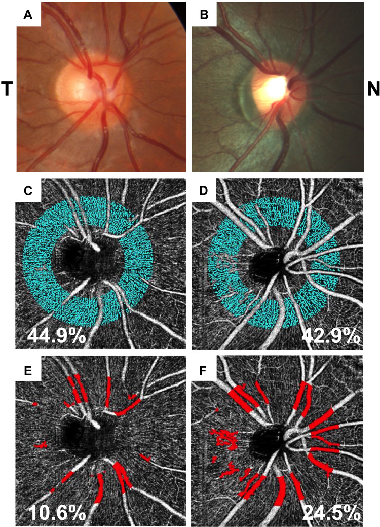
Purpose: Hemodynamic changes surrounding the optic nerve head are known to occur in thyroid-related orbitopathy (TRO). This pilot study explores the capillary and non-capillary peripapillary perfusion changes of the retina in TRO eyes without dysthyroid optic neuropathy (DON) using optical coherence tomography angiography (OCT-A).
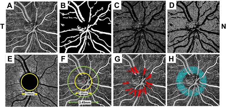
Methods: Non-capillary and capillary peripapillary perfusion densities were calculated using single 4.5 × 4.5mm en face "RPC layer" OCT-A scans of 8 TRO patients without DON (8 eyes, mean age 40.6 years, range 23-69 years). Results were compared to a previously published dataset of 133 healthy controls (133 eyes, mean 41.5 years, range 11-83 years). The strength of association was measured between OCT-A perfusion densities and clinical measures of TRO.
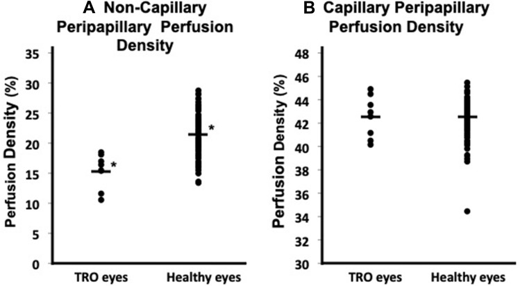
Results: Non-capillary peripapillary perfusion density in TRO eyes was found to be significantly decreased compared to healthy controls (TRO group 15.4 ± 2.9% vs controls 21.5 ± 3.1%; p < 0.0001). Capillary peripapillary perfusion densities showed no significant difference (TRO group 42.5 ± 1.8% vs controls 42.5 ± 1.5%; p = 1.0). Clinical measures of disease did not correlate well with OCT-A perfusion densities (p>0.05).
Conclusion: These findings may represent decreased blood flow and subclinical ischemia to the optic nerve. We discuss possible pathogenic mechanisms of thyroid-related vasculopathy, including vessel wall thickening due to immunologically-induced media enlargement.
Keywords: optical coherence tomography angiography; peripapillary microvasculature; thyroid-related orbitopathy; thyroid-related vasculopathy.
© 2022 Pinhas et al.
Conflict of interest statement
Richard B. Rosen M.D. is a paid consultant of Optovue, and has received personal fees and non-financial support from Optovue, and non-financial support from Topcon Medical. In addition, Dr. Rosen has a US patent - # WO 2016/109750 Al, 43625.140US01 issued to Optovue. Outside the submitted work, Dr. Rosen is a paid consultant of Guardion Health, and has received personal fees and stock from Boehringer-Ingelheim, non-financial support from Ocusciences, and equipment from CellView. Harsha S. Reddy M.D. is a paid advisory board member for Horizon Therapeutics. The authors report no other conflicts of interest in this work.
Figures
Similar articles
-
Peripapillary and Macular Vessel Density in Dysthyroid Optic Neuropathy: An Optical Coherence Tomography Angiography Study.Invest Ophthalmol Vis Sci. 2019 May 1;60(6):1863-1869. doi: 10.1167/iovs.18-25941. Invest Ophthalmol Vis Sci. 2019. PMID: 31042792
-
Optical coherence tomography measurements in compressive optic neuropathy associated with dysthyroid orbitopathy.Graefes Arch Clin Exp Ophthalmol. 2016 Aug;254(8):1617-1624. doi: 10.1007/s00417-016-3335-9. Epub 2016 May 12. Graefes Arch Clin Exp Ophthalmol. 2016. PMID: 27169807
-
Quantitative assessment of macular microvasculature and radial peripapillary capillary plexus in the fellow eyes of patients with retinal vein occlusion using OCT angiography.J Fr Ophtalmol. 2020 Nov;43(9):842-850. doi: 10.1016/j.jfo.2020.06.004. Epub 2020 Sep 11. J Fr Ophtalmol. 2020. PMID: 32928575
-
SWEPT-SOURCE OPTICAL COHERENCE TOMOGRAPHY ANGIOGRAPHY OF THE OPTIC DISK IN OPTIC NEUROPATHY.Retina. 2016 Dec;36 Suppl 1:S168-S177. doi: 10.1097/IAE.0000000000001259. Retina. 2016. PMID: 28005675
-
Peripapillary and parafoveal microvascular changes in eyes with optic neuritis and their fellow eyes measured by optical coherence tomography angiography: an Exploratory Study.Acta Ophthalmol. 2021 May;99(3):288-298. doi: 10.1111/aos.14577. Epub 2020 Aug 24. Acta Ophthalmol. 2021. PMID: 32833336
Cited by
-
Macular microvasculature in patients with thyroid-associated orbitopathy: a cross-sectional study.Thyroid Res. 2023 Aug 2;16(1):31. doi: 10.1186/s13044-023-00175-3. Thyroid Res. 2023. PMID: 37533056 Free PMC article.
-
Optical coherence tomography angiography in thyroid associated ophthalmopathy: a systematic review.BMC Ophthalmol. 2024 Jul 22;24(1):304. doi: 10.1186/s12886-024-03569-5. BMC Ophthalmol. 2024. PMID: 39039451 Free PMC article.
-
Optic nerve head optical coherence tomography angiography findings in patients with thyroid eye disease: a case-control study.Thyroid Res. 2022 Sep 21;15(1):17. doi: 10.1186/s13044-022-00134-4. Thyroid Res. 2022. PMID: 36127745 Free PMC article.
-
Thyroid-Associated Peripapillary Vascular Remodelling - A Novel Area for Research? [Letter].Clin Ophthalmol. 2022 May 12;16:1475-1476. doi: 10.2147/OPTH.S372789. eCollection 2022. Clin Ophthalmol. 2022. PMID: 35592669 Free PMC article. No abstract available.
References
-
- Khong JJ, McNab AA, Ebeling PR, et al. Pathogenesis of thyroid eye disease: review and update on molecular mechanisms. Br J Ophthalmol. 2016;100(1):142–150. - PubMed
-
- Bartley GB, Fatourechi V, Kadrmas EF, et al. The incidence of Graves’ ophthalmopathy in Olmsted County, Minnesota. Am J Ophthalmol. 1995;120(4):511–517. - PubMed
Grants and funding
LinkOut - more resources
Full Text Sources
Research Materials
