Review
. 2020 Sep 30;2(1):47-76.
doi: 10.1039/d0cb00151a. eCollection 2021 Feb 1.

DNA folds threaten genetic stability and can be leveraged for chemotherapy

Affiliations
Review

DNA folds threaten genetic stability and can be leveraged for chemotherapy

Joanna Zell et al. RSC Chem Biol. .

Abstract

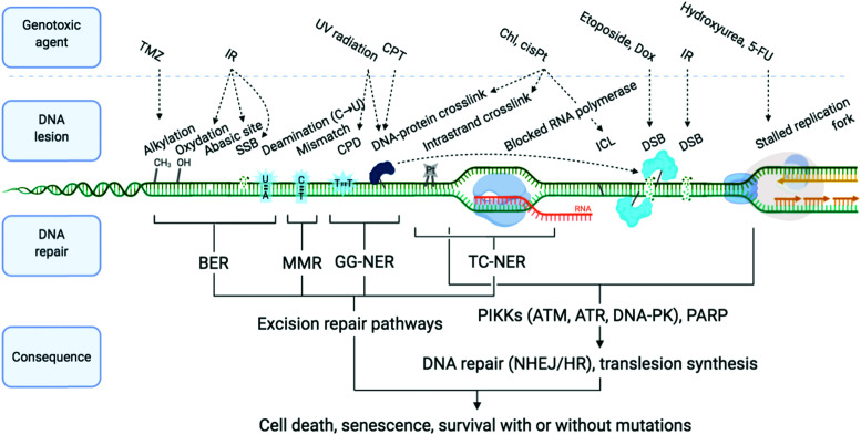
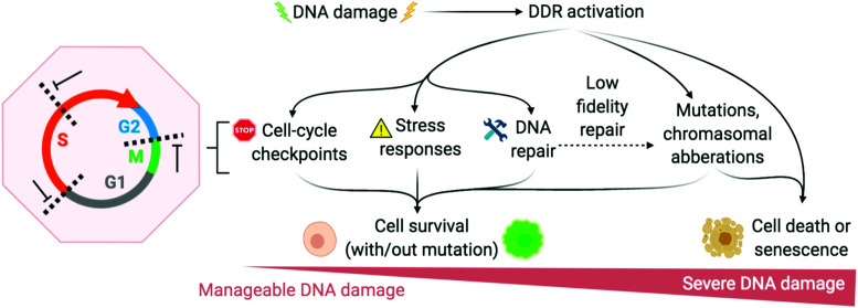
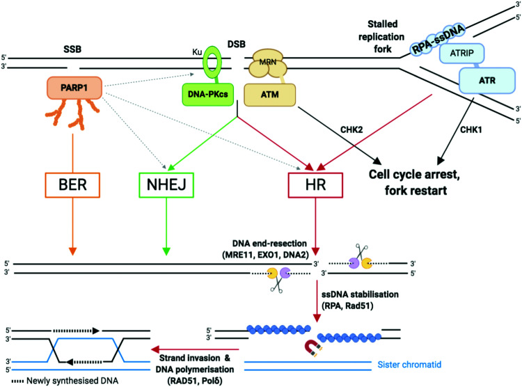
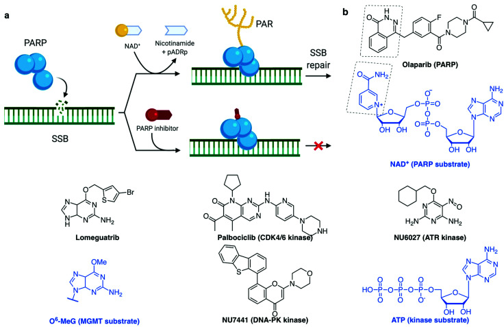
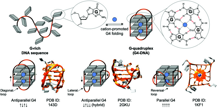
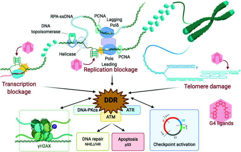
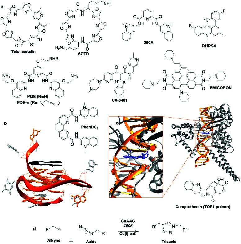
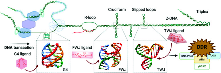
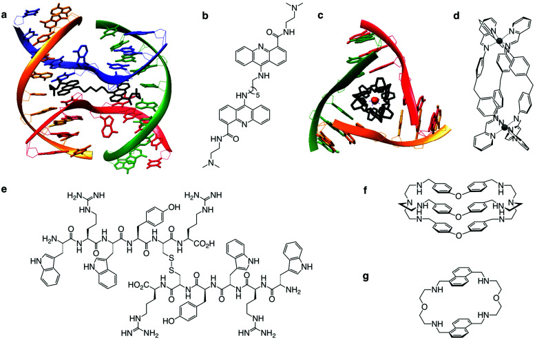
Damaging DNA is a current and efficient strategy to fight against cancer cell proliferation. Numerous mechanisms exist to counteract DNA damage, collectively referred to as the DNA damage response (DDR) and which are commonly dysregulated in cancer cells. Precise knowledge of these mechanisms is necessary to optimise chemotherapeutic DNA targeting. New research on DDR has uncovered a series of promising therapeutic targets, proteins and nucleic acids, with application notably via an approach referred to as combination therapy or combinatorial synthetic lethality. In this review, we summarise the cornerstone discoveries which gave way to the DNA being considered as an anticancer target, and the manipulation of DDR pathways as a valuable anticancer strategy. We describe in detail the DDR signalling and repair pathways activated in response to DNA damage. We then summarise the current understanding of non-B DNA folds, such as G-quadruplexes and DNA junctions, when they are formed and why they can offer a more specific therapeutic target compared to that of canonical B-DNA. Finally, we merge these subjects to depict the new and highly promising chemotherapeutic strategy which combines enhanced-specificity DNA damaging and DDR targeting agents. This review thus highlights how chemical biology has given rise to significant scientific advances thanks to resolutely multidisciplinary research efforts combining molecular and cell biology, chemistry and biophysics. We aim to provide the non-specialist reader a gateway into this exciting field and the specialist reader with a new perspective on the latest results achieved and strategies devised.


Conflict of interest statement

There are no conflicts to declare.

Figures

Fig. 1
Fig. 1. Examples of DNA-damaging agents, the lesion which they induce, and the downstream consequence. Temozolomide (TMZ), ionising radiation (IR), camptothecin (CPT), chlorambucil (Chl), cisplatin (cisPt), doxorubicin (Dox), single-strand break (SSB), double strand break (DSB), cyclobutane pyrimidine dimer (CPD), interstrand cross-link (ICL), base excision repair (BER), mismatch repair (MMR) and global genome nucleotide excision repair (GG-NER), transcription-coupled NER (TC-NER). Adapted from ref. 70, created with BioRender.
Fig. 2
Fig. 2. DNA damage response (DDR) is activated to varying degrees depending on the extremity of DNA damage. Adapted from ref. 72, created with BioRender.
Fig. 3
Fig. 3. Single-strand breaks (SSB) and double-strand breaks (DSB) repair pathways. Dotted arrows indicate that PARP1 can be activated by DSB and stimulate DSB repair by both HR and NHEJ. Adapted from ref. 37, created with BioRender.
Fig. 4
Fig. 4. (a) PARP recruitment to SSB and synthesis of poly(ADP-ribose) (PAR) to activate SSB repair. Inhibition of repair by PARP-trapping at the break site. (b) Examples of inhibitors (with their protein target) and the substrate they mimic (blue). (a) Created with BioRender.
Fig. 5
Fig. 5. Schematic representation of a G-rich sequence that folds into a G4 structure (upper panel), highlighting the structure of a guanine (G, upper panel, left) and a G-quartet (right). Topological diversity of G4s that can adopt parallel, hybrid and antiparallel conformation (lower panel, arrows indicate the polarity of the DNA strands), as elucidated by either NMR (PDB IDs 143D and 2GKU) or X-ray structure analysis (PDB ID 1KF1).
Fig. 6
Fig. 6. Schematic representation of the stabilisation of G4s, inducing replication and transcription blockages and telomere damage, producing a DDR response, which feeds back into damage signalling (γH2AX) and checkpoint inhibition, and eventually leads to recombination repair and/or cellular shutdown through apoptosis. Created with BioRender.
Fig. 7
Fig. 7. (a) Chemical structures of G4 ligands. (b) NMR structure of G4 with PhenDC ligand (PDB ID: 2MGN). (c) Crystal structure of TOP1cc with camptothecin (PDB ID: 1T8I). (d) Bioorthogonal copper catalysed click ligation performed in cells between PDSα and an azide-labelled fluorophore.
Fig. 8
Fig. 8. Alternative DNA structures form in the vicinity of DNA transactions. The junction point of a cruciform structure presents a four-way junction (FWJ, e.g. Holliday junction) and that of a slipped loop presents a three-way junction (TWJ). Stabilisation of these structures with chemicals (ligands) can impede DNA transactions and induce DDR. Adapted from ref. 35, created with BioRender.
Fig. 9
Fig. 9. (a and b) Crystal structure of FWJ with bisacridine ligand (PDB ID: 2GWA). (c and d) Crystal structure of TWJ with supramolecular iron cylinder (PDB ID: 2ET0), (e) FWJ ligand WRWYCR peptide in active disulfide form, TWJ ligands azacryptand TrisPOB (f) and azacyclophane 1,5-BisNP-O (g).
None
Joanna Zell
None
Francesco Rota Sperti
None
Sébastien Britton
None
David Monchaud

References

    1. Hanahan D. Weinberg R. A. Cell. 2000;107:57–70. doi: 10.1016/S0092-8674(00)81683-9. - DOI - PubMed
    1. Hanahan D. Weinberg R. A. Cell. 2011;144:646–674. doi: 10.1016/j.cell.2011.02.013. - DOI - PubMed
    1. Jackson S. P. Bartek J. Nat. Rev. 2009;461:1071–1078. - PMC - PubMed
    1. O’Connor M. J. Mol. Cell. 2015;60:547–560. doi: 10.1016/j.molcel.2015.10.040. - DOI - PubMed
    1. Minchom A. Aversa C. Lopez J. Ther. Adv. Med. Oncol. 2018;10:1–18. - PMC - PubMed