

Magnetic resonance image-based brain tumour segmentation methods: A systematic review
- PMID: 35340900
- PMCID: PMC8943308
- DOI: 10.1177/20552076221074122
Magnetic resonance image-based brain tumour segmentation methods: A systematic review
Abstract
Background: Image segmentation is an essential step in the analysis and subsequent characterisation of brain tumours through magnetic resonance imaging. In the literature, segmentation methods are empowered by open-access magnetic resonance imaging datasets, such as the brain tumour segmentation dataset. Moreover, with the increased use of artificial intelligence methods in medical imaging, access to larger data repositories has become vital in method development.
Purpose: To determine what automated brain tumour segmentation techniques can medical imaging specialists and clinicians use to identify tumour components, compared to manual segmentation.
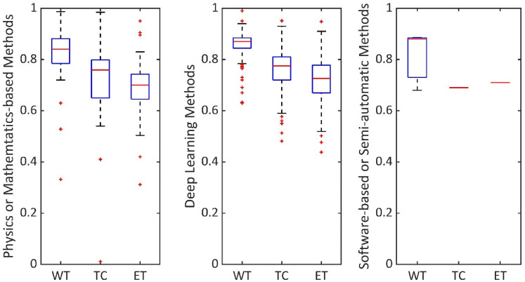
Methods: We conducted a systematic review of 572 brain tumour segmentation studies during 2015-2020. We reviewed segmentation techniques using T1-weighted, T2-weighted, gadolinium-enhanced T1-weighted, fluid-attenuated inversion recovery, diffusion-weighted and perfusion-weighted magnetic resonance imaging sequences. Moreover, we assessed physics or mathematics-based methods, deep learning methods, and software-based or semi-automatic methods, as applied to magnetic resonance imaging techniques. Particularly, we synthesised each method as per the utilised magnetic resonance imaging sequences, study population, technical approach (such as deep learning) and performance score measures (such as Dice score).
Statistical tests: We compared median Dice score in segmenting the whole tumour, tumour core and enhanced tumour.
Results: We found that T1-weighted, gadolinium-enhanced T1-weighted, T2-weighted and fluid-attenuated inversion recovery magnetic resonance imaging are used the most in various segmentation algorithms. However, there is limited use of perfusion-weighted and diffusion-weighted magnetic resonance imaging. Moreover, we found that the U-Net deep learning technology is cited the most, and has high accuracy (Dice score 0.9) for magnetic resonance imaging-based brain tumour segmentation.
Conclusion: U-Net is a promising deep learning technology for magnetic resonance imaging-based brain tumour segmentation. The community should be encouraged to contribute open-access datasets so training, testing and validation of deep learning algorithms can be improved, particularly for diffusion- and perfusion-weighted magnetic resonance imaging, where there are limited datasets available.
Keywords: Brain tumour; artificial intelligence; brain; magnetic resonance imaging; segmentation; systematic review.
© The Author(s) 2022.
Figures


Similar articles
-
CNN-based glioma detection in MRI: A deep learning approach.Technol Health Care. 2024;32(6):4965-4982. doi: 10.3233/THC-240158. Technol Health Care. 2024. PMID: 39031408 Free PMC article.
-
Deep-learning-based synthesis of post-contrast T1-weighted MRI for tumour response assessment in neuro-oncology: a multicentre, retrospective cohort study.Lancet Digit Health. 2021 Dec;3(12):e784-e794. doi: 10.1016/S2589-7500(21)00205-3. Epub 2021 Oct 20. Lancet Digit Health. 2021. PMID: 34688602 Clinical Trial.
-
Automated deep learning method for whole-breast segmentation in diffusion-weighted breast MRI.J Magn Reson Imaging. 2020 Feb;51(2):635-643. doi: 10.1002/jmri.26860. Epub 2019 Jul 13. J Magn Reson Imaging. 2020. PMID: 31301201 Free PMC article.
-
Accuracy of vestibular schwannoma segmentation using deep learning models - a systematic review & meta-analysis.Neuroradiology. 2025 Mar;67(3):729-742. doi: 10.1007/s00234-024-03449-1. Epub 2024 Aug 24. Neuroradiology. 2025. PMID: 39179652 Free PMC article.
-
Quantifying deep grey matter atrophy using automated segmentation approaches: A systematic review of structural MRI studies.Neuroimage. 2019 Nov 1;201:116018. doi: 10.1016/j.neuroimage.2019.116018. Epub 2019 Jul 15. Neuroimage. 2019. PMID: 31319182
Cited by
-
Clinical evaluation of two glioblastoma delineation methods based on neural networks.Tech Innov Patient Support Radiat Oncol. 2025 Aug 6;35:100330. doi: 10.1016/j.tipsro.2025.100330. eCollection 2025 Sep. Tech Innov Patient Support Radiat Oncol. 2025. PMID: 40822088 Free PMC article.
-
Whole Spine Segmentation Using Object Detection and Semantic Segmentation.Neurospine. 2024 Mar;21(1):57-67. doi: 10.14245/ns.2347178.589. Epub 2024 Feb 1. Neurospine. 2024. PMID: 38317546 Free PMC article.
-
Deep Learning Spinal Cord Segmentation Based on B0 Reference for Diffusion Tensor Imaging Analysis in Cervical Spondylotic Myelopathy.Bioengineering (Basel). 2025 Jun 28;12(7):709. doi: 10.3390/bioengineering12070709. Bioengineering (Basel). 2025. PMID: 40722401 Free PMC article.
-
Benchmarking commercial depth sensors for intraoperative markerless registration in neurosurgery applications.Int J Comput Assist Radiol Surg. 2025 Aug;20(8):1759-1769. doi: 10.1007/s11548-025-03416-y. Epub 2025 May 23. Int J Comput Assist Radiol Surg. 2025. PMID: 40407995 Free PMC article.
-
Performance and Robustness of Regional Image Segmentation Driven by Selected Evolutionary and Genetic Algorithms: Study on MR Articular Cartilage Images.Sensors (Basel). 2022 Aug 23;22(17):6335. doi: 10.3390/s22176335. Sensors (Basel). 2022. PMID: 36080793 Free PMC article.
References
-
- Lam WWM, Poon WS, Metreweli C. Diffusion MR imaging in glioma: does it have any role in the pre-operation determination of grading of glioma? Clin Radiol 2002; 57: 219–225. - PubMed
-
- Price SJ. The role of advanced MR imaging in understanding brain tumour pathology. Br J Neurosurg 2007; 21: 562–575. - PubMed
-
- Nilsson M, Englund E, Szczepankiewicz F, et al. Imaging brain tumour microstructure. Neuroimage 2018; 182: 232–250. - PubMed
Publication types
Grants and funding
LinkOut - more resources
Full Text Sources
