. 2022 Jan-Mar;39(1):30-36.
doi: 10.4103/joc.joc_129_21. Epub 2022 Feb 17.

How Accurately FNAC Reflects the Breast Papillary Lesions?

Affiliations

How Accurately FNAC Reflects the Breast Papillary Lesions?

Gülistan Gümrükçü et al. J Cytol. 2022 Jan-Mar.

Abstract

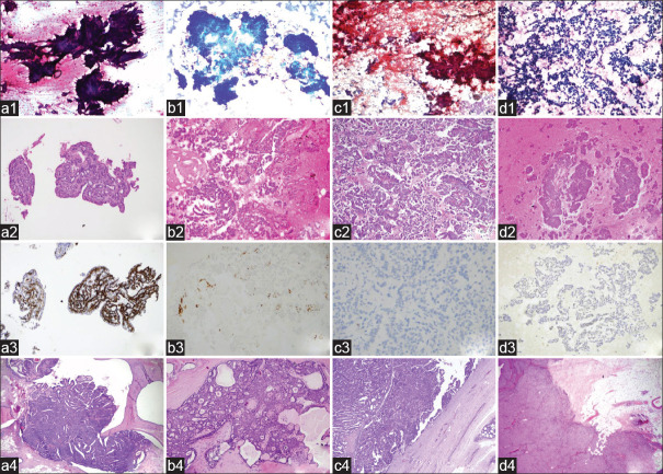
Context: Diagnosis of papillary lesions of the breast by fine needle aspiration cytology (FNAC) is problematic. For this reason, it is situated in the indeterminate zone in classification systems.

Aims: To ascertain the accuracy of cytological diagnosis of papillary lesions in distinguishing papillary lesions from non-papillary lesions and to determine whether papillomas can be reliably distinguished from malignant papillary lesions by FNAC.

Material and methods: A total of 346 cases with the diagnoses of breast papillary lesions were selected among 5112 breast FNAC procedures performed in our center. One hundred and thirty-nine cases with excised lesions were included in this study, and their corresponding histology was reviewed.

Results: Papillary lesion diagnosis was confirmed by histopathology in 103 (74.1%) of 139 patients. Cytology and histopathology results were not found to be compatible in 35 (25.2%) cases. The diagnostic accuracy of distinguishing papillary breast lesions as malignant or benign was assessed statistically. According to the cytology-histology comparison, one case was evaluated as false negative (FN) and twelve cases as false positive (FP). Overall accuracy, sensitivity, specificity, positive predictive value (PPV), and negative predictive value (NPV) of FNAC in distinguishing papillary lesions as benign or malignant were calculated as 87%, 97%, 83%, 72%, and 98%, respectively.

Conclusions: The diagnostic accuracy of papillary breast lesions classified by FNAC might be improved by careful evaluation together with cytological, radiological, and clinical findings (triple test). Cell block may allow more accurate evaluation of the papillary lesion and can be applied to immunohistochemical examination. It may also facilitate the differentiation of benign/malignant papillary lesions.

Keywords: Breast; FNAC; papillary carcinoma; papillary lesion; papilloma.


Conflict of interest statement

There are no conflicts of interest.

Figures

Figure 1
Figure 1
a: Intraductal papilloma: three-dimensional papillary fragment of epithelial cells, papanicolaou (PAP) x200 (a1); papillary structure in the cell block, H and E x200 (a2); positive staining with CK14, IHC x200 (a3); tissue section showing the intraductal papillary proliferation, H and E x40 (a4). b: Papillary ductal carcinoma in situ: crowded cell groups, May Grunwald-Giemsa (MGG) x100 (b1); uniform tumor cells overlying papillary structures H and E x100 (b2); foci with loss of myoepithelial in cell block shown with CK14, IHC x100 (b3); monotonous cell foci within the papilloma in the resection material, H and E x100 (b4). c: Encapsulated papillary carcinoma: hypercellular aspirate, large papillary structures, and individually dispersed atypical cells, some of which show columnar morphology, papanicolaou x100 (c1); well-formed papillary structures in cell block H and E x100 (c2); absence of basal layer staining with p63 in encapsulated papillary carcinoma consisting of monoclonal proliferation, IHC x200 (c3); tumor tissue completely surrounded by a thick fibrous capsule, H and E x100 (c4); d: Solid papillary carcinoma: cell groups consisting of a few cells, some of them columnar, showing significant loss of cohesion in the ground, MGG x40 (d1); three-dimensional papillary cell groups of varying sizes in the cell block, H and E x100 (d2); monotonous appearance with CK5/6, IHC x200 (d3); well-circumscribed, solid tumor in resection material H and E x100 (d4)
Figure 2
Figure 2
a: Proliferative fibrocystic changes and papillomatosis: Three-dimensional, cohesive, benign ductal epithelial cell clusters in papillary configuration on the ground of few bipolar bare-nucleated myoepithelial cells, PAP x40 (a1); apocrine metaplastic cell groups and foamy histiocytes, PAP x40 (a2); proliferative fibrocystic changes (papillomatosis, apocrine hyperplasia, and moderate epithelial hyperplasia and microcysts) in resection specimen H and E x100 (a3). b: Fibroadenoma with epithelial hyperplasia: hypercellular aspirate PAP x100 (b1); cohesive benign ductal epithelium clusters with three-dimensional papillary configuration PAP x100 (b2); florid epithelial hyperplasia focus in typical FA in tissue section H and E x100 (b3). c: Tubular carcinoma: cohesive, large and small, monotonous-looking epithelial clusters and cells showing loss of cohesiveness on the ground PAP x40 (c1); monotonous cell groups forming papilla-like structures, tubules, clefts in the cell block H and E x200 (c2); carcinoma consisting of well-formed glandular structures, with diffuse protrusions on the apical surface of the cytoplasm, H and E x100 (c3). d: Invasive ductal carcinoma on the background of papilloma: hypercellular aspirate. Numerous clumps of adherent and three-dimensional epithelial clusters, as well as atypical cells, most of which are individually shed and isolated PAP x200 (d1); papillary structures and dyshesive cells in the cell block H and E x100 (d2); surgical follow-up invasive ductal carcinoma with ductal carcinoma in situ H and E x100 (d3)

References

    1. Rosen PP, editor. 2nd ed. Philadelphia: Lippincott, Williams & Wilkins; 2001. Rosen's breast pathology.
    1. WH Classification of Tumors Editorial Board. Breast tumours. International Agency for Research on Cancer. 5th ed. Vol. 2. Lyon (France): 2019. WHO Classification of Tumours Series.
    1. Rakha EA, Ellis IO. Diagnostic challenges in papillary lesions of the breast. Pathology. 2018;50:100–10. - PubMed
    1. Masood S, Loya A, Khalbuss W. Is core needle biopsy superior to fine-needle aspiration biopsy in the diagnosis of papillary breast lesions? Diagn Cytopathol. 2003;28:329–34. - PubMed
    1. Field AS, Raymond WA, Schmitt FC. The International Academy of Cytology Yokohama System for reporting breast fine needle aspiration biopsy cytopathology. ActaCytol. 2019;63:257–73. - PubMed

LinkOut - more resources