Review
. 2022 May;59(3):399-414.
doi: 10.1177/03009858221083038. Epub 2022 Mar 26.

Canine and murine models of osteosarcoma

Affiliations
Review

Canine and murine models of osteosarcoma

Jessica Beck et al. Vet Pathol. 2022 May.

Abstract

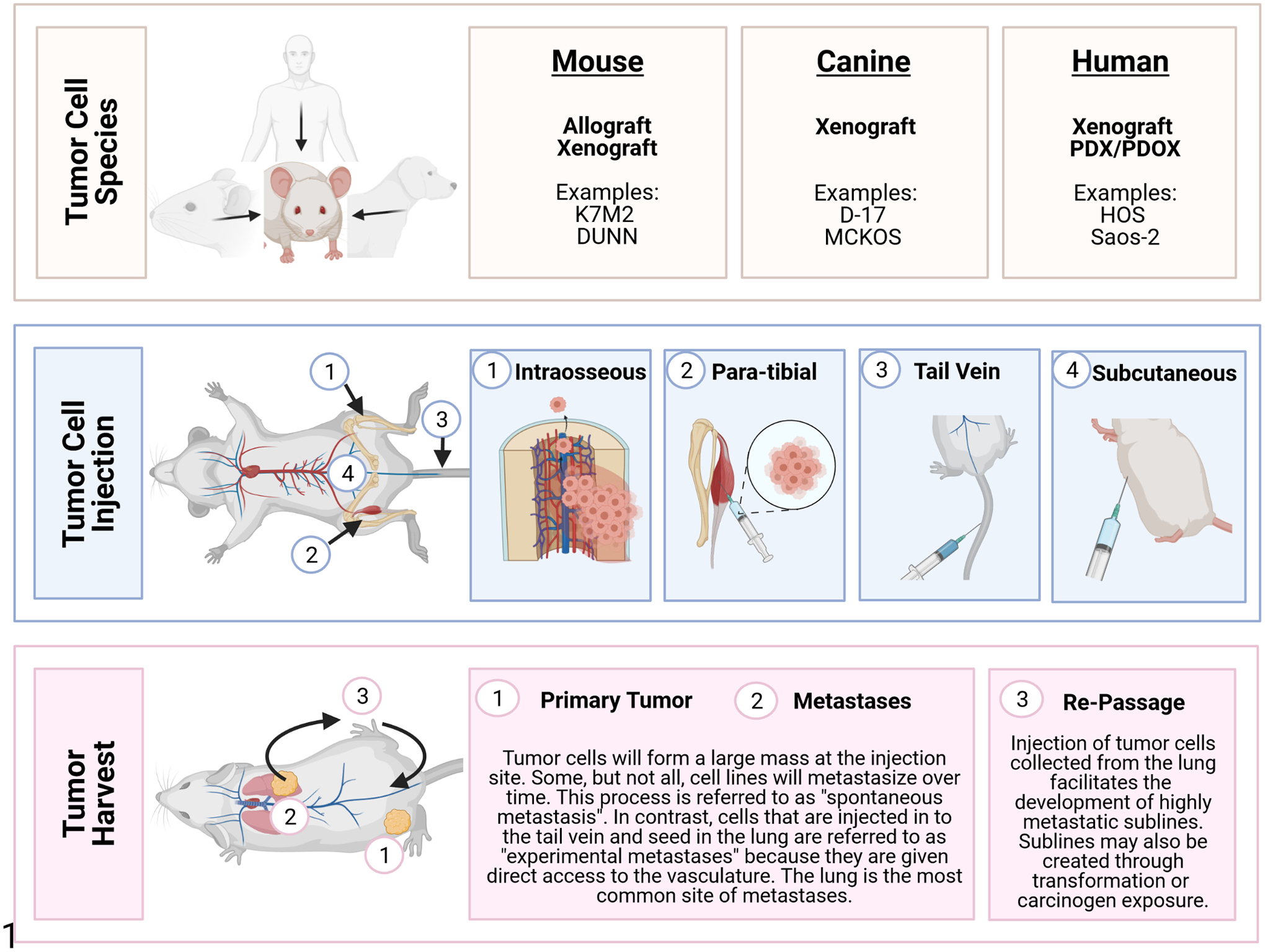
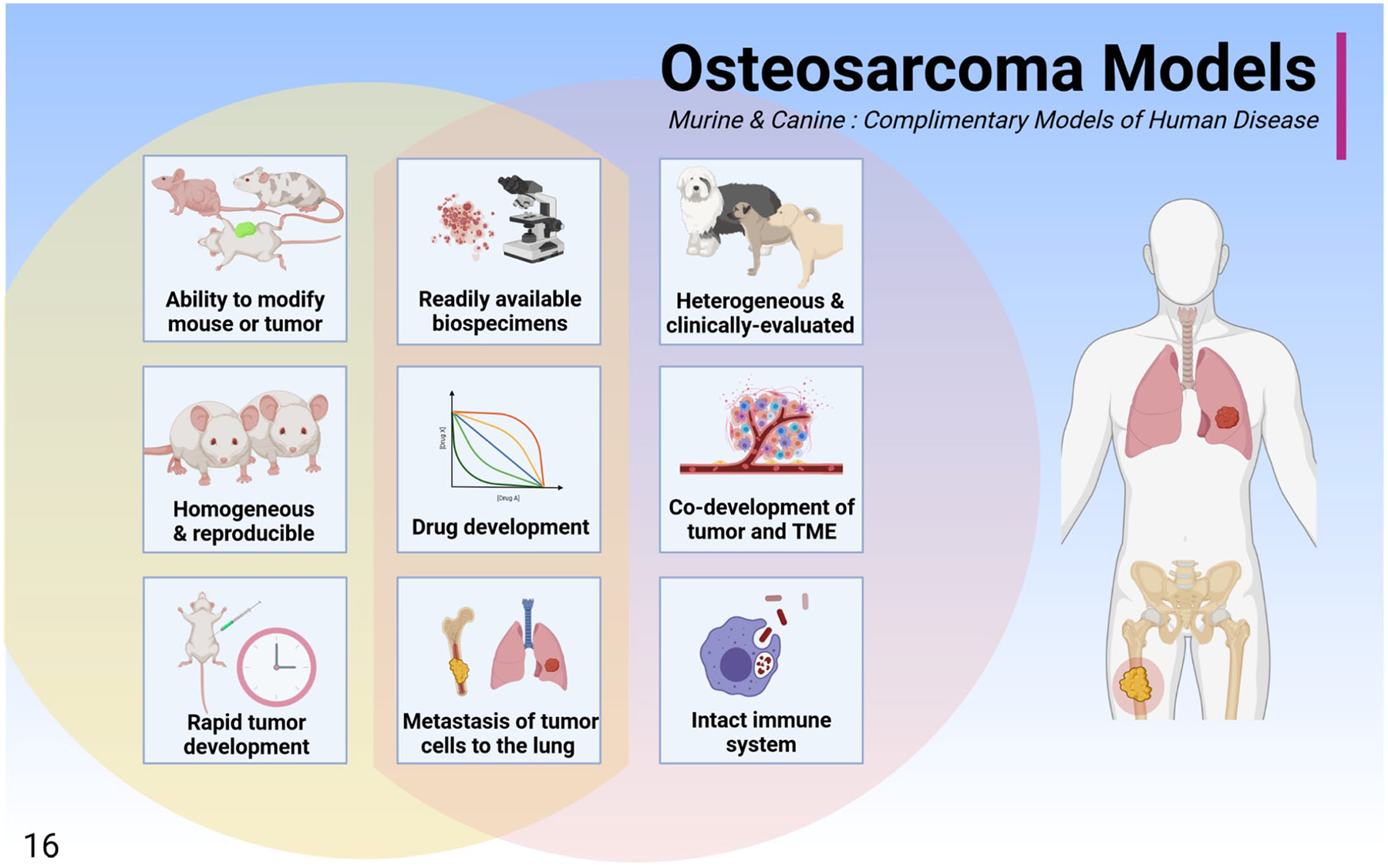
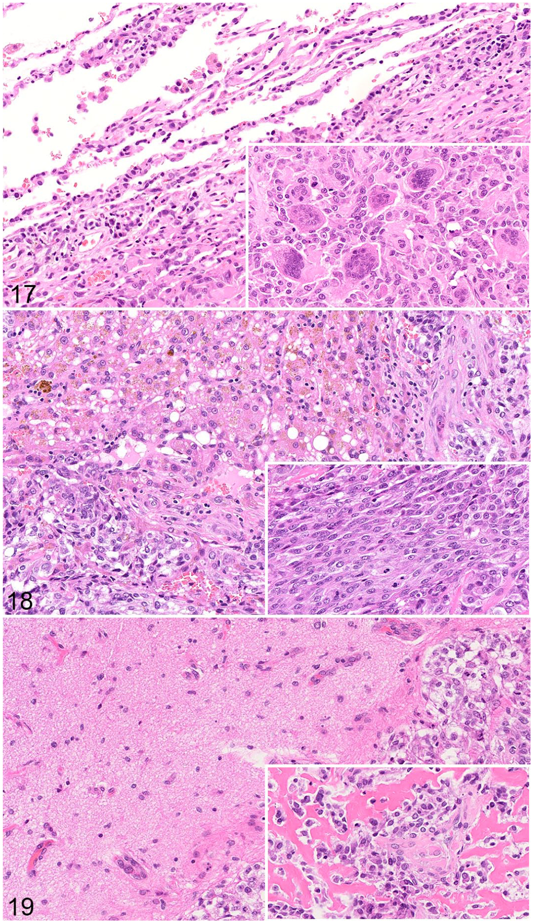
Osteosarcoma (OS) is the most common malignant bone tumor in children. Despite efforts to develop and implement new therapies, patient outcomes have not measurably improved since the 1980s. Metastasis continues to be the main source of patient mortality, with 30% of cases developing metastatic disease within 5 years of diagnosis. Research models are critical in the advancement of cancer research and include a variety of species. For example, xenograft and patient-derived xenograft (PDX) mouse models provide opportunities to study human tumor cells in vivo while transgenic models have offered significant insight into the molecular mechanisms underlying OS development. A growing recognition of naturally occurring cancers in companion species has led to new insights into how veterinary patients can contribute to studies of cancer biology and drug development. The study of canine cases, including the use of diagnostic tissue archives and clinical trials, offers a potential mechanism to further canine and human cancer research. Advancement in the field of OS research requires continued development and appropriate use of animal models. In this review, animal models of OS are described with a focus on the mouse and tumor-bearing pet dog as parallel and complementary models of human OS.

Keywords: canine; comparative oncology; dogs; experimental animal models; metastasis; mice; murine models; osteosarcoma; review; veterinary clinical trials.


Conflict of interest statement

Declaration of Conflicting Interests

The author(s) declared no potential conflicts of interest with respect to the research, authorship, and/or publication of this article.
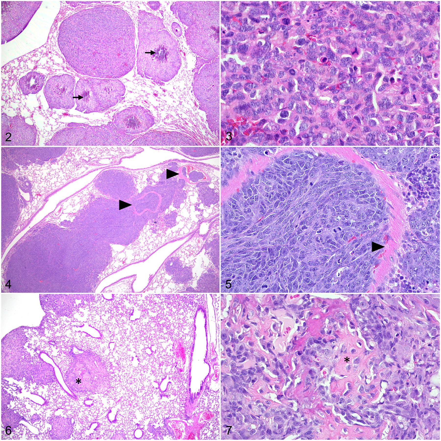
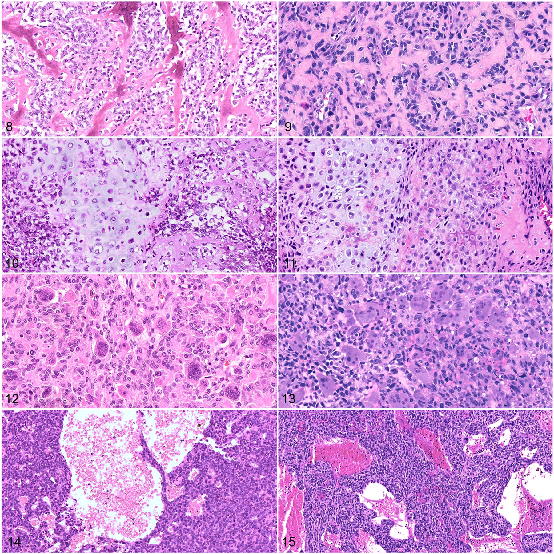
Figures

Figure 1.
Figure 1.
Osteosarcoma (OS) mouse models. OS cell lines may be derived from spontaneous primary OS or metastatic tumors that develop in humans, mice, and dogs (Tables 1 and 3). Once cell lines are established, they can be introduced into mouse models by several injection techniques including intraosseous, para-tibial, tail vein, or subcutaneous. Tail vein injection is associated with production of pulmonary tumors. In contrast, the other 3 methods lead to the development of a large neoplastic nodule at the injection site. These can progress to metastatic disease which primarily affects the lung but may also involve other tissues or organs. Highly metastatic sublines can be developed by collecting and re-passaging tumor cells through the lung.
Figures 2–7.
Figures 2–7.
Osteosarcoma, lung, mouse. Hematoxylin and eosin. Figure 2. Following para-tibial injection, SaOS-2 cells form pulmonary nodules with central mineralization (arrows). Figure 3. Lung metastasis of SaOS-2 cells with mitotic figures. Figure 4. Following tail vein injection, DLM8 murine OS cells form numerous pulmonary nodules. Tumor emboli are observed within pulmonary vessels (arrowheads). Figure 5. The remnants of a vessel wall (arrowhead) within a DLM8 cell tumor nodule. Figure 6. Para-tibial injection of MC-KOS canine OS cells forms multiple pulmonary tumors. Few tumor nodules contain central deposits of eosinophilic matrix (asterisk). Figure 7. Lung metastasis of MC-KOS cells with production of variably mineralized osteoid (asterisk).
Figures 8–15.
Figures 8–15.
Osteosarcoma, dog (left column) and human (right column). Hematoxylin and eosin. Figures 8 and 9. Osteoid is a distinguishing tumor feature observed in canine (Figure 8) and human (Figure 9) osteosarcomas. Figures 10 and 11. Some canine (Figure 10) and human (Figure 11) osteosarcomas also contain chondroid matrix (chondroblastic subtype). Figures 12 and 13. Giant cell-rich osteosarcomas are an uncommon histologic variant reported in both dogs (Figure 12) and humans (Figure 13). Figures 14 and 15. Telangiectatic osteosarcomas are histologically characterized by blood-filled spaces lined by tumor cells (dog, Figure 14; human, Figure 15).
Figure 16.
Figure 16.
The murine and canine osteosarcoma models are complimentary models of human disease that can be used separately or in parallel to interrogate osteosarcoma biology. These models share several benefits including readily available biospecimens, the ability to develop drugs, and tumor biology that is similar to that observed in humans, including the development of pulmonary metastases. While the mouse model is rapid, reproducible, and can be experimentally modulated, the dog may be more representative of human clinical disease due to natural co-development of the tumor and tumor microenvironment, shared environmental exposures, a diverse genetic background, and the presence of an intact immune system.
Figures 17–19.
Figures 17–19.
Metastatic osteosarcoma, dog. Hematoxylin and eosin. Figure 17. Lung. Alveoli along the edge of the metastasis are compressed; many contain erythrocytes. Inset: multinucleated cells are observed throughout the mass. Figure 18. Liver. There is mild lymphocytic infiltration of the hepatic parenchyma adjacent to the tumor. Many hepatocytes are vacuolated and contain brown pigment. Inset: The metastasis is comprised of polygonal to spindle-shaped cells. Figure 19. Brain. There is a well-demarcated OS metastasis with compression of the adjacent parenchyma. Inset: Osteoid is abundant within the tumor.

References

    1. Abarrategi A, Gambera S, Alfranca A, et al. c-Fos induces chondrogenic tumor formation in immortalized human mesenchymal progenitor cells. Sci Rep. 2018;8:15615–15613. - PMC - PubMed
    1. Al-Khan AA, Nimmo JS, Day MJ, et al. Fibroblastic subtype has a favourable prognosis in appendicular osteosarcoma of dogs. J Comp Pathol. 2020;176:133–144. - PubMed
    1. Angstadt AY, Motsinger-Reif A, Thomas R, et al. Characterization of canine osteosarcoma by array comparative genomic hybridization and RT-qPCR: signatures of genomic imbalance in canine osteosarcoma parallel the human counterpart. Genes Chromosom Cancer. 2011;50:859–874. - PubMed
    1. Angstadt AY, Thayanithy V, Subramanian S, et al. A genome-wide approach to comparative oncology: high-resolution oligonucleotide aCGH of canine and human osteosarcoma pinpoints shared microaberrations. Cancer Genet. 2012;205:572–587. - PubMed
    1. Asai T, Ueda T, Itoh K, et al. Establishment and characterization of a murine osteosarcoma cell line (LM8) with high metastatic potential to the lung. Int J Cancer. 1998;76:418–422. - PubMed

Publication types