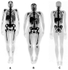
Assessing Bone Marrow Activity with [ 18F]FLT PET in Patients with Essential Thrombocythemia and Prefibrotic Myelofibrosis: A Proof of Concept
- PMID: 35341409
- PMCID: PMC8966096
- DOI: 10.1177/15330338221086396
Assessing Bone Marrow Activity with [ 18F]FLT PET in Patients with Essential Thrombocythemia and Prefibrotic Myelofibrosis: A Proof of Concept
Abstract
Objectives: This study aims to assess the value of FLT-PET as a non-invasive tool to differentiate between patients with ET and Pre-PMF. This study is a pilot study to have a proof of concept only. Methods: This is a prospective, interventional study where a total of 12 patients were included. Each patient underwent FLT PET imaging as well as bone marrow examination (gold standard). In addition, semi-quantitative (SUVmax and SUVmean) measurements of FLT uptake in the liver, spleen, and Lspine, SUVmean, as well as the Total Lesion Glycolysis (TLG) of the Lspine were performed. Results from the two patient cohorts were compared using = Kruskal-Wallis statistical test. A P-value of <.05 is considered to be statistically significant. Results: The differences in FLT SUVmax and SUVmean measurements in the three organs (liver, spleen, and LSpine) between the ET and Pre-PMF patients were not statistically significant (P > .05). In contrast, TLG measurements in the LSpine were statistically different (P = .013), and therefore, compared to gold standard bone marrow results, TLG can separate ET and Pre-PMF patients. Conclusion: This study is a proof of concept about the potential to discriminate between ET and pre-PMF patients in a non-invasive way. TLG of the LSpine in FLT PET images is a potential quantitative parameter to distinguish between ET and pre-PMF patients.
Keywords: essential thrombocythemia; fluorothymidine F-18; positron emission tomography; prefibrotic myelofibrosis.
Conflict of interest statement
Figures


References
-
- Agool A, Schot BW, Jager PL, Vellenga E. 18F-FLT PET in hematologic disorders: a novel technique to analyze the bone marrow compartment. J Nucl Med. 2006. Oct 1;47(10):1592-1598. - PubMed
-
- Arber DA, Orazi A, Hasserjian R, et al. The 2016 revision to the World Health Organization classification of myeloid neoplasms and acute leukemia. Blood. 2016 May 19;127(20):2391-2405. - PubMed
-
- Thiele J, The KH. WHO Diagnostic criteria for polycythemia vera, essential thrombocythemia, and primary myelofibrosis. Curr Hematol Malig Rep. 2009((Jan 4(1):33-40. - PubMed
Publication types
MeSH terms
Substances
LinkOut - more resources
Full Text Sources

