. 2022 May 15;434(9):167559.
doi: 10.1016/j.jmb.2022.167559. Epub 2022 Mar 24.

Acquired Disorder and Asymmetry in a Domain-Swapped Model for γ-Crystallin Aggregation

Affiliations

Acquired Disorder and Asymmetry in a Domain-Swapped Model for γ-Crystallin Aggregation

Vatsala Sagar et al. J Mol Biol. .

Abstract

Misfolding and aggregation of proteins occur in many pathological states. Because of the inherent disorder involved, these processes are difficult to study. We attempted to capture aggregation intermediates of γS-crystallin, a highly stable, internally symmetrical monomeric protein, by crystallization under mildly acidic and oxidizing conditions. Here we describe novel oligomerization through strained domain-swapping and partial intermolecular disulfide formation. This forms an octamer built from asymmetric tetramers, each of which comprises an asymmetric pair of twisted, domain-swapped dimers. Each tetramer shows patterns of acquired disorder among subunits, ranging from local loss of secondary structure to regions of intrinsic disorder. The octamer ring is tied together by partial intermolecular disulfide bonds, which may contribute to strain and disorder in the octamer. Oligomerization in this structure is self-limited by the distorted octamer ring. In a more heterogeneous environment, the disordered regions could serve as seeds for cascading interactions with other proteins. Indeed, solubilized protein from crystals retain many features observed in the crystal and are prone to further oligomerization and precipitation. This structure illustrates modes of loss of organized structure and aggregation that are relevant for cataract and for other disorders involving deposition of formerly well-folded proteins.

Keywords: aggregation; asymmetry; disorder; domain-swapping; unfolding.


Conflict of interest statement

Declaration of interests

The authors declare that they have no known competing financial interests or personal relationships that could have appeared to influence the work reported in this paper.

Figures

Figure 1.
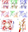
Figure 1.. An asymmetric octameric structure for oxidized mouse L16-γS.
A) Ribbon structure of a typical monomeric γS-crystallin (PDB: 5VH1) showing N- and C-domains. B) Arrangement of N (red)and C-domains (blue) and partial disulfide bonds (yellow) in the octamer (viewed down the axis of the N-domain assembly). C) Orthogonal view of octamer showing two layers of domains and partial disulfides. D) Heat map of B-factors for the octamer, showing asymmetry. The core is better folded than the periphery and one side is more disordered than the other. Color panel shows gradation of B factor values. E) A view of the octamer colored by subunits (s1: red; s2: orange; s3: green; s4: blue) down the axis of the stacked N-domains. F) Orthogonal view of the octamer colored by subunit as in E.
Figure 2.
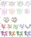
Figure 2.. Domain-swapping in the octamer
A) Electron density (2FO-FC; 1σ) for the connecting peptides of s1–4. Side chains are poorly defined, but the direction and connectivity of the backbone is clear. ;B) The domain arrangement for one the s1/s2 domain-swapped dimer of the γS octamer is compared with the relaxed domain-swap seen in crystals of βB2-crystallin. In the γS octamer, the dimer is twisted compared with the relaxed βB2 version. C) Alignment of the main chain ribbon for the four connecting peptides (colored as in A) showing different degrees of twist and a shorter span than that in βB2 (violet). D) Main chain traces for s1–s4 with N-domains aligned. The four C-domains have slightly different relative orientations. E) Four domain swapped dimers (each pair colored differently) assemble to form the octamer.
Figure 3.
Figure 3.. N-domain contacts in the octamer.
A) Electron density (2FO-FC; 1σ) for partial C24-C24 disulfide bond. The paired cysteines for s1 are close but do not form a disulfide. B) The arrangement of the octamer layer interface. The C24-C24 pairs are flanked by H83 of the connecting peptides and the N-terminal (N-) arms. C) Other N domain interactions. In each layer of the octamer, four N-domains form contacts centered on L16. D) Details of interaction in C.
Figure 4.
Figure 4.. Secondary structure changes and disorder in the octamer.
A) Electron density for the surface exposed β-sheet of the C-domain in s1–s4. The b-a-d-c’ order of β-strands is shown for s1. At 1σ density, several side chains are missing as well as parts of backbone in s4. B) Apparent loss of hydrogen bonds in the exposed C-domain β-sheets in s1–s4. Asterisks indicate lost hydrogen-bonds, as judged by distance and geometry or by loss of density. Viewed from a slightly different angle from A) to make sheet structure clearer. C) Alignment of chain trace for N- and C-domains of s1–s4 (colored as before) showing differences in the backbone path. Some major loop shifts are indicated by stars. D) Loss of a Tyr corner in the C-domain of s2. Left: The expected arrangement in s1; right: main chain deviation in s2. Asterisk shows position of K153 which is either up or down depending on whether the bend is V-shaped or broadened. E) Electron density (2FO-FC; 1σ) for C22, 24, 26, 82 in s1–4 (colored as in other figures) showing evidence for partial C22–82 disulfide.
Figure 5:
Figure 5:. Multimerization and disorder in dissolved octamer crystals
A) CD spectra of uncrystallized monomer and dissolved crystals of γS-L16. Evidence for a loss of organized secondary structure in the crystal as the β-sheet minimum broadens with a shoulder at 205nm. Spectra were acquired at room temperature in 0.3 mg/ml in 50 mM sodium acetate pH 5.0, buffer using a 1-mm path length cuvette. Crystal samples were diluted tenfold (from 30% glycerol stock) and both spectra were acquired in 3% glycerol (v/v), which was added to the uncrystallized sample. B) SEC-MALS of uncrystallized (left panel) and dissolved crystals of γS-L16 (right panel) at 1 week. Red trace shows light scattering (LS); green shows UV absorbance at 280nm. Monomer shows a single major peak consistent with a size of 22kDa. Dissolved crystal shows two major peaks consistent with dimer (48kDa) and octamer (152kDa) size ranges in solution. Molecular mass scale is shown as kDa. C) Non-reducing SDS PAGE of crystallized protein gives a 44kDa band consistent with a disulfide linked dimer. Lane 1 shows uncrystallized protein; 2 shows dissolved crystal under non-reducing conditions; 3 shows dissolved crystal under reducing conditions. The minor band apparently running below 20kDa in both native and crystallized protein was analyzed by mass spectroscopy and determined to be full-length protein containing an internal disulfide bond (as seen in figure 4E). D) Non-reducing SDS PAGE of a time course (0–90 mins) of glutaraldehyde cross-linking of uncrystallized monomer and dissolved crystals of γS-L16. For uncrystallized protein (left side of gel) monomer is predominant. In contrast, the dissolved crystal shows patterns consistent with increasing multimer formation up to octamer. E) Mass photometry (ISCAMS) of dissolved crystal solution. The MP data was plotted as kernel density estimate (KDE) distributions (blue histograms) with Gaussian function fit (red) for average molecular mass of each distribution component. Mass estimates are shown. This technique is performed at relatively low concentration which favors dissociation of the larger complexes.
Fig 6.
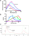
Fig 6.. The crystallized protein is aggregation-prone.
A) Trp fluorescence spectra of native protein and dissolved crystal (2 weeks old) in buffer and in 7M GdnCl. Unfolding the native protein red-shifts the maximum intensity. The dissolved crystal gives a red-shifted spectrum consistent with partial unfolding. Uncrystallized (U) and crystallized (C) samples in buffer are shown by red and blue lines respectively; the same preparation in 7M GdnCl are shown in pink and light blue. B) Turbidity of solutions at 600nm. While uncrystallized monomer remains stable in solution at 60C, the dissolved crystal rapidly forms light scattering particles and precipitates under the same conditions. Results are shown for two preparations of crystal, both freshly dissolved (red and green traces, day (D) 1) and after a further 24 hrs at 20C, the same as the crystallization conditions (dark and light blue traces, day (D) 2), Turbidity increases more rapidly on day 2 (D2). All samples contained a total of 3% glycerol (v/v). C) SEC-MALS of 3-week-old dissolved crystals. Compared with data in Fig 5B, there is evidence for an increase in formation of larger species, up to 4×10⁶ Da, consistent with increasing tendency towards aggregation. Light scattering (LS) and UV (280nm) traces are indicated. Calculated size ranges for LS peaks are shown in kDa.

Similar articles

Cited by

References

    1. Chiti F & Dobson CM (2006). Protein misfolding, functional amyloid, and human disease. Annu Rev Biochem 75, 333–66. - PubMed
    1. Lee S, Mahler B, Toward J, Jones B, Wyatt K, Dong L, Wistow G & Wu Z (2010). A single destabilizing mutation (F9S) promotes concerted unfolding of an entire globular domain in gammaS-crystallin. J Mol Biol 399, 320–30. - PMC - PubMed
    1. Mahler B, Doddapaneni K, Kleckner I, Yuan C, Wistow G & Wu Z (2011). Characterization of a transient unfolding intermediate in a core mutant of gammaS-crystallin. J Mol Biol 405, 840–50. - PMC - PubMed
    1. Dyson HJ & Wright PE (2005). Intrinsically unstructured proteins and their functions. Nat Rev Mol Cell Biol 6, 197–208. - PubMed
    1. Ferina J & Daggett V (2019). Visualizing Protein Folding and Unfolding. J Mol Biol 431, 1540–1564. - PubMed

Publication types

LinkOut - more resources