. 2022 Jul;102(1):96-107.
doi: 10.1016/j.kint.2022.02.029. Epub 2022 Mar 24.

Non-canonical Wnt/calcium signaling is protective against podocyte injury and glomerulosclerosis

Affiliations

Non-canonical Wnt/calcium signaling is protective against podocyte injury and glomerulosclerosis

Dong Zhou et al. Kidney Int. 2022 Jul.

Abstract

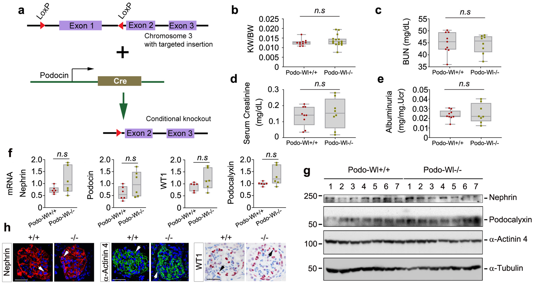
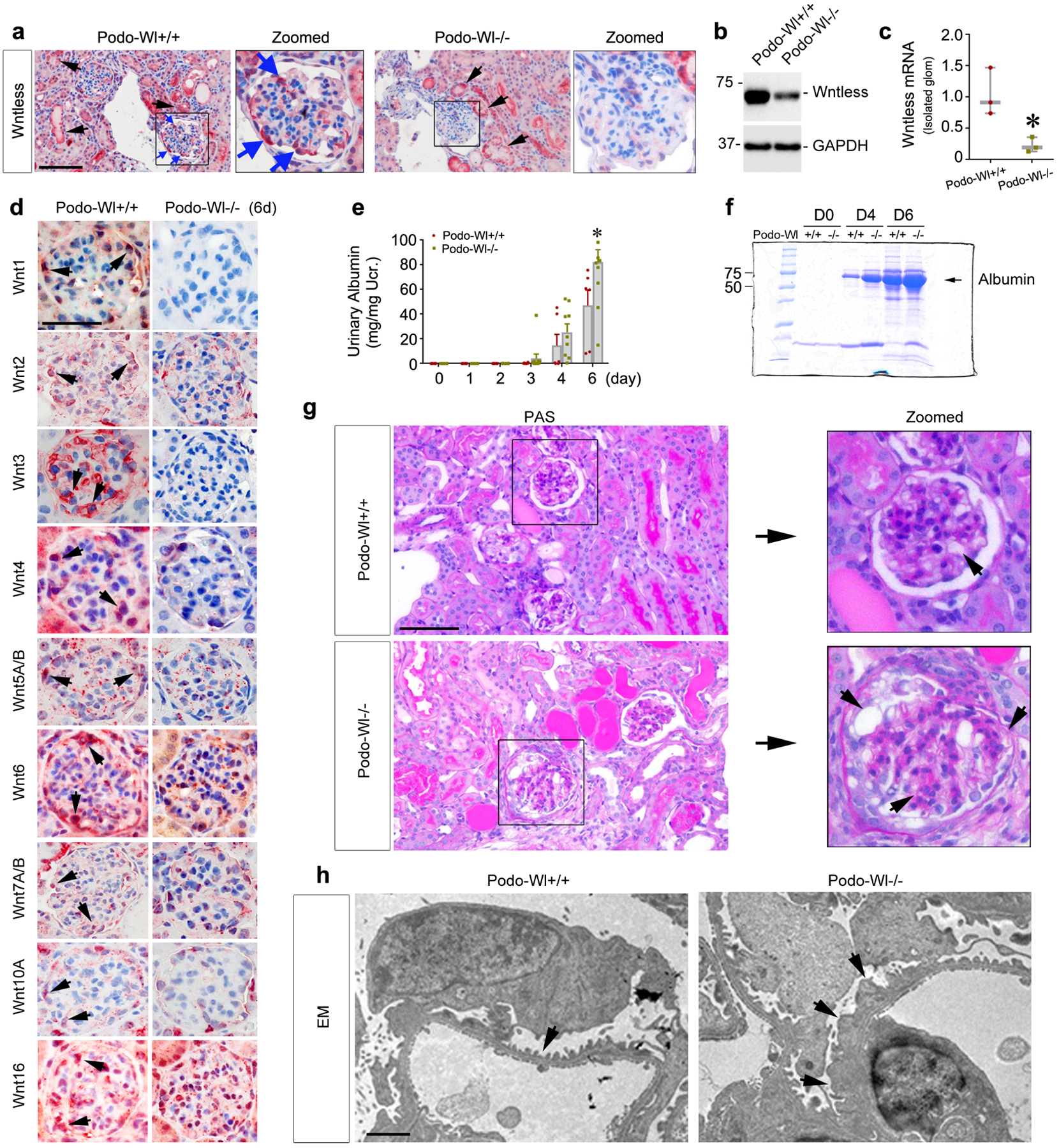
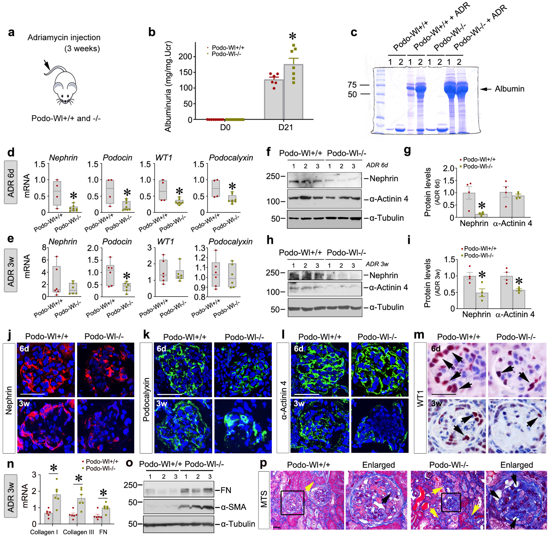
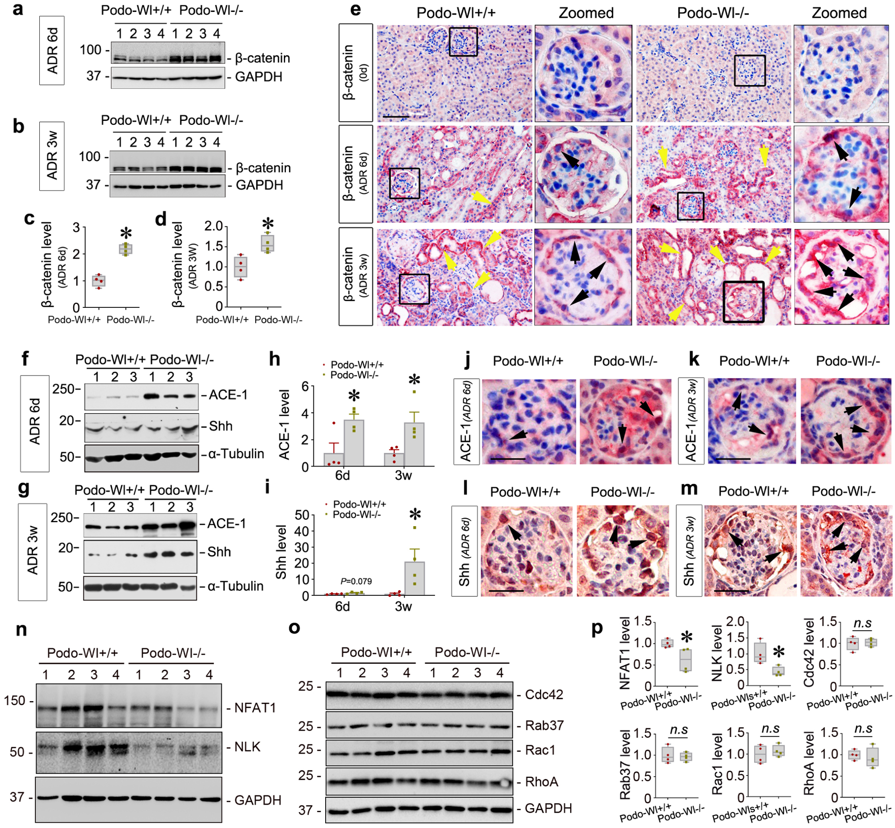
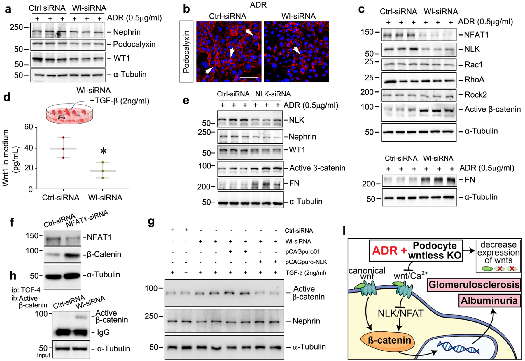
Activation of canonical Wnt signaling has been implicated in podocyte injury and proteinuria. As Wnts are secreted proteins, whether Wnts derived from podocytes are obligatory for promoting proteinuria remains unknown. To address this, we generated conditional knockout mice where Wntless, a cargo receptor protein required for Wnt secretion, was specifically deleted in glomerular podocytes. Mice with podocyte-specific ablation of Wntless (Podo-Wntless-/-) were phenotypically normal. However, after inducing kidney damage with Adriamycin for six days, Podo-Wntless-/- mice developed more severe podocyte injury and albuminuria than their control littermates. Surprisingly, ablation of Wntless resulted in upregulation of β-catenin, accompanied by reduction of nephrin, podocin, podocalyxin, and Wilms tumor 1 proteins. In chronic injury induced by Adriamycin, increased albuminuria, aggravated podocyte lesions and extracellular matrix deposition were evident in Podo-Wntlessl-/- mice, compared to wild type mice. Mechanistically, specific ablation of Wntless in podocytes caused down-regulation of the nuclear factor of activated T cell 1 (NFAT1) and Nemo-like kinase (NLK), key downstream mediators of non-canonical Wnt/calcium signaling. In vitro, knockdown of either NFAT1 or NLK induced β-catenin activation while overexpression of NLK significantly repressed β-catenin induction and largely preserved nephrin in glomerular podocytes. Thus, our results indicate that podocyte-derived Wnts play an important role in protecting podocytes from injury by repressing β-catenin via activating non-canonical Wnt/calcium signaling.

Keywords: Wnt signaling; Wntless; glomerulosclerosis; nemo-like kinase; nuclear factor of activated T cell; podocytes; proteinuria.


Conflict of interest statement

DISCLOSURE

All the authors declared no competing interests.

Figures

Figure 1.
Figure 1.
Podocyte-specific deletion of Wntless has no effects on glomerular structure and function. (a) Schematic diagram shows the strategy of crossbreeding Wl floxed mice (129S-Wlstm1.1Lan/J) with podocin-Cre transgenic mice. The Wl floxed mutant mice possess a loxP site before the ATG start site in the 5’ untranslated region and another upstream of exon 2 of the Wl gene. (b-e) Ablation of Wl in podocytes did not affect the ratio of kidney weight (KW)/body weight (BW) (b), blood urea nitrogen (BUN) (c), serum creatinine levels (d), and urinary albumin levels (albuminuria) (e) compared with control mice (n = 8–15). Age (8–10 weeks)- and sex-matched mice littermates were used. (f) qPCR analyses showed that there was little change in renal mRNA levels of nephrin, podocin, podocalyxin, and WT1 between Podo-Wl+/+ and Podo-Wl−/− mice (n = 6). (g) Western blot analyses demonstrated no significant changes in renal expression of nephrin, podocalyxin, and α-actinin 4 proteins between Podo-Wl+/+ and Podo-Wl−/− mice kidneys (n = 7). (h) Representative micrographs showed protein expression of nephrin, α-actinin 4, and WT1 in Podo-Wl+/+ and Podo-Wl−/− kidneys, as indicated. Four mice were assessed (n = 4) with similar results. Scale bar, 25 μm.
Figure 2.
Figure 2.
Mice with podocytes-specific ablation of Wntless are more susceptible to glomerular injury induced by Adriamycin (ADR). Podo-Wl+/+ and Podo-Wl−/− mice were treated with ADR and euthanized at 6 days after injection. (a) Immunohistochemical staining showed Wl expression in Podo-Wl+/+ and Podo-Wl−/− mice diseased kidneys. Blue arrows indicate positive staining in podocytes. Black arrows indicate positive staining in tubules. Scale bar, 50 μm. (b) Western blot analyses revealed kidney protein levels of Wl in Podo-Wl+/+ and Podo-Wl−/− mice whole kidney lysates. (c) qPCR analyses of Wl mRNA in isolated glomeruli from Podo-Wl+/+ and Podo-Wl−/− mice. *P < 0.05 (n = 3). (d) Immunohistochemical staining demonstrated multiple Wnts including Wnt1, 2, 3, 4, 5A/B, 6, 7A/B, 10A, and 16 were reduced in Podo-Wl−/− mice diseased glomeruli, compared to Podo-Wl+/+ mice. Arrows indicate positive staining. Scale bar, 25 μm. (e) Urinary albumin levels were increased in Podo-Wl−/− mice, compared with Podo-Wl+/+ mice. *P < 0.05 (n = 6–11). (f) Representative SDS–polyacrylamide gel electrophoresis showed the urinary proteins after normalization to urinary creatinine in Podo-Wl+/+ and Podo-Wl−/− mice as indicated at day (D) 0, 4, and 6. Urine proteins after separation were stained with Coomassie blue R-250. (g) Periodic acid–Schiff (PAS) staining showed kidney histologic changes in Podo-Wl+/+ and Podo-Wl−/− mice at 6 days after ADR. Boxed areas are enlarged. Arrows indicate damaged area. Scale bar, 50 μm. (h) Electron microscopy (EM) shows the ultrastructure of glomerular filtration apparatuses in Podo-Wl+/+ and Podo-Wl−/− kidneys. Representative EM images demonstrated podocyte foot process effacement and altered glomerular basement membrane after ADR injection. Scale bar, 1 μm.
Figure 3.
Figure 3.
Blockade of Wnt secretion aggravates podocytes injury and glomerulosclerosis induced by Adriamycin. (a) Schematic diagram. (b) Urinary albumin levels were increased in Podo-Wl−/− mice, compared with Podo-Wl+/+ controls, at day (D) 21 after ADR injection. *P < 0.05 (n = 6–7). (c) Representative Coomassie-stained SDS–polyacrylamide gels showing urinary proteins after normalization to urinary creatinine in Podo-Wl+/+ and Podo-Wl−/− mice as indicated. (d-e) qPCR analyses showed renal mRNA expression of podocyte-specific markers nephrin, podocin, WT1, and podocalyxin in Podo-Wl+/+ and Podo-Wl−/− mice at 6 days (d) and 3 weeks (e) after ADR. *P < 0.05 (n = 4–8 for 6 days, n=6–7 for 3 weeks). (f-i) Western blot analyses showed renal protein levels of nephrin and α-actinin 4 in Podo-Wl+/+ and Podo-Wl−/− mice at 6 days and 3 weeks after ADR injection. Representative western blot (f, h) and quantitative data (g, i) are shown. *P < 0.05 (n = 4). (j-m) Representative micrographs show immunostaining of nephrin (j), podocalyxin (k), α-actinin 4 (l), and WT1 (m) in Podo-Wl+/+ and Podo-Wl−/− kidneys at 6 days and 3 weeks after ADR. Scale bar, 25 μm. (n) qPCR revealed increased mRNA expression of collagen I, collagen III, and fibronectin (FN) in Podo-Wl−/− mice, compared with Podo-Wl+/+ mice at 3 weeks after ADR. *P < 0.05 (n = 6). (o) Western blot analyses showed renal FN and α-SMA levels in Podo-Wl+/+ and Podo-Wl−/− mice at weeks after ADR. (p) Masson’s trichrome staining (MTS) showed collagen deposition in Podo-Wl+/+ and Podo-Wl−/− kidneys at 3 weeks after ADR. Scale bar, 25 μm. Yellow arrows indicate collagen deposited in interstitial compartment. Black arrows indicate collagen deposited in glomeruli.
Figure 4.
Figure 4.
Podocytes-specific ablation of Wntless induces β-catenin activation. (a-d) Western blot analyses show renal β-catenin protein in Podo-Wl−/− mice at 6 days (6d) and 3 weeks (3w) after ADR. Representative western blot (a, b) and quantitative data on β-catenin (c, d) are presented. *P < 0.05 (n = 4). (e) Representative micrographs show β-catenin expression at day 0 (0d), 6 days, and 3 weeks after ADR injection in Podo-Wl+/+ and Podo-Wl−/− mice. Boxed areas are enlarged (Zoomed). Yellow arrows indicate positive staining in tubules. Black arrows indicate positive staining in podocytes. Scale bar, 25 μm. (f, g) Representative western blots show ACE-1 and Shh expression in Podo-Wl+/+ and Podo-Wl−/− kidneys at 6 days or 3 weeks after ADR. (h, i) Graphs of quantitative data for renal ACE-1 (h) and Shh (i) levels after ADR. *P < 0.05 (n = 4). (j, k) Representative micrographs show ACE1 expression at 6 days and 3 weeks after ADR in Podo-Wl+/+ and Podo-Wl−/− glomeruli. Arrows indicate positive staining. Scale bar, 20 μm. (l, m) Representative micrographs show Shh protein expression in Podo-Wl+/+ and Podo-Wl−/− mice diseased glomeruli after ADR at 6 days or 3 weeks. Arrows indicate positive staining. Scale bar, 20 μm. (n-p) Representative western blots (n, o) and quantitative data (p) show NFAT1, NLK, Cdc42, Rab37, Rac1, and RhoA levels in Podo-Wl+/+ and Podo-Wl−/− kidneys after ADR. *P < 0.05 (n = 4).
Figure 5.
Figure 5.
Blockade of Wnt secretion activates β-catenin via repression of non-canonical signaling. (a) Western blot analyses demonstrated knockdown of Wl (Wl-siRNA) in cultured podocytes reduced nephrin, podocalyxin, and WT1 expression under Adriamycin (ADR) stress for 24 hours, compared with control (Ctrl siRNA). (b) Immunofluorescence staining revealed diminished expression of podocalyxin in Wl-knockdown podocytes after stimulation with ADR, compared with control. Arrows indicate positive staining. Scale bar, 25 μm. (c) Blockade of Wnt secretion by knocking down Wl inhibited non-canonical Wnt signaling. Representative western blot analyses show that knockdown of Wl significantly reduced NFAT1 and NLK levels (but not Rac1, RhoA, Rab37, and ROCK2) and activated β-catenin when podocytes were presented with ADR at 0.5μg/ml, compared with control scrambled siRNA. (d) ELISA revealed Wnt1 concentration in the Wl-knockdown podocytes conditioned medium after incubation with TGF-β. (e) Western blot analyses show that knockdown of NLK repressed nephrin and WT1 and induced active β-catenin and fibronectin (FN) in cultured podocytes after incubation with ADR. (f) Western blot analyses showed that knockdown of NFAT1 induced β-catenin activation in cultured podocytes. (g) Western blot analyses demonstrated that overexpression of NLK repressed β-catenin activation and preserved nephrin expression in Wl-knockdown podocytes after incubation with TGF-β in vitro. (h) β-catenin functionally binds to TCF-4 to activate downstream gene transcription. Podocyte Wl knockdown (Wl-siRNA) was immunoprecipitated (IP) with TCF-4 antibody, followed by immunoblotting (IB) with antibody against active β-catenin. (i) Our model shows that ablation of Wl in podocytes represses noncanonical Wnt/calcium signaling, which leads to β-catenin activation, proteinuria, and glomerulosclerosis.

Similar articles

Cited by

References

    1. Tan RJ, et al. Tubular injury triggers podocyte dysfunction by beta-catenin-driven release of MMP-7. JCI Insight 4; e122399 (2019). - PMC - PubMed
    1. Greka A & Mundel P Cell biology and pathology of podocytes. Annu Rev Physiol 74, 299–323 (2012). - PMC - PubMed
    1. Torban E, et al. From podocyte biology to novel cures for glomerular disease. Kidney Int 96, 850–861 (2019). - PubMed
    1. Assady S, Wanner N, Skorecki KL & Huber TB New Insights into Podocyte Biology in Glomerular Health and Disease. J Am Soc Nephrol 28, 1707–1715 (2017). - PMC - PubMed
    1. Zhou L & Liu Y Wnt/beta-catenin signalling and podocyte dysfunction in proteinuric kidney disease. Nat Rev Nephrol 11, 535–545 (2015). - PMC - PubMed

Publication types