. 2021 Apr 8;12(16):5787-5795.
doi: 10.1039/d1sc01331a. eCollection 2021 Apr 28.

Light-guided intrabodies for on-demand in situ target recognition in human cells

Affiliations

Light-guided intrabodies for on-demand in situ target recognition in human cells

Eike F Joest et al. Chem Sci. .

Abstract

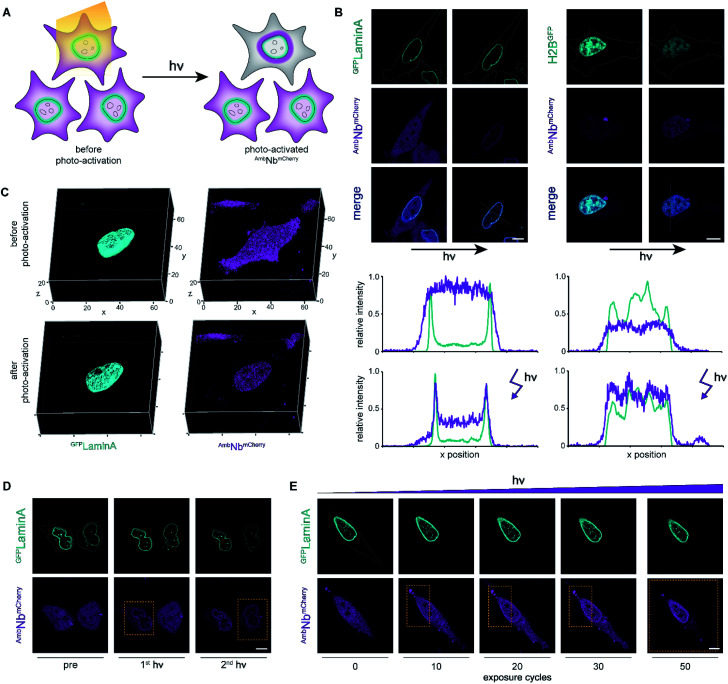
Due to their high stability and specificity in living cells, fluorescently labeled nanobodies are perfect probes for visualizing intracellular targets at an endogenous level. However, intrabodies bind unrestrainedly and hence may interfere with the target protein function. Here, we report a strategy to prevent premature binding through the development of photo-conditional intrabodies. Using genetic code expansion, we introduce photocaged amino acids within the nanobody-binding interface, which, after photo-activation, show instantaneous binding of target proteins with high spatiotemporal precision inside living cells. Due to the highly stable binding, light-guided intrabodies offer a versatile platform for downstream imaging and regulation of target proteins.


Conflict of interest statement

The authors declare no conflicts of interest.

Figures

Fig. 1
Fig. 1. On-demand tracing of intracellular targets by photo-activatable intrabodies. (A) The X-ray structure of the nanobody-GFP complex (pdb: 3K1K) reveals Tyr37 as a key residue in the binding region to GFP (turquoise). Nanobody, magenta. (B) Tyr37 is replaced by ortho-nitrobenzyl- or nitropiperonyl-caged tyrosines (ONBY or NPY, respectively). (C) Schematic illustration of stable cell line generation for expression of photo-activated intrabodies by genetic code expansion. Mammalian cells are co-transfected with plasmids encoding the recombinase and either wild-type (WTNbmCherry) or amber-containing nanobodies (AmbNbmCherry).
Fig. 2
Fig. 2. Expression of photo-activatable intrabodies by stable cell lines. (A) Wild-type intrabody WTNbmCherry is expressed after tetracycline induction. For intracellular binding analysis, a plasmid encoding a GFP-tagged target is transiently transfected; tet, tetracycline. (B) WTNbmCherry expression analyzed by flow cytometry without (−) or with (+) tetracycline induction. (C) Live-cell imaging showing expression of WTNbmCherry and binding of mEGFPLaminA (relative PMT gain settings from the image acquisition in grey). (D) Intrabody AmbNbmCherry expression by amber suppression with photocaged tyrosines. AmbNbmCherry-positive cells are transiently transfected with plasmids encoding the optimized NPYRS/tRNA pair and a target protein, respectively. After transfection, AmbNbmCherry expression was induced when cells were supplied with photocaged tyrosines. (E) Expression of WTNbmCherry and AmbNbmCherry monitored by flow cytometry. WTNbmCherry expression was tetracycline-induced and AmbNbmCherry was expressed using amber suppression conditions. Cell counts were normalized to mode (n = 3). (F) Amber suppression conditions enabling AmbNbmCherry expression with photocaged amino acids. Mean fluorescence of monoclonal AmbNbmCherry-positive cells analyzed by flow cytometry (n = 3). Normalized to expression with amber suppression conditions in the presence of NPY. (G) Live-cell imaging of AmbNbmCherry revealing blocking of binding to GFPLaminA through incorporation of photocaged ONBY or NPY at Tyr37. In (F) and (G): (−) no supplements, (+) transient transfection of NPYRS/tRNA, (++) transient transfection of NPYRS/tRNA and tetracycline induction. In (C) and (G): scale bar = 10 μm.
Fig. 3
Fig. 3. Photo-activation of intrabodies with high spatiotemporal precision. (A) Light exposure unleashes the intrabody and hence allows instant binding to the target protein. (B) Live-cell imaging and photo-activation of AmbNbmCherry. After photo-activation, AmbNbmCherry and target protein colocalization was observed. Corresponding line scans highlight intrabody binding. (C) 3D imaging revealing almost background-free target binding of AmbNbmCherry after illumination. (D) Single-cell intrabody photo-activation with high local precision. (E) Fine-tuned AmbNbmCherry photo-activation by local energy dosage. In (D) and (E), yellow boxes indicate the light-exposed area. Maximum light exposure corresponds to a bleaching function with 250 iterations and 50 cycles using a 405 nm diode laser (4.5 mW μm−2). The whole region of interest (ROI) or the indicated rectangular selection was illuminated, respectively. Cells were supplemented with NPY.

References

    1. Hamers-Casterman C. Atarhouch T. Muyldermans S. Robinson G. Hammers C. Songa E. B. Bendahman N. Hammers R. Nature. 1993;363:446–448. doi: 10.1038/363446a0. - DOI - PubMed
    1. Greenberg A. S. Avila D. Hughes M. Hughes A. McKinney E. C. Flajnik M. F. Nature. 1995;374:168–173. doi: 10.1038/374168a0. - DOI - PubMed
    1. Riechmann L. Muyldermans S. J. Immunol. Methods. 1999;231:25–38. doi: 10.1016/S0022-1759(99)00138-6. - DOI - PubMed
    1. Kubala M. H. Kovtun O. Alexandrov K. Collins B. M. Protein Sci. 2010;19:2389–2401. doi: 10.1002/pro.519. - DOI - PMC - PubMed
    1. Kunz P. Zinner K. Mücke N. Bartoschik T. Muyldermans S. Hoheisel J. D. Sci. Rep. 2018;8:7934. doi: 10.1038/s41598-018-26338-z. - DOI - PMC - PubMed