. 2021;5(4):423-442.
doi: 10.20517/jtgg.2021.31. Epub 2021 Dec 5.

Evaluation of immunologic parameters in canine glioma patients treated with an oncolytic herpes virus

Affiliations

Evaluation of immunologic parameters in canine glioma patients treated with an oncolytic herpes virus

M R Chambers et al. J Transl Genet Genom. 2021.

Abstract

Aim: To molecularly characterize the tumor microenvironment and evaluate immunologic parameters in canine glioma patients before and after treatment with oncolytic human IL-12-expressing herpes simplex virus (M032) and in treatment naïve canine gliomas.

Methods: We assessed pet dogs with sporadically occurring gliomas enrolled in Stage 1 of a veterinary clinical trial that was designed to establish the safety of intratumoral oncoviral therapy with M032, a genetically modified oncolytic herpes simplex virus. Specimens from dogs in the trial and dogs not enrolled in the trial were evaluated with immunohistochemistry, NanoString, Luminex cytokine profiling, and multi-parameter flow cytometry.

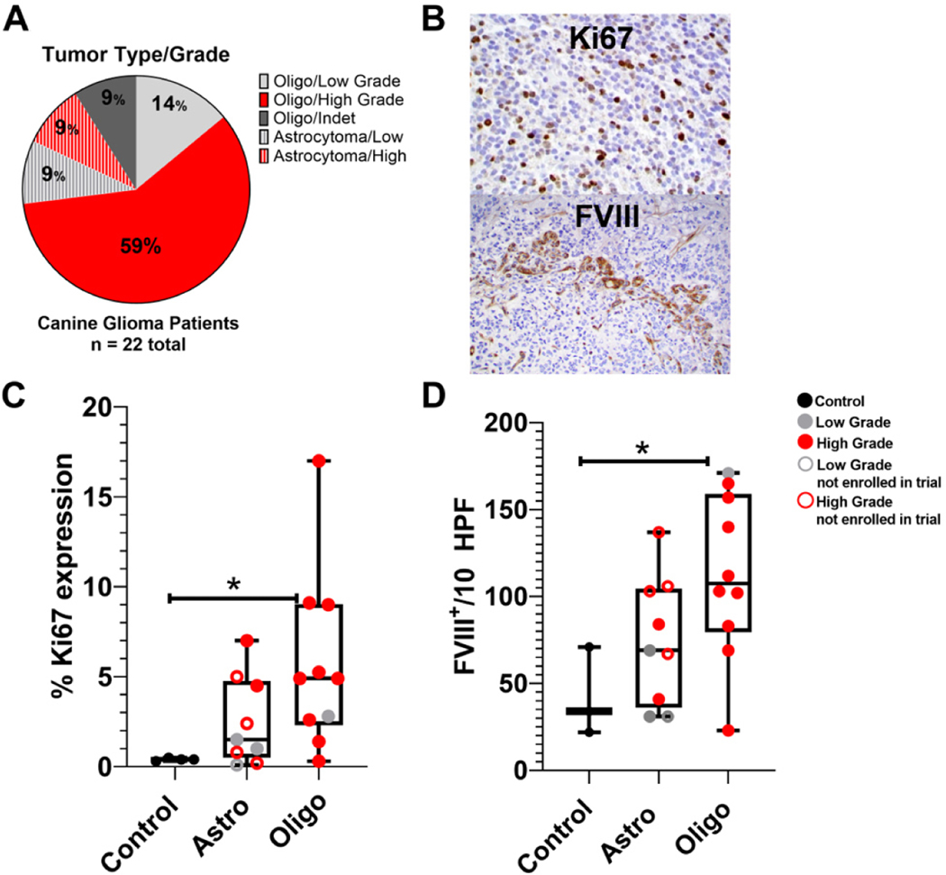
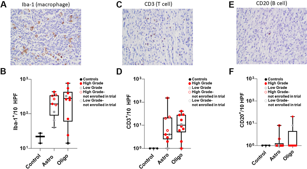
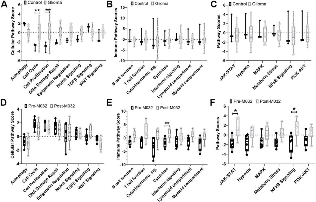
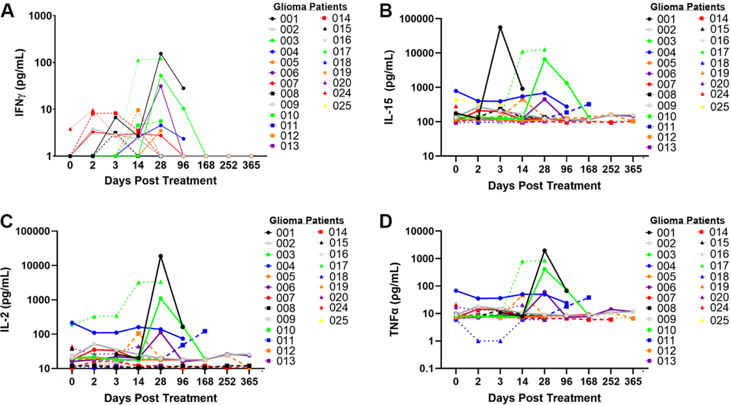
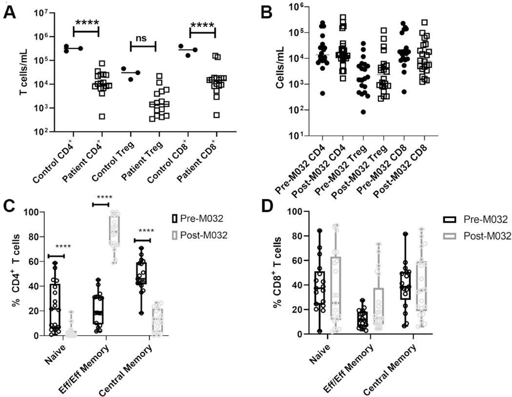
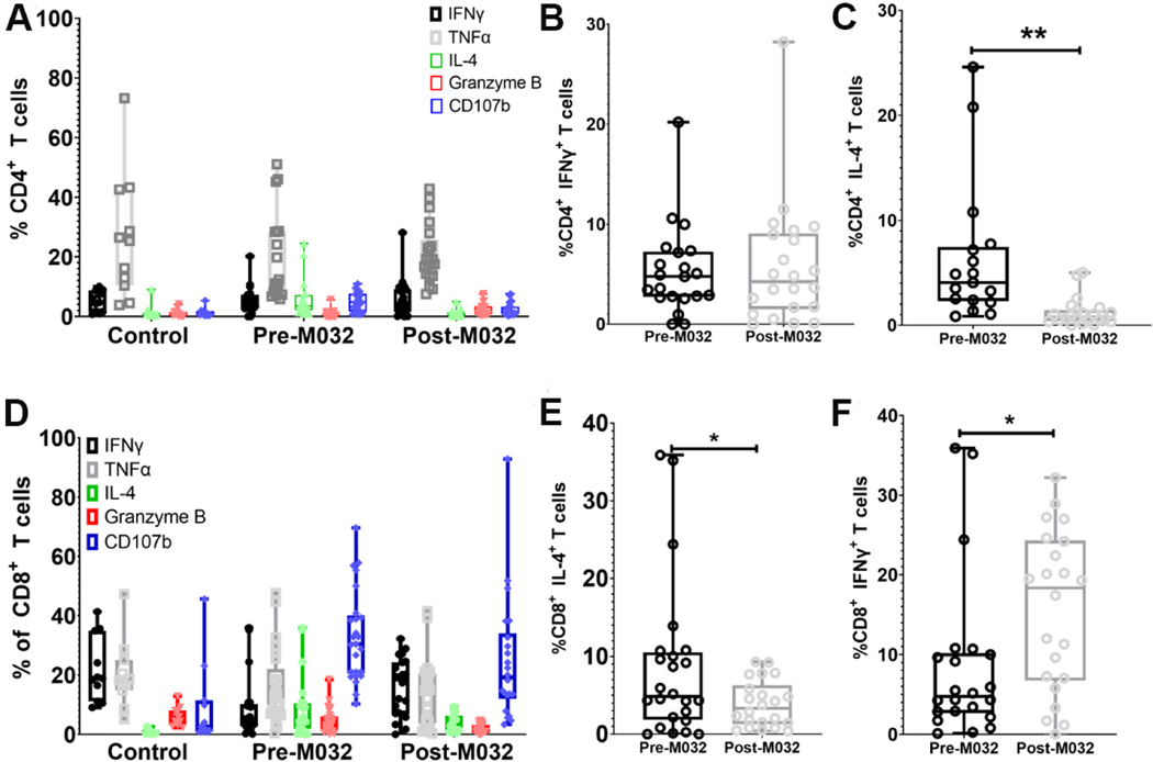
Results: Treatment-naive canine glioma microenvironment had enrichment of Iba1 positive macrophages and minimal numbers of T and B cells, consistent with previous studies identifying these tumors as immunologically "cold". NanoString mRNA profiling revealed enrichment for tumor intrinsic pathways consistent with suppression of tumor-specific immunity and support of tumor progression. Oncolytic viral treatment induced an intratumoral mRNA transcription signature of tumor-specific immune responses in 83% (5/6) of canine glioma patients. Changes included mRNA signatures corresponding with interferon signaling, lymphoid and myeloid cell activation, recruitment, and T and B cell immunity. Multiplexed protein analysis identified a subset of oligodendroglioma subjects with increased concentrations of IL-2, IL-7, IL-6, IL-10, IL-15, TNFα, GM-CSF between 14 and 28 days after treatment, with evidence of CD4+ T cell activation and modulation of IL-4 and IFNγ production in CD4+ and CD8+ T cells isolated from peripheral blood.

Conclusion: These findings indicate that M032 modulates the tumor-immune microenvironment in the canine glioma model.

Keywords: M032; NanoString; Oncolytic herpes virus; canine glioma; large animal model; tumor microenvironment.


Conflict of interest statement

Conflicts of interest Markert JM and Gillespie GY are founders of and own stock and stock options (< 8% interest) in Aettis Inc., a biotech company developing other oHSVs that are not the subject of this current investigation. Gillespie GY currently serves as one of five unpaid members of the Board of Directors for Aettis Inc. Markert JM and Gillespie GY are founders of and own stock and stock options (< 25%) in Treovir, LLC, which has licensed G207 HSV that is not the subject of the current investigation. Gillespie GY has served as a paid advisor to the Program Project (Brigham and Women’s Hospital, Boston, MA) that seeks to find improved methods for the application of distinct oHSV to treat localized and metastatic cancers. This is generally, but not specifically, related to the subject matter of this investigation. Markert JM also holds intellectual property in another oHSV not the subject of this current investigation that has been licensed by Mustang Biotech Inc., and reports being an equity owner in Catherex (< 8%), which underwent a structured buyout by Amgen and no longer exists; and has served as a consultant for Imugene.

Figures

Figure 1.
Figure 1.
Tumor characteristics. (A) Clinical trial patient tumor types and grades. (B) Representative intra-nuclear Ki67 and FVIII-related antigen expression in canine glioma tumor microenvironment. (C) Percentages of Ki67 expression in controls (n = 3), astrocytomas {n = 9 [4 patient astrocytomas and 5 astrocytomas from dogs not in the clinical trial (A1–5)]}; and oligodendrogliomas (n = 10). (D) Numbers of FVIII-related antigen positive endothelial cells/10 cumulative high power fields in the same tumors. One-way analysis of variance (column analyses) with Sidak’s correction for multiple comparisons was applied with *P < 0.05 indicative of statistical significance for oligodendroglioma vs. control samples.
Figure 2.
Figure 2.
Immunostaining of treatment-naïve canine gliomas. (A, C, E) Representative immunostaining for (A) Iba1, (C) CD3, and (E) CD20 (40× magnification). (B, D, F) Numbers of (B) Iba1+ cells, (D) CD3+ lymphocytes, and (F) CD20+ lymphocytes within control (n = 3), astrocytoma (n = 9) and oligodendroglioma (n = 10) specimens. Using ordinary one-way ANOVA (column analyses) with Sidak’s multiple comparison test, statistical significance was calculated to correct the false discovery rate. Statistically significant differences were not noted despite obvious trends based on mean values for Astro and Oligo.
Figure 3.
Figure 3.
mRNA expression. Identification of differential expression of mRNA gene signatures and associated (A) cellular, (B) cell signaling, and (C) immune pathways, comparing initial biopsy specimens (n = 19) to brain tissue controls (n = 3). Identification of differential expression of mRNA gene signatures and associated with (D) cellular, (E) cell signaling, and (F) immune pathways, comparing the six matched patient samples before and after M032 treatment [Table 3]. Gene expression is grouped according to mRNA function and represented as pathway scores where > 0 indicates upregulation in tumor tissue and < 0 indicates downregulation in tumor tissue compared to normal. Statistical significance between groups was evaluated by two-way ANOVA with correction for multiple comparisons using Holm-Sidak *P < 0.05 and **P < 0.01.
Figure 4.
Figure 4.
Expression of TH1 associated cytokines in canine glioma patients treated with M032. Expression of (A) IFNγ, (B) IL-15, (C) IL-2, and (D) TNFα prior to surgery (day 0), after surgery (days 2 and 3), and at various time points after intracranial infusion with M032 (days 14 through 365).
Figure 5.
Figure 5.
CD4+ T cells in canine glioma patients before and after treatment with M032. (A, B) Numbers of CD4+ Foxp3 , CD4+ Foxp3+ (Treg), and CD8+ T cells in the peripheral blood of (A) healthy controls vs. pre-M032 treatment canine glioma patients and (B) pre- vs. day 14 post-M032 treatment canine glioma patients. (C, D) The proportions of naïve (CD44lo CD45RAhi), central memory (CD44hi CD45RAhi), and effector/effector memory (CD44hi CD45RAlo) in (C) CD4+ and (D) CD8+ T cells. Two-way ANOVA for grouped analyses and Dunnet’s correction for multiple comparisons was used to calculate statistical significance. ****P < 0.0001, ***P < 0.001, **P <0.01.
Figure 6.
Figure 6.
IL-4 and IFNγ production in recirculating T cell subsets in canine glioma patients before and after treatment with M032. (A) Percentages of IFNγ, TNFα, IL-4, Granzyme B, and CD107 expression in CD4+ T cells after PMA ionomycin stimulation in healthy controls, canine glioma patients pre-M032 treatment, and at day 14 post-M032 treatment. Comparison between pre-M032 (black) and day 14 post-M032 (gray) revealed (B) no significant change in IFNγ and (C) a significant decrease in IL-4 post-M032 treatment. (D) Percentages of IFNγ, TNFα, IL-4, Granzyme B, and CD107B expression in CD8+ T lymphocytes after PMA ionomycin stimulation in healthy controls, pre-M032 treatment canine glioma patients and day 14 post-M032 treatment patients. Comparison between pre-M032 (black) and day 14-post M032 (gray) revealed a significant (E) decrease in IL-4 and (F) increase in IFNγ production at 14 days after treatment. Statistical significance (B-E) was evaluated using an unpaired student’s t test (Welch’s correction for unequal variance). *P < 0.05, **P < 0.01, ***P < 0.001, ****P < 0.

Similar articles

Cited by

References

    1. Weller M, Wick W, Aldape K, et al. Glioma. Nat Rev Dis Primers 2015;1:15017. DOI PubMed - PubMed
    1. Wen PY, Kesari S. Malignant gliomas in adults. N Engl J Med 2008;359:492–507. DOI PubMed - PubMed
    1. Siegel RL, Miller KD, Fuchs HE, Jemal A. Cancer statistics, 2021. CA Cancer J Clin 2021;71:7–33. DOI PubMed - PubMed
    1. Ostrom QT, Patil N, Cioffi G, Waite K, Kruchko C, Barnholtz-Sloan JS. CBTRUS statistical report: primary brain and other central nervous system tumors diagnosed in the United States in 2013–2017. Neuro Oncol 2020;22:iv1-iv96. DOI PubMed PMC - PMC - PubMed
    1. Park JS, Withers SS, Modiano JF, et al. Canine cancer immunotherapy studies: linking mouse and human. J Immunother Cancer 2016;4:97. DOI PubMed PMC - PMC - PubMed

LinkOut - more resources