Review
. 2022 Jun 15;86(2):e0002621.
doi: 10.1128/mmbr.00026-21. Epub 2022 Mar 28.

Molecular Virology of SARS-CoV-2 and Related Coronaviruses

Affiliations
Review

Molecular Virology of SARS-CoV-2 and Related Coronaviruses

Yu-An Kung et al. Microbiol Mol Biol Rev. .

Abstract

Coronavirus disease 2019 (COVID-19) is caused by severe acute respiratory syndrome coronavirus 2 (SARS-CoV-2). The global COVID-19 pandemic continues to threaten the lives of hundreds of millions of people, with a severe negative impact on the global economy. Although several COVID-19 vaccines are currently being administered, none of them is 100% effective. Moreover, SARS-CoV-2 variants remain an important worldwide public health issue. Hence, the accelerated development of efficacious antiviral agents is urgently needed. Coronavirus depends on various host cell factors for replication. An ongoing research objective is the identification of host factors that could be exploited as targets for drugs and compounds effective against SARS-CoV-2. In the present review, we discuss the molecular mechanisms of SARS-CoV-2 and related coronaviruses, focusing on the host factors or pathways involved in SARS-CoV-2 replication that have been identified by genome-wide CRISPR screening.

Keywords: SARS-CoV-2; coronavirus.


Conflict of interest statement

The authors declare no conflict of interest.

Figures
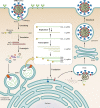
FIG 1
FIG 1
SARS-CoV-2 life cycle. SARS-CoV-2 interacts with the ACE2 receptor, and the spike (S) protein is cleaved by TMPRSS2, after which fusion between the viral and host membranes occurs. SARS-CoV-2 can also enter host cells by endocytosis, and the S protein is then activated by endosomal cathepsins. After the viral (+)ssRNA genome is released into the host cytoplasm, it is translated and produces the polyproteins pp1a and pp1ab, which are autoproteolytically processed into the nonstructural proteins nsp1 to -16. The nsps assemble the coronavirus replicase-transcriptase complex (RTC) and remodel the membranes to form organelles for viral RNA synthesis. Viral replication and transcription occur in double-membrane vesicles (DMVs) derived from the ER. Newly synthesized viral genomic RNA is exported from the DMV interior via the pore channel and is then encapsidated by the nucleocapsid (N) protein. The nested transcribed subgenomic RNAs (sgRNAs) are translated into the structural proteins S, envelope (E), membrane (M), and N and accessory proteins. S, E, and M are anchored to the ER membrane and migrate to the virion assembly site known as the endoplasmic reticulum-Golgi intermediate compartment (ERGIC). The viral ribonucleoprotein (vRNP) complexes migrate to the ERGIC and bud into the lumen. The enveloped virion is then released from the cells via lysosomes.
FIG 2
FIG 2
SARS-CoV-2 spike protein structure, activation, and host cell entry. (A) Schematic drawing of the coronavirus spike three-dimensional (3D) structure and domain structure, including the signal peptide (SP), N-terminal domain (NTD), receptor-binding domain (RBD), fusion peptide (FP), heptad repeat 1 (HR1), heptad repeat 2 (HR2), transmembrane (TM), cytoplasmic (CP), and proteolytic (S1/S2 and S2′) cleavage sites. Arrowheads indicate the cleavage sites of furin and TMPRSS2. (B) SARS-CoV-2 can enter the host cell by TMPRSS2 or furin activation of fusion of the viral and host cell membranes (surface activation). SARS-CoV-2 can also enter the host cell via binding of the viral spike (S) protein to a cellular receptor and virion endocytosis. In the endosome, the pH-dependent cysteine protease cathepsin L (CatL) activates the S protein, causing fusion within the endosomal membrane (endosomal activation). The viral genome is released by TMPRSS2-mediated host cell entry or from the endosome and is partially and completely replicated and translated in the ER to form new SARS-CoV-2 virions.
FIG 3
FIG 3
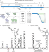
Genome structure and transcriptome architecture of SARS-CoV-2. (A) Schematic representation of the SARS-CoV-2 genome with annotations based on the reference sequence of the Wuhan-Hu-1 strain (GenBank accession number NC_045512.2). The distributions of open reading frames (ORFs) and coding regions of each nonstructural protein (nsp) across the genome are indicated. Both (+)gRNA and positive-stranded subgenomic RNA [(+)sgRNA] carry an identical leader sequence at the 5′ terminus (red). The (+)gRNA serves as the template for the synthesis of (−)gRNA as well as (−)sgRNAs that are subsequently used to synthesize (+)gRNA and (+)sgRNAs. The replication and transcription processes are carried out by nsps. Complexes with various functions are comprised of different nsps, as indicated in the schematic. (B) The 5′ end of the genomic sequence can fold into multiple stem-loops (SLs). The transcription/replication sequence (TRS) of SL3 is marked in red. The upstream region of the TRS encompassing SL1 and SL2 is defined as the leader sequence. Coding sequences located at the 5′ genomic region, including the upstream ORF in SL4, are represented by closed circles. (C) The secondary structures of the 3′ genomic sequences include an evolutionarily conserved bulged stem-loop (BSL) and a three-helix junction structure that is formed by base pairing between the hypervariable region (HVR) and the upstream/downstream sequences of SL1. The unwinding of the three-helix structure by the replicase-transcriptase complex (RTC) leads to a conformational change, allowing base pairing between the SL1 apical loop and the stem of the BSL to form a pseudoknot (PK).

References

    1. Nicola M, Alsafi Z, Sohrabi C, Kerwan A, Al-Jabir A, Iosifidis C, Agha M, Agha R. 2020. The socio-economic implications of the coronavirus pandemic (COVID-19): a review. Int J Surg 78:185–193. 10.1016/j.ijsu.2020.04.018. - DOI - PMC - PubMed
    1. Tao K, Tzou PL, Nouhin J, Gupta RK, de Oliveira T, Kosakovsky Pond SL, Fera D, Shafer RW. 2021. The biological and clinical significance of emerging SARS-CoV-2 variants. Nat Rev Genet 22:757–773. 10.1038/s41576-021-00408-x. - DOI - PMC - PubMed
    1. Huang C, Wang Y, Li X, Ren L, Zhao J, Hu Y, Zhang L, Fan G, Xu J, Gu X, Cheng Z, Yu T, Xia J, Wei Y, Wu W, Xie X, Yin W, Li H, Liu M, Xiao Y, Gao H, Guo L, Xie J, Wang G, Jiang R, Gao Z, Jin Q, Wang J, Cao B. 2020. Clinical features of patients infected with 2019 novel coronavirus in Wuhan, China. Lancet 395:497–506. 10.1016/S0140-6736(20)30183-5. - DOI - PMC - PubMed
    1. Gao YD, Ding M, Dong X, Zhang JJ, Kursat Azkur A, Azkur D, Gan H, Sun YL, Fu W, Li W, Liang HL, Cao YY, Yan Q, Cao C, Gao HY, Bruggen MC, van de Veen W, Sokolowska M, Akdis M, Akdis CA. 2021. Risk factors for severe and critically ill COVID-19 patients: a review. Allergy 76:428–455. 10.1111/all.14657. - DOI - PubMed
    1. Gold JAW, Rossen LM, Ahmad FB, Sutton P, Li Z, Salvatore PP, Coyle JP, DeCuir J, Baack BN, Durant TM, Dominguez KL, Henley SJ, Annor FB, Fuld J, Dee DL, Bhattarai A, Jackson BR. 2020. Race, ethnicity, and age trends in persons who died from COVID-19—United States, May-August 2020. MMWR Morb Mortal Wkly Rep 69:1517–1521. 10.15585/mmwr.mm6942e1. - DOI - PMC - PubMed

Publication types

Supplementary concepts