. 2022 Apr 26;13(2):e0040222.
doi: 10.1128/mbio.00402-22. Epub 2022 Mar 28.

Viral Mimicry of Interleukin-17A by SARS-CoV-2 ORF8

Affiliations

Viral Mimicry of Interleukin-17A by SARS-CoV-2 ORF8

Xin Wu et al. mBio. .

Abstract

Severe acute respiratory syndrome coronavirus 2 (SARS-CoV-2) infection triggers cytokine-mediated inflammation, leading to a myriad of clinical presentations in COVID-19. The SARS-CoV-2 open reading frame 8 (ORF8) is a secreted and rapidly evolving glycoprotein. Patients infected with SARS-CoV-2 variants with ORF8 deleted are associated with mild disease outcomes, but the molecular mechanism behind this is unknown. Here, we report that SARS-CoV-2 ORF8 is a viral cytokine that is similar to but distinct from interleukin 17A (IL-17A) as it induces stronger and broader human IL-17 receptor (hIL-17R) signaling than IL-17A. ORF8 primarily targeted blood monocytes and induced the heterodimerization of hIL-17RA and hIL-17RC, triggering a robust inflammatory response. Transcriptome analysis revealed that besides its activation of the hIL-17R pathway, ORF8 upregulated gene expression for fibrosis signaling and coagulation dysregulation. A naturally occurring ORF8 L84S variant that was highly associated with mild COVID-19 showed reduced hIL-17RA binding and attenuated inflammatory responses. This study reveals how SARS-CoV-2 ORF8 by a viral mimicry of the IL-17 cytokine contributes to COVID-19 severe inflammation. IMPORTANCE Patients infected with SARS-CoV-2 variants lacking open reading frame 8 (ORF8) have been associated with milder infection and disease outcome, but the molecular mechanism behind how this viral accessory protein mediates disease pathogenesis is not yet known. In our study, we revealed that secreted ORF8 protein mimics host IL-17 to activate IL-17 receptors A and C (IL-17RA/C) and induces a significantly stronger inflammatory response than host IL-17A, providing molecular insights into the role of ORF8 in COVID-19 pathogenesis and serving as a potential therapeutic target.

Keywords: COVID-19; inflammation; viral IL-17.


Conflict of interest statement

The authors declare no conflict of interest.

Figures
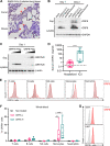
FIG 1
FIG 1
Secreted ORF8 binds primarily to CD14+ blood monocytes. (A) ORF8 is highly expressed in the lung tissues of SARS-CoV-2-infected ferrets and mice. Immunohistochemistry showed ORF8 expression after SARS-CoV-2 infection in lung sections of ferrets and mice. Scale bar, 1 mm. Arrowheads mark ORF8 expression. (B) ORF8 is detected in the supernatants of SARS-CoV-2-infected Vero cells. At 24 h or 48 h postinfection, cell lysates and culture supernatants (Sup.) were harvested, and ORF8 protein was detected by immunoblotting using ORF8 antibody. (C) ORF8 is secreted into the supernatants of transfected 293T cells. Plasmid DNA encoding the SARS-CoV-2 ORF7b or ORF8 with a Flag tag was transfected into 293T cells. At 48 h posttransfection with SARS-CoV-2 ORF7b-Flag or ORF8-Flag, cell lysates and supernatants (Sup.) were harvested, and ORF7b/ORF8 proteins were detected by immunoblotting using Flag antibody. (D) SARS-CoV-2 ORF8 concentrations (ng/mL) in the sera of hospitalized COVID-19 patients (n = 40) and ICU-admitted COVID-19 patients (n = 9). The horizontal line indicates the median value of the pre-COVID-19 pandemic sera of healthy subjects (n = 9). (E, F) Purified Flag-tagged ORF8 protein primarily binds to monocytes in human blood cells. Whole blood derived from healthy donors (n = 6) was incubated with purified ORF8-Flag for 2 h. Subsequently, granulocytes and PBMCs were isolated and subjected to flow cytometry with Flag antibody. Various immune cells were gated by specific antibodies: CD45+ CD3+ T cells, CD45+ CD19+ B cells, CD45+ CD14+ monocytes, CD45+ CD56+ NK cells, and CD45+ CD16+ CD11b+ neutrophils. (G) Purified ORF8-Flag binds to monocytic cell lines. THP-1 and U937 cells were incubated with purified ORF8-Flag for 2 h, and then ORF8-Flag binding activity was analyzed by flow cytometry. P values were calculated by Mann-Whitney U test (D) and two-way ANOVA with Dunnett test for multiple comparisons (F). *, P < 0.05; **, P < 0.01; ***, P < 0.005; ****, P < 0.0001.
FIG 2
FIG 2
ORF8 interacts with hIL-17RA to activate inflammatory responses. (A) Hypothesis model of ORF8-mediated IL-17RA signaling. (B) Purified ORF8-Flag interacts with 293T-expressing hIL-17RA. At 24 h posttransfection with vector or hIL-17RA expression vector, 293T cells were subsequently incubated for 30 min with Flag-tagged ORF8 or untreated, followed by flow cytometry with anti-Flag antibody. (C) THP-1 cells express high levels of surface hIL-17RA. THP-1 cells were stained with hIL-17RA antibody and followed by flow cytometry. (D) Purified ORF8-Flag protein interacts with endogenous IL-17RA in THP-1 cells. THP-1 cells were incubated with purified ORF8-Flag proteins for 2 h, and cell lysates were immunoprecipitated with anti-Flag, followed by immunoblotting with indicated antibodies. (E) ORF8 induces the phosphorylation of p65 and IκΒα. THP-1 cells were treated with or without ORF8 (50 ng/mL) for the indicated times, followed by immunoblots with the indicated antibodies. (F) ORF8 treatment increases inflammatory cytokine gene expression. THP-1 cells were treated with ORF7b or ORF8 at the indicated concentrations for 16 h, followed by RT-qPCR. (G) ORF8 neutralizing antibody blocks ORF8 function. THP-1 cells were treated with purified ORF8 (20 ng/mL) and its antibody at the indicated concentrations for 16 h, followed by RT-qPCR. (H) IL-17RA neutralizing antibody blocks ORF8 function. THP-1 cells were treated with control IgG or IL-17RA neutralizing antibody at the indicated concentrations for 2 h and then stimulated with ORF8 for 16 h, followed by RT-qPCR. P values were calculated by two-way ANOVA with Tukey’s test in panel F or Bonferroni’s test in panels G and H for multiple comparisons. *, P < 0.05; **, P < 0.01; ***, P < 0.005; ****, P < 0.0001.
FIG 3
FIG 3
ORF8 is a potent viral mimic of hIL-17A. After 2 h of mock treatment or treatment with ORF8 or hIL-17A, human monocytes and plasma were isolated from whole blood of male and female donors (n = 4), and their RNAs were subjected to RNA-Seq analysis. (A) Volcano plot analysis of differential gene expressions in ORF8 and hIL-17A treatment compared to mock treatment (n = 4). (B) Comparison of significantly increased or decreased gene expression between monocytes treated with ORF8 or hIL-17A is presented as a Venn diagram. (C) Comparison analysis of IPA canonical pathways significantly enriched in the ORF8- or hIL-17A-treated group compared to the control group (left panel). A heat map of the top 10 transcription factors commonly up- or downregulated by ORF8 and hIL-17A is shown in the middle panel, and a heat map of NF-κB-regulated target genes commonly affected by ORF8 or hIL-17A treatment is shown in the right panel. A negative Z score means the inactivation of genes or pathways compared to the control group, and a positive Z score means the activation of genes or pathways compared to the control group. (D) NF-κB-regulated genes (IL12B, CSF3, IL6, CCL20, CXCL1, and CXCL2) were normalized to GAPDH and expressed as fold change relative to mock controls. (E) CXCL1 and IL-6 ELISA of plasma of ORF8- or hIL-17A-treated whole blood, with horizontal lines indicating median values of mock-treated whole blood observed. (F) IPA of uniquely ORF8-regulated pathways. (G) Immune profiling of ORF8-upregulated unique genes. (H) RT-qPCR analysis of ORF8-upregulated unique genes (COL17A1, MMP10, and SERPINB2) in primary monocytes that were normalized to GAPDH and expressed as fold change relative to mock controls. (I) RT-qPCR analysis of ORF8-upregulated unique genes (COL17A1, MMP10, and SERPINB2) in THP-1 cells were normalized to GAPDH and expressed as fold change relative to mock controls. P values were calculated by one-way ANOVA with Tukey’s posttest in panels D, H, and I and multiple t test in panel E. *, P < 0.05; **, P < 0.01; ***, P < 0.005; ****, P < 0.0001.
FIG 4
FIG 4
ORF8 binding enhances the heterodimerization of hIL-17RA/C. (A) Purified ORF8-Flag interacts with hIL-17RA and hIL-17RC. At 24 h posttransfection of the IL-17 receptor construct, 293T cells were subsequently incubated for 30 min with mock-treated or purified ORF8-Flag, followed by staining with anti-Flag antibody for flow cytometry. (B) Purified ORF8-Flag protein interacts with endogenous IL-17RA and IL-17RC in THP-1 cells. THP-1 cells were incubated with purified ORF8-Flag for 2 h. Cell lysates were immunoprecipitated with anti-Flag, followed by immunoblotting with the indicated antibodies. (C) Effects of IL-17RA/B/C deficiency on the ORF8-induced phosphorylation of p65 and IκBα. IL-17RA, IL-17RB, or IL-17RC knockout (KO) THP-1 cells were generated by the CRISPR-Cas9 method. IL-17RA KO, IL-17RB KO, IL-17RC KO, or control THP-1 cells were mock treated or treated with ORF8 for the indicated times, followed by immunoblotting with the indicated antibodies. (D) Effects of IL-17RA/B/C deficiency on ORF8-mediated cytokine gene expression. IL-17RA KO, IL-17RB KO, IL-17RC KO, or control THP-1 cells were mock treated or treated with ORF8 for 16 h, followed by qRT-PCR to measure the mRNA levels of indicated genes. (E) Proximity ligation assay (PLA) of IL-17RA/C heterodimerization in NIH 3T3 cells. At 24 h posttransfection with HA-hIL-17RA and Myc-hIL-17RC, NIH 3T3 cells were treated with hIL-17A or purified ORF8 for 30 min, followed by PLA. P values were calculated by two-way ANOVA with Dunnett’s posttest in panel D or one-way ANOVA with Tukey’s posttest in panel E. *, P < 0.05; **, P < 0.01; ***, P < 0.005; ****, P < 0.0001.
FIG 5
FIG 5
ORF8 mutational analysis for hIL-17RA and hIL-17RC interaction. (A) Y42, I71, I76, and E106 resides in SARS-CoV-2 ORF8 structure. (B) Binding activity of ORF8 mutants to THP-1 cells. THP-1 cells were incubated with purified ORF8 WT or mutants for 2 h, followed by flow cytometry with anti-Flag antibody. (C, D) Binding activity of ORF8 mutants to hIL-17RA (C) or hIL-17RC (D). At 24 h posttransfection with hIL-17RA (C) or hIL-17RC (D) expression plasmids, 293T cells were subsequently incubated for 30 min with mock treatment or purified ORF8 WT or mutants, followed by flow cytometry with anti-Flag antibody. (E) Signaling activity of ORF8 mutant to induce inflammatory cytokine gene expressions. THP-1 cells were mock treated or treated with ORF8 WT or mutants for 16 h, followed by RT-qPCR to measure the mRNA levels of the indicated genes, with horizontal lines indicating median values of the mRNA levels of the indicated genes in mock-treated THP1 cells observed. (F) PLA of ORF8 mutant-mediated hIL-17RA/C heterodimerization in NIH 3T3 cells. At 24 h posttransfection with HA-hIL-17RA and Myc-hIL-17RC, NIH 3T3 cells were treated with hIL-17A or purified ORF8 WT or mutants for 30 min, followed by PLA. P values were calculated by one-way ANOVA with Dunnett’s posttest in panels E and F. *, P < 0.05; **, P < 0.01; ***, P < 0.005; ****, P < 0.0001.
FIG 6
FIG 6
ORF8 natural variants for hIL-17R binding and inflammatory signaling activity. (A) S24, V62, and L84 residues in SARS-CoV-2 ORF8 structure. (B) Binding activity of ORF8 natural variants to THP-1 cells. THP-1 cells were incubated with mock treatment or ORF8 WT or variants for 2 h, followed by flow cytometry with anti-Flag antibody. (C, D) Binding activity of ORF8 natural variants to hIL-17RA (C) or hIL-17RC (D). At 24 h posttransfection with hIL-17RA (C) or hIL-17RC (D) expression plasmids, 293T cells were subsequently incubated for 30 min with mock treatment or purified ORF8 WT or variants, followed by flow cytometry with anti-Flag antibody. (E) Signaling activity of ORF8 variants to induce inflammatory cytokine gene expressions. THP-1 cells were mock treated or treated with ORF8 WT or variants for 16 h, followed by RT-qPCR to measure the mRNA levels of indicated genes, with horizontal lines indicating median values of the mRNA levels of indicated genes in mock-treated THP1 cells observed. (F) L84S variant shows the loss of IL-17RA/RC heterodimerization. At 24 h posttransfection with HA-hIL-17RA and Myc-hIL-17RC, NIH 3T3 cells were treated with the ORF8 WT or L84S variant for 30 min, followed by PLA. (G) Predicted structural representation of the Y42, Y106, and L84 residues at the binding interface between ORF8 and IL-17RA. P values were calculated by one-way ANOVA with Dunnett’s posttest in panels E and F. *, P < 0.05; **, P < 0.01; ***, P < 0.005; ****, P < 0.0001.

References

    1. Cascella M, Rajnik M, Aleem A, Dulebohn S, Di Napoli R. 2021. Features, evaluation, and treatment of coronavirus (COVID-19). StatPearls https://www.statpearls.com/articlelibrary/viewarticle/52171/. - PubMed
    1. Wu F, Zhao S, Yu B, Chen Y-M, Wang W, Song Z-G, Hu Y, Tao Z-W, Tian J-H, Pei Y-Y, Yuan M-L, Zhang Y-L, Dai F-H, Liu Y, Wang Q-M, Zheng J-J, Xu L, Holmes EC, Zhang Y-Z. 2020. A new coronavirus associated with human respiratory disease in China. Nature 579:265–269. doi:10.1038/s41586-020-2008-3. - DOI - PMC - PubMed
    1. Fajgenbaum DC, June CH. 2020. Cytokine storm. N Engl J Med 383:2255–2273. doi:10.1056/NEJMra2026131. - DOI - PMC - PubMed
    1. Zhou P, Yang X-L, Wang X-G, Hu B, Zhang L, Zhang W, Si H-R, Zhu Y, Li B, Huang C-L, Chen H-D, Chen J, Luo Y, Guo H, Jiang R-D, Liu M-Q, Chen Y, Shen X-R, Wang X, Zheng X-S, Zhao K, Chen Q-J, Deng F, Liu L-L, Yan B, Zhan F-X, Wang Y-Y, Xiao G-F, Shi Z-L. 2020. A pneumonia outbreak associated with a new coronavirus of probable bat origin. nature 579:270–273. doi:10.1038/s41586-020-2012-7. - DOI - PMC - PubMed
    1. Rouvier E, Luciani M, Mattei M, Denizot F, Golstein P. 1993. CTLA-8, cloned from an activated T cell, bearing AU-rich messenger RNA instability sequences, and homologous to a herpesvirus saimiri gene. J Immunol 150:5445–5456. - PubMed

Supplementary concepts