Review
. 2022 Jul 1;102(3):1495-1552.
doi: 10.1152/physrev.00015.2021. Epub 2022 Mar 28.

Salivary gland function, development, and regeneration

Affiliations
Review

Salivary gland function, development, and regeneration

Alejandro M Chibly et al. Physiol Rev. .

Abstract

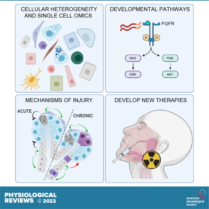
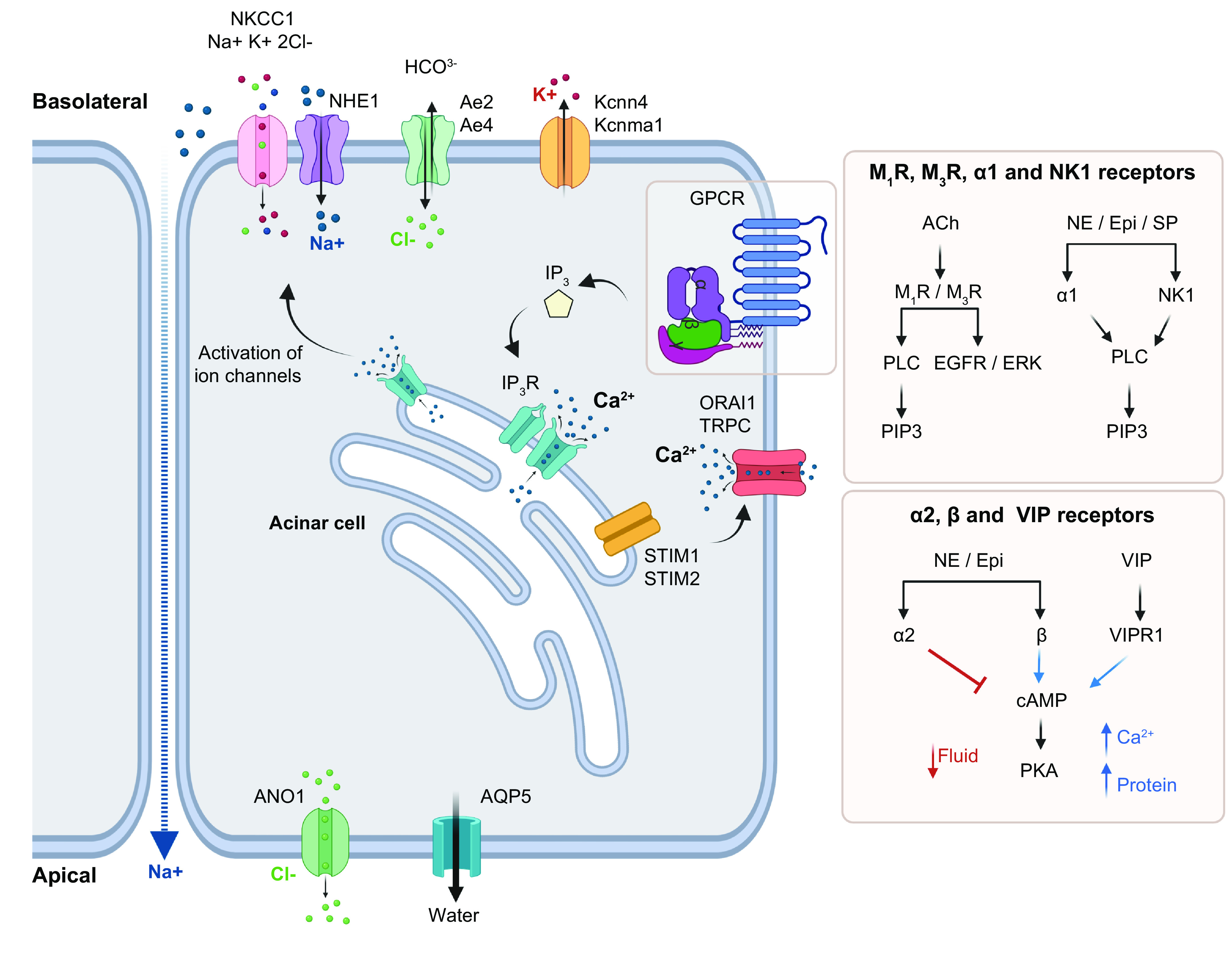
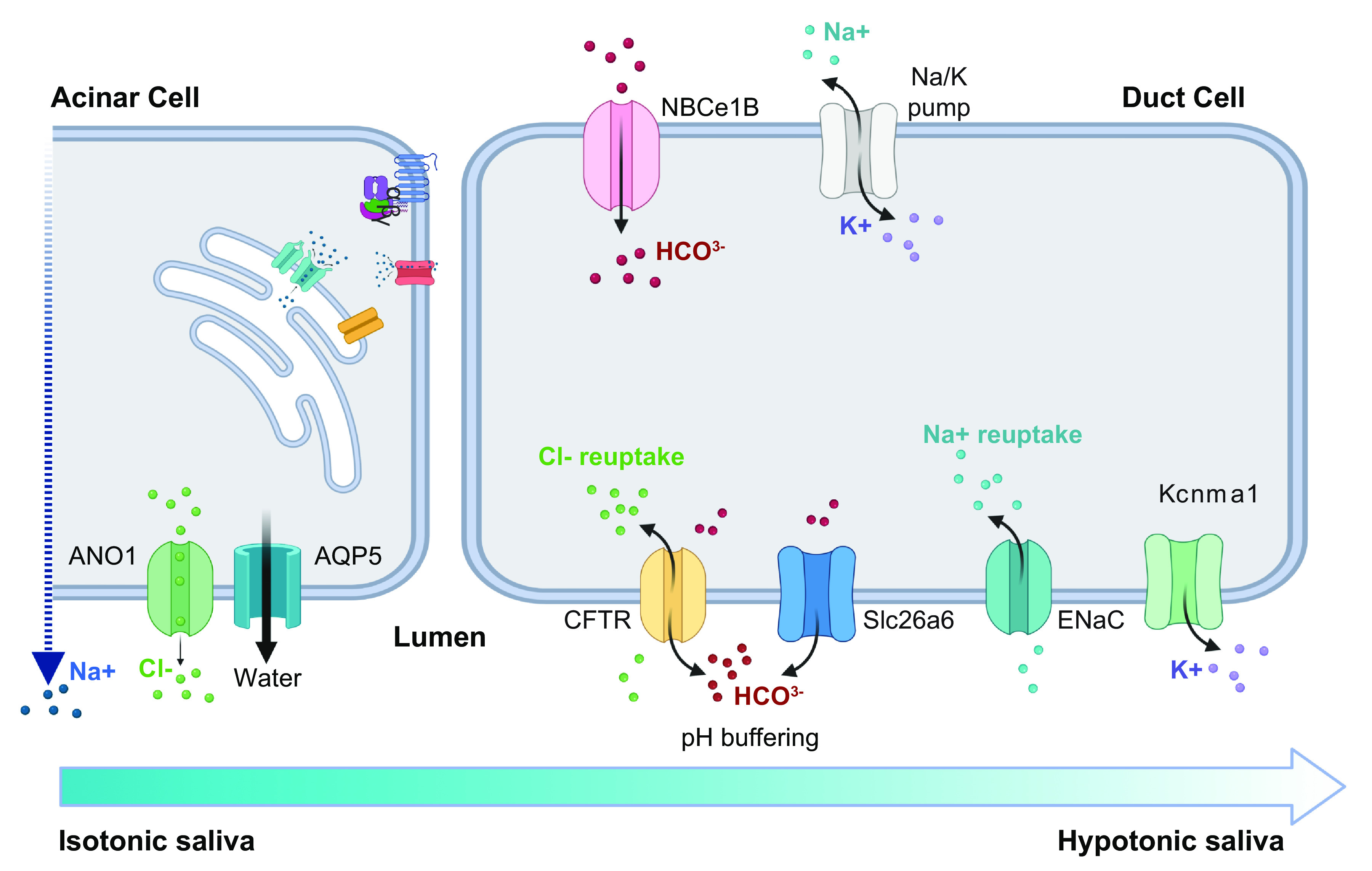
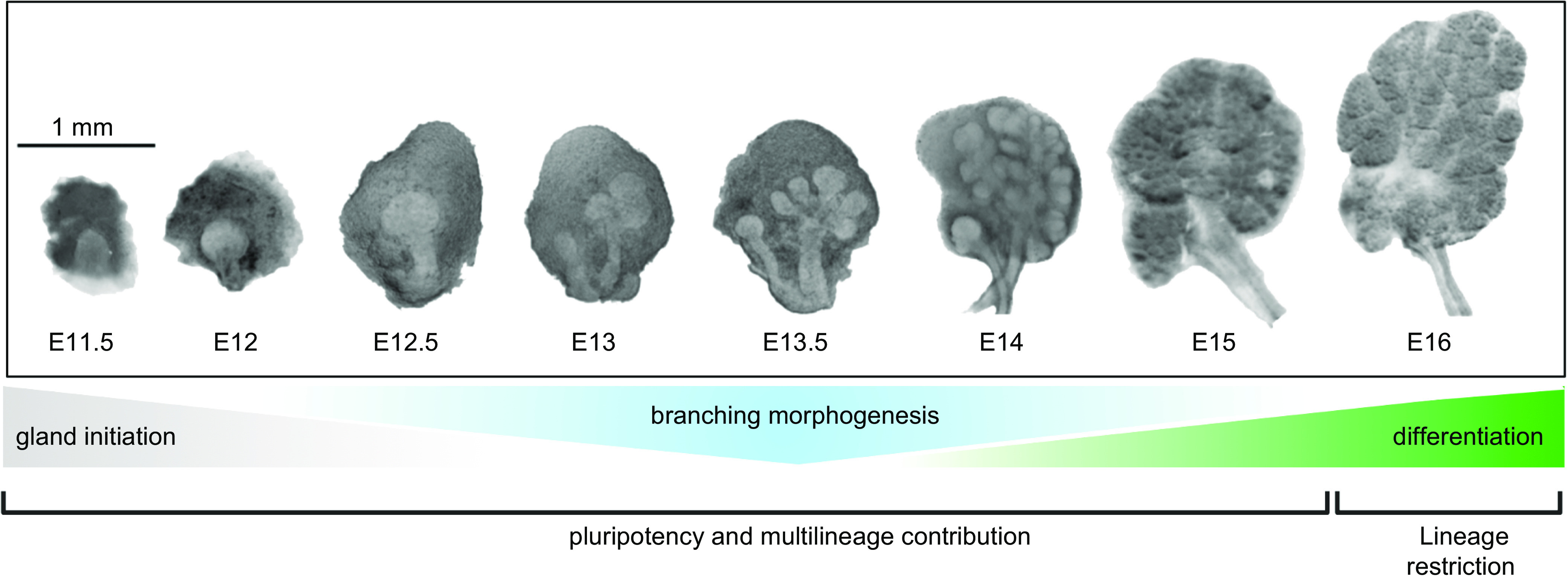
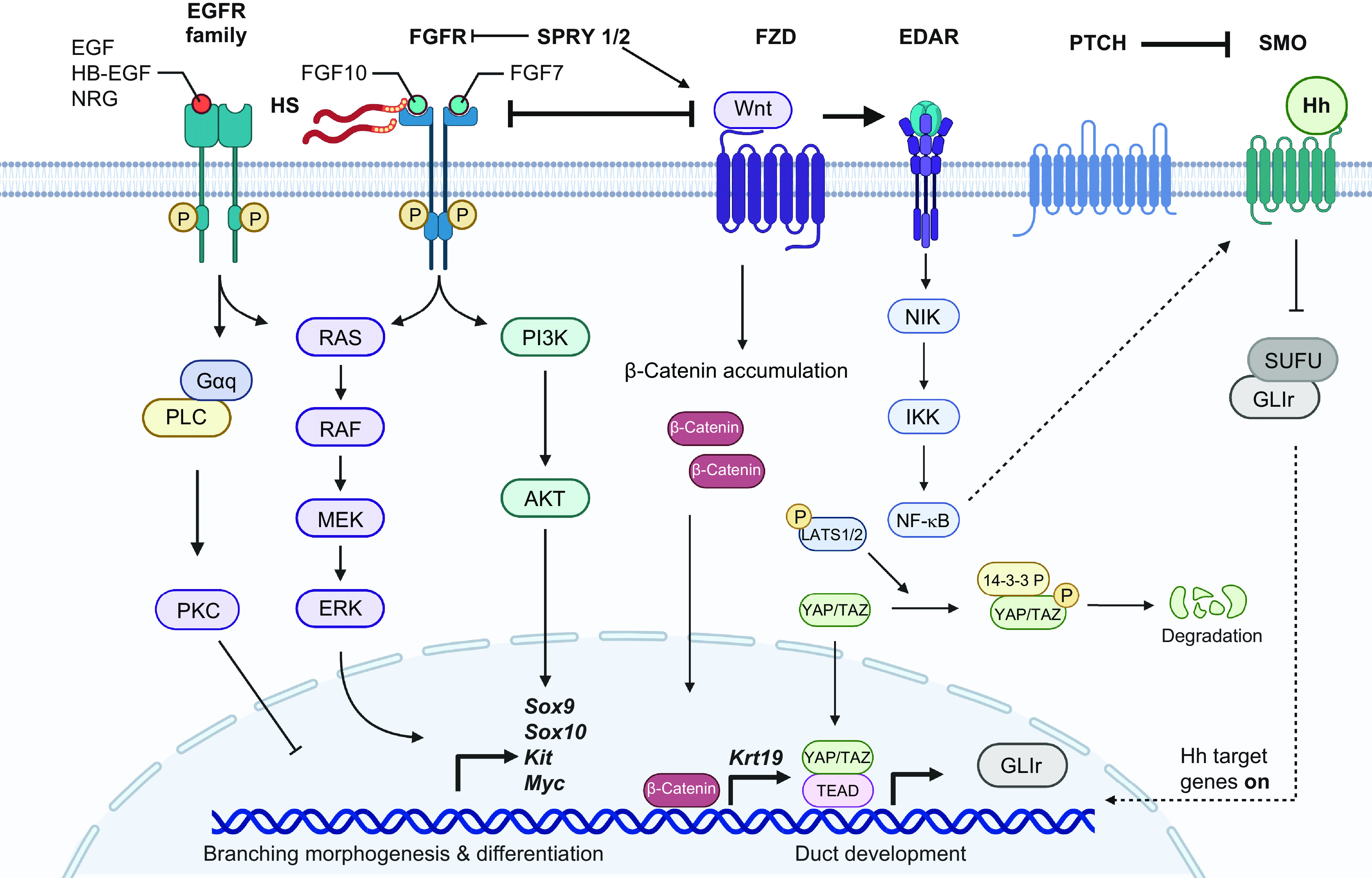
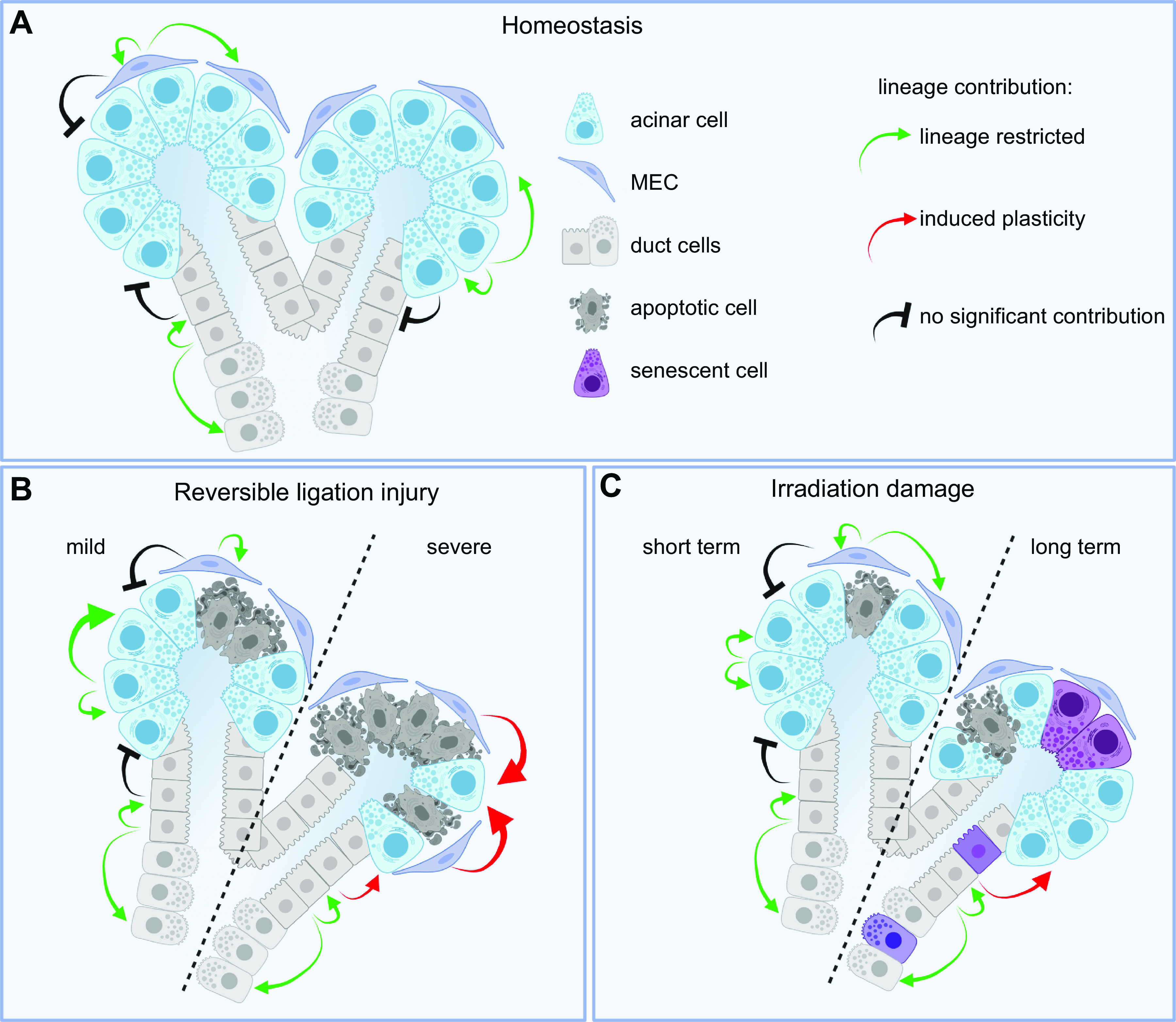
Salivary glands produce and secrete saliva, which is essential for maintaining oral health and overall health. Understanding both the unique structure and physiological function of salivary glands, as well as how they are affected by disease and injury, will direct the development of therapy to repair and regenerate them. Significant recent advances, particularly in the OMICS field, increase our understanding of how salivary glands develop at the cellular, molecular, and genetic levels: the signaling pathways involved, the dynamics of progenitor cell lineages in development, homeostasis, and regeneration, and the role of the extracellular matrix microenvironment. These provide a template for cell and gene therapies as well as bioengineering approaches to repair or regenerate salivary function.

Keywords: exocrine secretion; gene therapy; progenitor cell; salivary gland; xerostomia.


Conflict of interest statement

No conflicts of interest, financial or otherwise, are declared by the authors.

Figures

None
Graphical abstract
FIGURE 1.
FIGURE 1.
Salivary glands in human and mouse. A: the major pairs of SGs in human are the parotid, submandibular, and sublingual glands. B: hundreds of minor SGs are distributed throughout the oral cavity and include labial glands. C: an additional pair of SGs recently described are tubarial SGs localized near the torus tubarius. D and E: cellular heterogeneity of human and mouse SGs is evidenced by single-cell RNAseq. See glossary for abbreviations. Created with BioRender.com, with permission.
FIGURE 2.
FIGURE 2.
Model of major cell types of the mouse submandibular gland (SMG). SMG epithelium is divided into secretory acini and ducts. Serous and seromucous acinar cells are surrounded by MECs. Ducts are divided into sections containing specific duct cell types. Non-epithelial cell types in the surrounding ECM include fibroblasts, immune cells, blood vessels, and nerves. See glossary for abbreviations. Created with BioRender.com, with permission.
FIGURE 3.
FIGURE 3.
Cell types of the mouse SMG. A: secretory acinar cells (MUC10, yellow) secrete saliva into the intercalated duct (ID) (SMGC, magenta) and nuclei (DAPI, cyan). B: intercalated duct subpopulations include Gstt1+/Smgc+ (GSTT1, magenta) and Gfra3/Kit+ (GFRA3, yellow) cells and nuclei (DAPI, cyan). C: myoepithelial cells (KRT5, magenta), imaged in a thick section to highlight their stellate morphology, surround the acini and duct with long cellular processes. D: ionocytes (Fgf10Cre:TdTomatofl, yellow) with long cellular processes are located within the duct and nuclei (DAPI, cyan). See glossary for abbreviations.
FIGURE 4.
FIGURE 4.
Model of stage 1 of saliva secretion. Secretion is initiated by activation of muscarinic receptors M1 or M3 by acetylcholine (ACh) or adrenergic receptors (α1) by norepinephrine (NE) and epinephrine (Epi) via specific pathways involving IP3 release or cAMP modulation. IP3 binds to its receptor in the ER to promote Ca2+ release, which is sensed by STIM1/2 in the ER membrane (EM). STIM1 recruits Orai1/TRPC to stimulate Ca2+ reentry into the cell. Ca2+ depletion from the ER recruits AQP5 to the apical membrane for water expulsion and activates NKCC1, NHE1, Ae2, Ae4, Kcnma1, and Kcnn4 in the basolateral membrane to import Na+ and Cl into the cellular space while exporting K+ and HCO3. In turn, ANO1 is activated in the luminal membrane to secrete Cl. Na+ also undergoes transepithelial transport toward the lumen, resulting in NaCl-rich isotonic saliva. See glossary for abbreviations. Created with BioRender.com, with permission.
FIGURE 5.
FIGURE 5.
Model of stage 2 of saliva secretion. Isotonic saliva secreted by acinar cells is rich in NaCl. As it travels through the ductal system, duct cells reabsorb NaCl via CFTR and ENaC while exporting HCO3 via both CFTR and Slc26a6. The resulting saliva is hypotonic and near neutral pH. See glossary for abbreviations. Created with BioRender.com, with permission.
FIGURE 6.
FIGURE 6.
Embryonic development of mouse SMGs. Gland initiation starts around E11.5, with an epithelial thickening growing into a condensing neural crest-derived mesenchyme. Epithelial cells are pluripotent and undergo branching morphogenesis from ∼E13. Lineage restriction occurs once differentiation starts between E15 and E16, although branching morphogenesis continues and secretory and MEC differentiation begin. See glossary for abbreviations.
FIGURE 7.
FIGURE 7.
Developmental signaling pathways in mouse SMG. FGF and EGF receptor signaling are major drivers of branching morphogenesis and epithelial differentiation, particularly through induction of Sox9, Sox10, Kit, and Myc expression, which delineate embryonic progenitors in the end bud. Wnt, EDAR, Shh, and Hippo signaling effector Yap are involved in duct development and lumen formation. Wnt and FGF receptor signaling have a mutual inhibitory relationship to balance branching morphogenesis and differentiation with lumen formation and duct maturation. See glossary for abbreviations. Created with BioRender.com, with permission.
FIGURE 8.
FIGURE 8.
Models of epithelial lineage relationships during homeostasis and in models of damage. A and B: epithelial lineages are self-restricted, and cell types regenerate themselves during homeostasis (A) and mild reversible ligation injury (B). During severe ligation injury, cell plasticity of multiple populations becomes evident, particularly MECs and duct cells, which can regenerate all cell types, including acinar cells. C: after IR damage, duct cells can partially differentiate into acinar cells. See glossary for abbreviations. Created with BioRender.com, with permission.
FIGURE 9.
FIGURE 9.
Model of gene therapy to restore salivary function. Intraductal delivery of an AAV2-based gene of interest (GOI) or a control vector. Immunostaining highlights IR-induced alterations in acinar morphology. The immunostaining shows the aquaporin water channel (AQP5, red) on the luminal surface of acinar cells and IDs that are surrounded by MECs (SMA, green) and nuclei (DAPI, magenta). Immunofluorescence image is a maximum-intensity projection of confocal sections. In the model the AAV2-GOI is expressed in the gland and prevents or repairs IR-induced epithelial damage. See glossary for abbreviations. Created with BioRender.com, with permission.

References

    1. Azuma N, Katada Y, Sano H. Deterioration in saliva quality in patients with Sjogren’s syndrome: impact of decrease in salivary epidermal growth factor on the severity of intraoral manifestations. Inflamm Regen 38: 6, 2018. doi:10.1186/s41232-018-0062-0. - DOI - PMC - PubMed
    1. Bergdahl M, Bergdahl J. Low unstimulated salivary flow and subjective oral dryness: association with medication, anxiety, depression, and stress. J Dent Res 79: 1652–1658, 2000. doi:10.1177/00220345000790090301. - DOI - PubMed
    1. Lan X, Chan JY, Pu JJ, Qiao W, Pang S, Yang WF, Wong KC, Kwong DL, Su YX. Saliva electrolyte analysis and xerostomia-related quality of life in nasopharyngeal carcinoma patients following intensity-modulated radiation therapy. Radiother Oncol 150: 97–103, 2020. doi:10.1016/j.radonc.2020.06.016. - DOI - PubMed
    1. Turner MD. Hyposalivation and xerostomia: etiology, complications, and medical management. Dent Clin North Am 60: 435–443, 2016. doi:10.1016/j.cden.2015.11.003. - DOI - PubMed
    1. Xu F, Laguna L, Sarkar A. Aging-related changes in quantity and quality of saliva: where do we stand in our understanding? J Texture Stud 50: 27–35, 2019. doi:10.1111/jtxs.12356. - DOI - PubMed

Publication types

LinkOut - more resources