Review
. 2022 Sep;51(9):1743-1764.
doi: 10.1007/s00256-022-04022-8. Epub 2022 Mar 28.

Society of Skeletal Radiology- white paper. Guidelines for the diagnostic management of incidental solitary bone lesions on CT and MRI in adults: bone reporting and data system (Bone-RADS)

Affiliations
Review

Society of Skeletal Radiology- white paper. Guidelines for the diagnostic management of incidental solitary bone lesions on CT and MRI in adults: bone reporting and data system (Bone-RADS)

Connie Y Chang et al. Skeletal Radiol. 2022 Sep.

Abstract

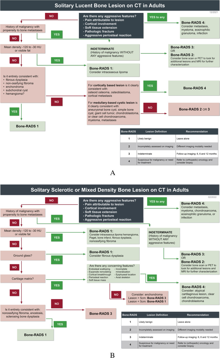
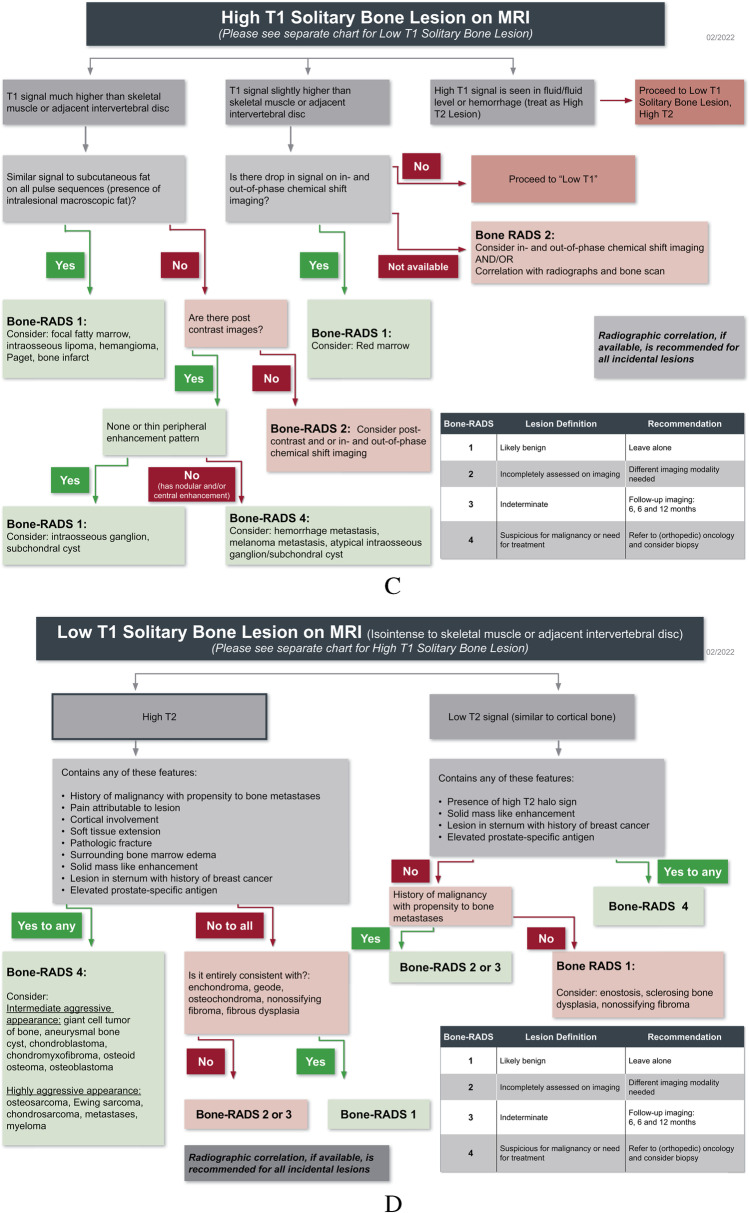
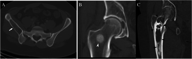
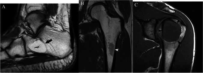
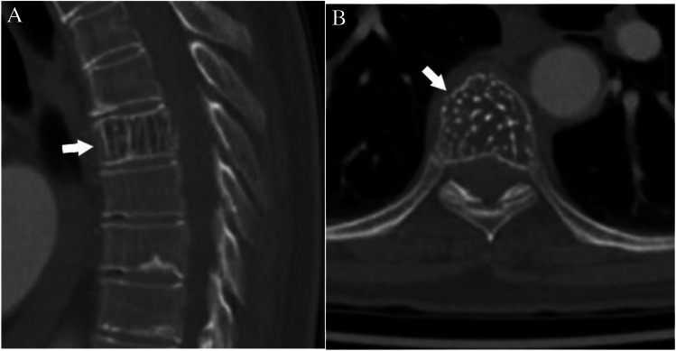
The purpose of this article is to present algorithms for the diagnostic management of solitary bone lesions incidentally encountered on computed tomography (CT) and magnetic resonance (MRI) in adults. Based on review of the current literature and expert opinion, the Practice Guidelines and Technical Standards Committee of the Society of Skeletal Radiology (SSR) proposes a bone reporting and data system (Bone-RADS) for incidentally encountered solitary bone lesions on CT and MRI with four possible diagnostic management recommendations (Bone-RADS1, leave alone; Bone-RADS2, perform different imaging modality; Bone-RADS3, perform follow-up imaging; Bone-RADS4, biopsy and/or oncologic referral). Two algorithms for CT based on lesion density (lucent or sclerotic/mixed) and two for MRI allow the user to arrive at a specific Bone-RADS management recommendation. Representative cases are provided to illustrate the usability of the algorithms.

Keywords: Bone tumors; CT; MRI; Management.


Conflict of interest statement

The authors declare no competing interests.

Figures

Fig. 1
Fig. 1
Flowcharts for evaluating: A solitary lucent bone lesions on CT, B solitary sclerotic and mixed density bone lesions on CT, C High T1 solitary bone lesion on MRI, and D low T1 solitary bone lesion on MRI
Fig. 1
Fig. 1
Flowcharts for evaluating: A solitary lucent bone lesions on CT, B solitary sclerotic and mixed density bone lesions on CT, C High T1 solitary bone lesion on MRI, and D low T1 solitary bone lesion on MRI
Fig. 2
Fig. 2
CT classification of bone lesions by density. CT images show A lucent (arrow), B sclerotic (pointed arrow), and C mixed density (arrowheads) lesions in 3 patients with giant cell tumor, prostate cancer metastasis, and fibrous dysplasia, respectively
Fig. 3
Fig. 3
MRI lesions based on T1 signal intensity. T1-weighted MRI images show A a calcaneal intraosseous lipoma (arrow) that has signal intensity much higher than skeletal muscle with the same signal intensity as fat, B a focus of red marrow (arrowhead) in the proximal femur that is slightly hyperintense to skeletal muscle, and C a chondroblastoma (arrowheads) in the humeral that has signal isointense to skeletal muscle
Fig. 4
Fig. 4
MRI lesions based on T2 signal intensity. A Axial T2-weighted MRI image of the hip shows a uniformly hypointense lesion (arrow) in the femoral head consistent with an enostosis. B Sagittal T2-weighted fat-suppressed MRI images show a lobulated hyperintense lesion in the humeral head consistent with a low-grade cartilage lesion/enchondroma
Fig. 5
Fig. 5
Chemical shift imaging. 56-year-old man with incidental lesion in the right sacral ala. The lesion (arrows) is isointense to skeletal muscle on the A in-phase T1-weighted gradient echo MR image and has uniform loss (drop) in signal on the B oppose phase T1-weighted gradient echo MR image indicating the presence of internal fat. The lesion is compatible with a focus of red marrow
Fig. 6
Fig. 6
CT assessment of concerning features. A Coronal CT image shows cortical involvement (arrow) of the anterior femoral cortex from an enchondroma. B Axial soft tissue window CT image shows a destructive lung cancer metastasis in the ilium with large soft tissue component (pointed arrows). C Axial CT image shows a pathologic fracture (arrowheads) in the femoral neck due to a giant cell tumor of bone. D Axial CT image shows periosteal reaction (thin arrows) along the femoral cortex from presumed stress fracture
Fig. 7
Fig. 7
Characteristic CT lucent lesions. A Coronal CT image shows a lucent lesion (arrow) in the proximal femur consistent with fibrous dysplasia, B coronal CT image shows a cortically based lucent non-ossifying fibroma in the proximal tibia, C coronal CT image shows an enchondroma with punctate calcifications in the distal femur, D coronal CT image shows a subchondral cyst abutting the right hip joint with degenerative changes, and E axial CT image shows a lucent lesion with a “polka dot” appearance of calcifications in T12 consistent with a hemangioma
Fig. 8
Fig. 8
Assessment of lesion density with ROI. We recommend placing a ROI in the largest area that is most representative of the lesion to obtain mean Hounsfield unit (HU) values. A Incidental sclerotic focus in the right iliac bone has mean HU = 1104 consistent with an enostosis. B Lesion in the proximal femur has mean HU =  − 21 consistent with fat in an intraosseous lipoma
Fig. 9
Fig. 9
T1 hyperintense hemorrhagic lesions can mask underlying lesions and should follow the “High T2” algorithm. There is a lucent lesion (arrows) in the proximal humerus on the radiographs (A). The lesion has high signal (from hemorrhage) and a fluid level on the axial T1- (B) and axial T2- (C) weighted images. Lesion was was solitary bone cyst at definitive treatment
Fig. 10
Fig. 10
MRI assessment of concerning features. A Sagittal T1-weighted MR image shows a giant cell tumor in the proximal tibia with anterior and posterior cortical involvement (arrows). B Sagittal T2-weighted fat-suppressed MRI image shows a renal cell metastasis in the proximal tibia with soft tissue extension (pointed arrow) posteriorly. C Coronal T2-weighted MR image shows a lung cancer metastasis in the proximal humerus with pathologic fracture (arrowhead) of the lateral cortex. D Axial T2-weighted fat-suppressed MR image shows a low signal prostate cancer metastasis in the distal femur with peripheral halo (thin arrow) of high T2 signal. E Sagittal T1-weighted fat-suppressed post-contrast MRI image shows an enhancing breast cancer metastasis (fat arrow) in the mid shaft of the femur. F Axial T2-weighted fat-suppressed MR image shows a hyperintense metastasis (outlined arrow) in the sternum in a patient with breast cancer
Fig. 11
Fig. 11
Case 1. Intraosseous lipoma. 23-year-old female runner presents with 3 months of left hip pain radiating to the groin with incidental lesion (arrows) detected in the proximal femur on MRI. The lesion is hyperintense (similar to subcutaneous fat) on the A coronal T1-weighted MR image and is hypointense on the B coronal T2-weighted fat-suppressed MR image. C Radiograph shows a lucent lesion in the proximal femur
Fig. 12
Fig. 12
Case 2. Enostosis. 68-year-old female presents with right pelvic pain after a fall. Coronal CT image shows a densely sclerotic lesion (arrow) with spiculated margins in the proximal femur with mean density = 1644 HU
Fig. 13
Fig. 13
Case 3. Breast cancer metastasis. 57-year-old female runner presents with thigh pain. There is a lesion (arrows) in the midshaft of the femur that is isointense to skeletal muscle on the A axial T1-weighted MR image and hyperintense on the B axial T2-weighted fat-suppressed MR image
Fig. 14
Fig. 14
Case 4. Cartilaginous neoplasm. There is an expansile mixed lesion (arrows) expanding the posterior right iliac bone (axial location). The lesion has central punctate foci of calcifications consistent with a cartilaginous lesion and concerning endosteal scalloping. Definitive surgery revealed low-grade chondrosarcoma
Fig. 15
Fig. 15
Case 5. Fibrous dysplasia. 84-year-old female with right hip pain and clinical suspicion for hip osteoarthritis who underwent right hip MRI and subsequent CT for incidentally discovered lesion (arrows) in the right ischium. The lesion is isointense to muscle on the A coronal T1-weighted MR image and hyperintense on the B coronal T2-weighted fat-suppressed MR image. The lesion has a sclerotic rim and ground glass appearance on the C coronal CT image
Fig. 16
Fig. 16
Case 6. Non-ossifying fibroma. 21-year-old man with medial knee pain. A Sagittal and B axial CT images show a mixed density cortically based lesion (arrows) with in the distal femur without concerning features
Fig. 17
Fig. 17
Case 7. A 27-year-old female presents with vague thigh pain for 1 year. Coronal CT image reveals a solitary focal lucent lesion in the left proximal femoral metaphysis with a well-defined sclerotic margin, cortical involvement (arrow), and mean HU of 18 units; a simple bone cyst at surgery. B and C 18-year-old female presents with knee pain worse over last 3 months. MRI of the left knee reveals a cystic lesion (arrowheads) involving the distal femoral metaphysis with fluid levels (thin arrows) due to internal blood products. The lesion has a well-defined sclerotic margin and demonstrates mild expansile remodeling. The lesion was aneurysmal bone cyst at treatment
Fig. 18
Fig. 18
Case 8. Red marrow. 39-year-old female presents with knee pain suspicious for meniscal tear. There is an incidentally discovered lesion (arrows) in the distal femoral shaft that is slightly hyperintense to skeletal muscle on the A sagittal T1-weighted MR image and hyperintense on the B sagittal T2 fat-suppressed MR image. This finding is compatible with focal red marrow
Fig. 19
Fig. 19
Case 9. Subchondral cyst. 30-year-old man presents with 1 month of pleuritic chest pain. There is an asymptomatic incidental lucent lesion (arrows) in the glenoid which abuts the articular surface on the A axial and B sagittal CT images. Lesion has mean density of 25 HU
Fig. 20
Fig. 20
Case 10. Hemangioma. 73-year-old female with chest pain. Chest CT reveals an incidental T7 lesion. The lesion (arrows) has vertical striations on the A sagittal CT image and results in a “polka dot” appearance on the B axial CT image

Similar articles

Cited by

References

    1. Musculoskeletal Tumor Society: systematic literature review on the use of imaging prior to referral to a musculoskeletal oncologist. Rosemont, IL, Musculoskeletal Tumor Society, February 2018. [Internet]. Available from: http://msts.org/view/download.php/education/pdfs/use-of-imaging-prior-to...
    1. Miller TT. Bone tumors and tumorlike conditions: analysis with conventional radiography. Radiology. 2008;246:662–674. doi: 10.1148/radiol.2463061038. - DOI - PubMed
    1. Lodwick GS, Wilson AJ, Farrell C, Virtama P, Dittrich F. Determining growth rates of focal lesions of bone from radiographs. Radiology. 1980;134:577–583. doi: 10.1148/radiology.134.3.6928321. - DOI - PubMed
    1. Madewell JE, Ragsdale BD, Sweet DE. Radiologic and pathologic analysis of solitary bone lesions. Part I: internal margins. Radiol Clin North Am. 1981;19:715–48. - PubMed
    1. Ragsdale BD, Madewell JE, Sweet DE. Radiologic and pathologic analysis of solitary bone lesions. Part II: periosteal reactions. Radiol Clin North Am. 1981;19:749–83. - PubMed