. 2022 Mar 27;79(4):211.
doi: 10.1007/s00018-022-04242-0.

Fine-tuning of epithelial taste bud organoid to promote functional recapitulation of taste reactivity

Affiliations

Fine-tuning of epithelial taste bud organoid to promote functional recapitulation of taste reactivity

Anish Ashok Adpaikar et al. Cell Mol Life Sci. .

Abstract

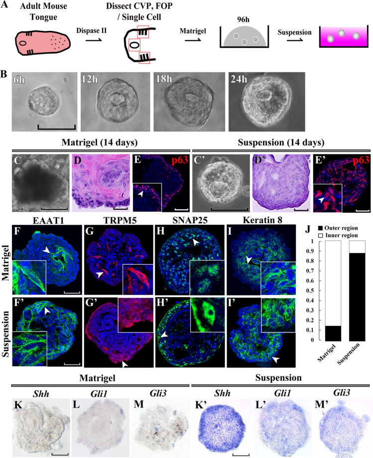
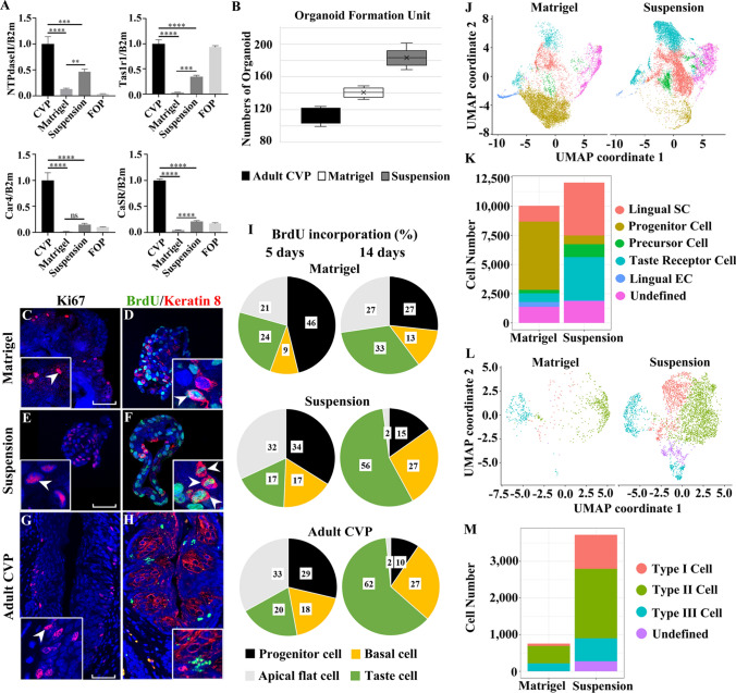
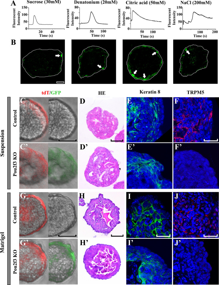
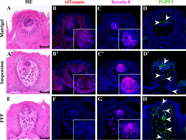
Taste stem/progenitor cells from posterior mouse tongues have been used to generate taste bud organoids. However, the inaccessible location of taste receptor cells is observed in conventional organoids. In this study, we established a suspension-culture method to fine-tune taste bud organoids by apicobasal polarity alteration to form the accessible localization of taste receptor cells. Compared to conventional Matrigel-embedded organoids, suspension-cultured organoids showed comparable differentiation and renewal rates to those of taste buds in vivo and exhibited functional taste receptor cells and cycling progenitor cells. Accessible taste receptor cells enabled the direct application of calcium imaging to evaluate the taste response. Moreover, suspension-cultured organoids can be genetically altered. Suspension-cultured taste bud organoids harmoniously integrated with the recipient lingual epithelium, maintaining the taste receptor cells and gustatory innervation capacity. We propose that suspension-cultured organoids may provide an efficient model for taste research, including taste bud development, regeneration, and transplantation.

Keywords: Apicobasal polarity; Calcium imaging; Genetic alteration; Single-cell RNA sequencing; Suspension-culture; Taste bud organoids; Taste receptor cells; Transplantation.


Conflict of interest statement

The authors have no relevant financial or non-financial interests to disclose.

Figures

Fig. 1
Fig. 1
Suspension-cultured organoids exhibit reversal of apicobasal polarity and contain Shh-expressing/-responsive cells. A Schematic of suspension-culture method for taste bud organoids. Single cell-derived taste bud organoids are formed in Matrigel and later transferred to low-attachment plates for suspension-culture with the same culture medium. B Time-lapse imaging of suspension-cultured organoids under a bright-field microscope. The size of the organoid increased gradually to roughly 50 μm after 24 h. C, C′ Bright-field images show that Matrigel-embedded organoids are larger than suspension-cultured organoids after a 14-day culture. D, D′ An eosin-stained keratinizing inner core is observed in the Matrigel-group but not in the suspension-group. E The basal cell marker p63 is detected in the outer region of Matrigel-embedded organoids. E′ After suspension-culture, p63-positive cells are scattered and found in the inner region. FI′ 14-day cultured organoids are immunostained using anti-EAAT1, TRPM5, SNAP25 and Keratin 8 antibodies. F Type I taste receptor cells marked by EAAT1 (green) are observed inside organoids (arrowhead). F′ EAAT1 positive cells are found in the outer region of organoids (arrowhead). G Type II taste receptor cells marked by TRPM5 (red) are found in the inner region of organoids (arrowhead). G′ TRPM5 positive cells are detected in the outer region contacting the medium (arrowhead). H Type III taste receptor cells marked by SNAP25 (green) are randomly distributed in the inner region of organoids (arrowhead). H′ SNAP25 positive cells are detected in the outer region of suspension-cultured organoids. I K8 positive cells, a pan taste receptor cells marker, are observed in the inner region of Matrigel-embedded organoids (arrowhead). I′ K8 positive cells are found in the outer region of organoids (arrowhead). J Most of K8 positive cells are localized internally in the Matrigel-embedded group but are localized in the outer region in the suspension-cultured group. Weak expressions of Shh (K), Gli1 (L) and Gli3 (M) in Matrigel-embedded organoids are partially detected by whole-mount in situ hybridization. K′ Shh is detected in several cells inside the suspension-cultured organoids. L′ Gli1 is broadly expressed in the inner region. M′ Gli3 positive cells are detected inside the suspension-cultured taste bud organoids. Scale bars in (BC′, E, FM) = 50 μm; (D, D′, E′, K′M′) = 25 μm. Arrowheads indicate localization of ZO-1, β-catenin and p63. Red squares indicate CVP and FOP (CVP: circumvallate papilla, FOP: foliate papillae). All panels represent data from at least five independent specimens (n > 5) (p < 0.05)
Fig. 2
Fig. 2
Suspension-cultured organoids show higher similarity to taste buds. A Taste receptor cell markers are significantly higher in suspension-cultured organoids after 14 days. CVP and FOP are used as technical and topological control, respectively. B The number of single cell-derived organoid forming units is larger in the suspension group than in the Matrigel and CVP epithelium groups after 7 days of culture. C Representative Ki67 staining (red) shows proliferating cells in the outer region of Matrigel-embedded organoids (arrowhead). D Double staining of BrdU (green) and K8 (red) shows that proliferative cells are partially differentiated into taste receptor cells (arrowhead) 5 days after BrdU incorporation. E Ki67 positive proliferating cells are broadly located in suspension-cultured organoids (arrowhead). F A higher number of BrdU/K8-positive cells are found in suspension-cultured organoids 5 days after incorporation (arrowhead). G Ki67 positive cells are found in the perigemmal region (arrowhead). H BrdU-incorporated cells (green) are found in the intragemmal and perigemmal regions 5 days after BrdU incorporation. I BrdU label retaining cells are categorized as progenitor, precursor, apical flat, and taste cell populations based on double staining with anti-K14/SHH/K8 and anti-BrdU antibodies. Pie charts show the cell populations at 5 and 14 days after BrdU incorporation. Numbers indicate the percentage of each cell population among BrdU label retaining cells. The proportion of each cell population is similar among the suspension group and adult CVP epithelium. J Uniform manifold approximation projection (UMAP) of single cell transcriptome data obtained from Matrigel-embedded and suspension-cultured organoids. The colors attribute to the different cell populations identified. K Suspension-cultured organoids show a higher cell number of lingual stem, precursor, and taste receptor cells. L UMAP plot comparing the subtypes of taste receptor cells from Matrigel-embedded and suspension-cultured organoids. M Suspension-cultured organoid showing a higher number of all types of taste receptor cells compared to a Matrigel-embedded organoid. Scale bars = 50 μm. Arrowheads indicate Ki67-positive cells or BrdU labeled cells. At least five independent batches of organoids were analyzed for RT-qPCR and organoid formation assays (n > 5). Matrigel/suspension organoids and adult mice were labeled with BrdU and processed for cell categorization experiments (n > 5). Data represented are mean ± SD; RT–qPCR data were compared by Tukey's multiple comparison test. **, *** and **** indicate p < 0.01, p < 0.001 and p < 0.0001, respectively, ns—not significant
Fig. 3
Fig. 3
Tastant-evoked responses and genetic alteration potential in suspension-cultured organoids. A Graph representing fluorescence intensity changes in response to different tastant solutions. B Fluorescence intensity peaks are observed after sweet, bitter, salty, or sour tastant treatments to the taste bud organoids. C, C′, G, G′ 48 h after electroporation, organoids displayed wide areas containing tdTomato-positive (control) or tdTomato-/GFP-positive cells (Pou2f3 KO). D, D′, H, H′ No morphological differences are observed between the control and the Pou2f3 knockout (KO) organoids by hematoxylin and eosin (HE) staining in suspension-cultured and Matrigel-embedded organoids. E, E′, I, I′ K8 (green) positive cells are broadly localized throughout the control and Pou2f3 KO organoids. F, F′, J, J′ The number of TRPM5-positive Type II taste receptor cells are notably reduced in Pou2f3 KO organoids when compared to the control. Scale bars in (C, C′, D, D′, G, G′, H, H′) = 50 μm; (E, E′, F, F′, I, I′, J, J′) = 25 μm. At least 10 organoids were analyzed for taste-evoked calcium response (n > 10). At least five organoids were analyzed for immunostaining (n > 5)
Fig. 4
Fig. 4
Integration of transplanted organoids in the lingual epithelium. A Subcutaneously transplanted Matrigel-embedded organoids integrated within the lingual epithelium of recipient mice. B tdTomato-positive cells are found inside and surrounding the transplanted organoids. C K8-positive taste receptor cells are found in the transplanted organoids. D Low PGP9.5 staining shows that there are few neuronal components surrounding transplanted Matrigel-embedded organoids. A′ A cluster of spindle-shaped cells is observed in transplanted suspension-cultured organoids within the lingual epithelium. B′ tdTomato-positive cells are found around the transplanted organoids. C′ Cluster of cells with strong K8 staining are observed. D′ PGP9.5 is detected inside the transplanted organoid and its surrounding mesenchyme. EH Fungiform papilla showing taste bud and stroma core. Cells with strong K8 and PGP9.5 staining are detected, while a tdTomato-positive cell is not observed. PGP9.5-positive nerve fibers are clearly detected through taste cells to underlying mesenchyme. Scale bars = 50 μm. Arrowheads indicate PGP9.5 reactive nerve fibers. Transplantation was performed on five mice for each group (n = 5)

Similar articles

Cited by

References

    1. Barlow LA, Klein OD. Current topics in developmental biology. Amsterdam: Elsevier; 2015. Developing and regenerating a sense of taste; pp. 401–419. - PMC - PubMed
    1. Chaudhari N, Roper SD. The cell biology of taste. J Cell Biol. 2010;190:285–296. doi: 10.1083/jcb.201003144. - DOI - PMC - PubMed
    1. Kinnamon SC, Dionne VE, Beam KG. Apical localization of K+ channels in taste cells provides the basis for sour taste transduction. Proc Natl Acad Sci. 1988;85:7023–7027. doi: 10.1073/pnas.85.18.7023. - DOI - PMC - PubMed
    1. Liman ER, Zhang YV, Montell C. Peripheral coding of taste. Neuron. 2014;81:984–1000. doi: 10.1016/j.neuron.2014.02.022. - DOI - PMC - PubMed
    1. Chandrashekar J, Hoon MA, Ryba NJ, Zuker CS. The receptors and cells for mammalian taste. Nature. 2006;444:288–294. doi: 10.1038/nature05401. - DOI - PubMed