. 2022 May;36(5):e22279.
doi: 10.1096/fj.202200008R.

Ornithine decarboxylase deficiency critically impairs nitrogen metabolism and survival in Aedes aegypti mosquitoes

Affiliations

Ornithine decarboxylase deficiency critically impairs nitrogen metabolism and survival in Aedes aegypti mosquitoes

Jun Isoe et al. FASEB J. 2022 May.

Abstract

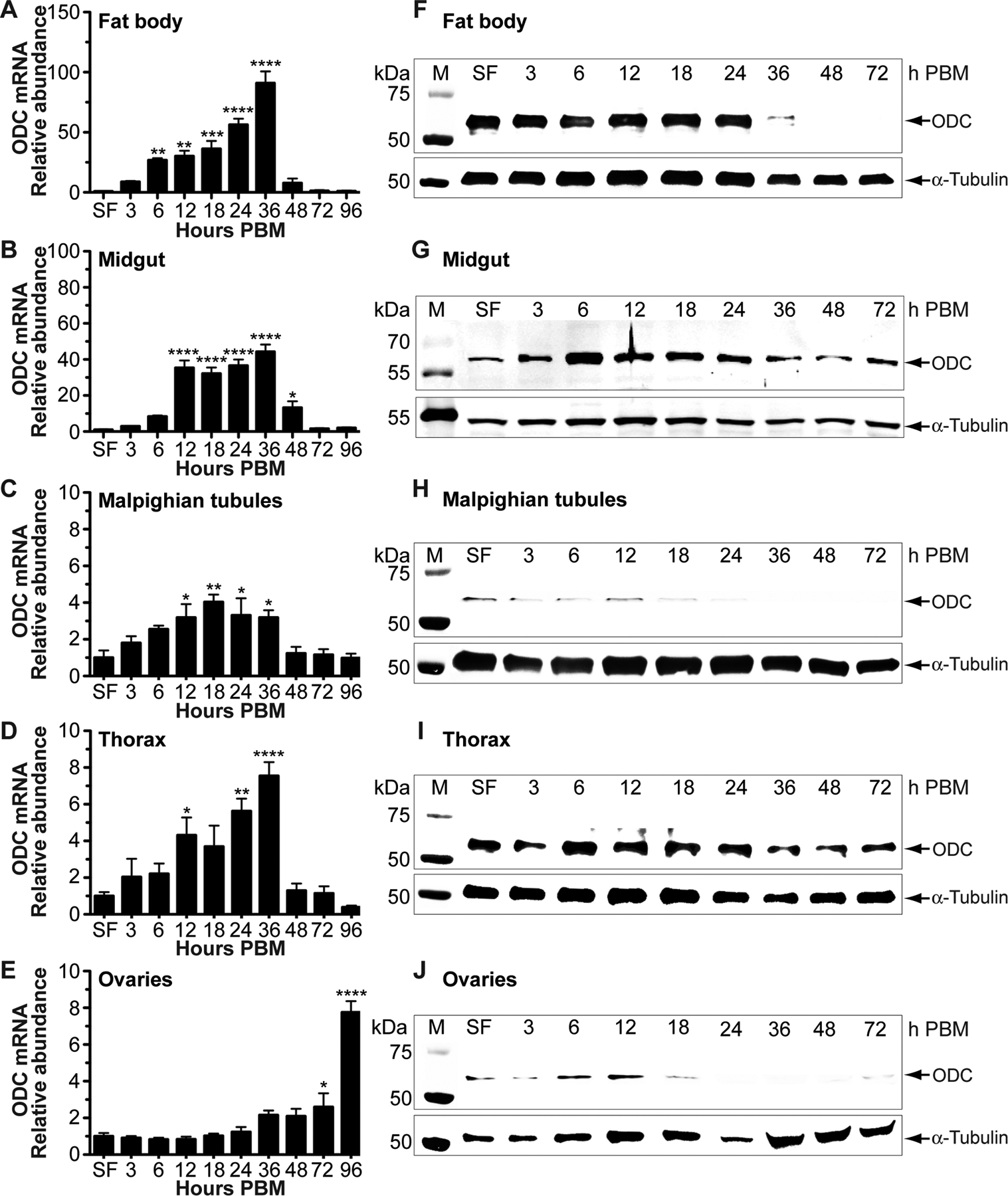
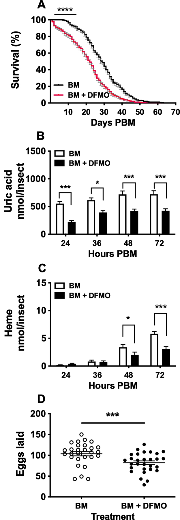
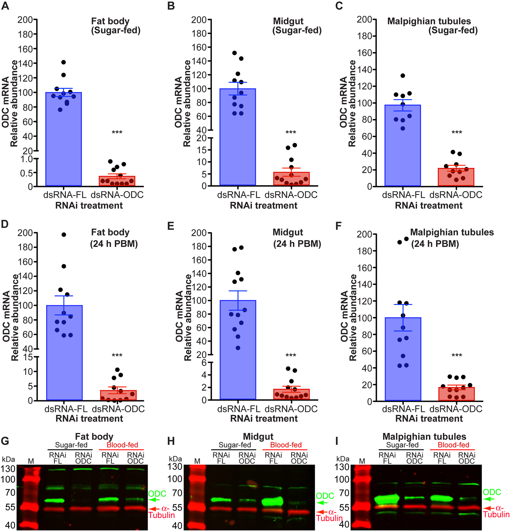
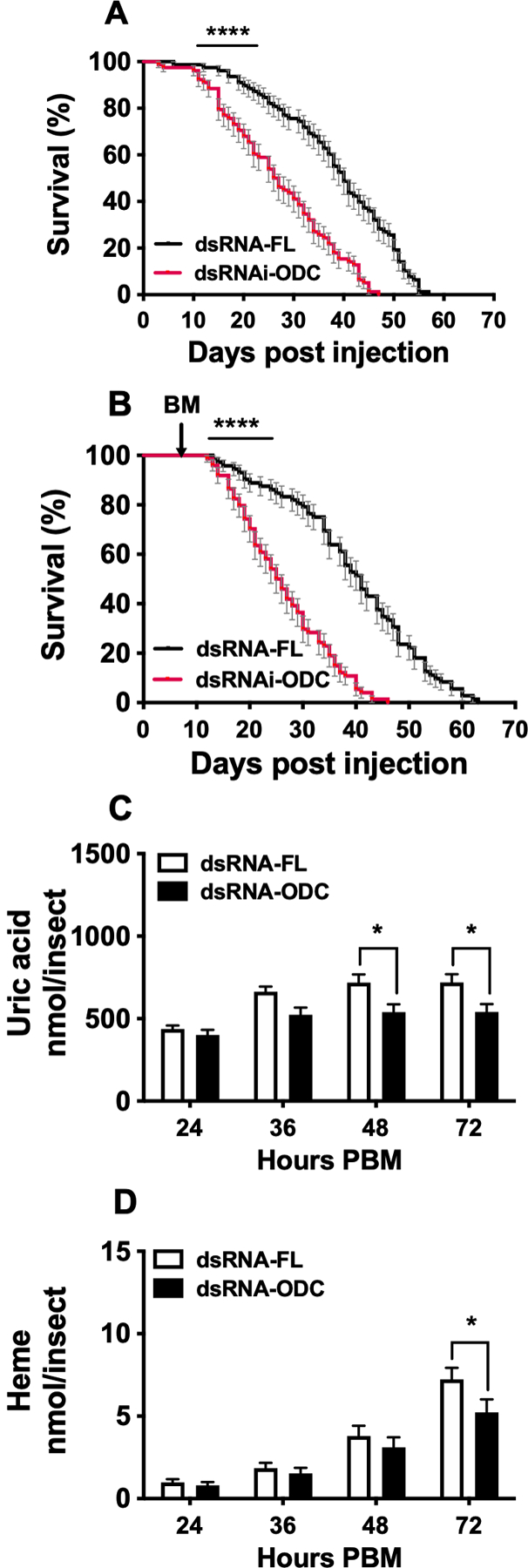
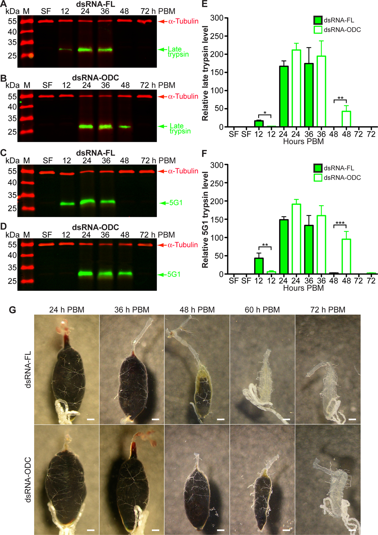
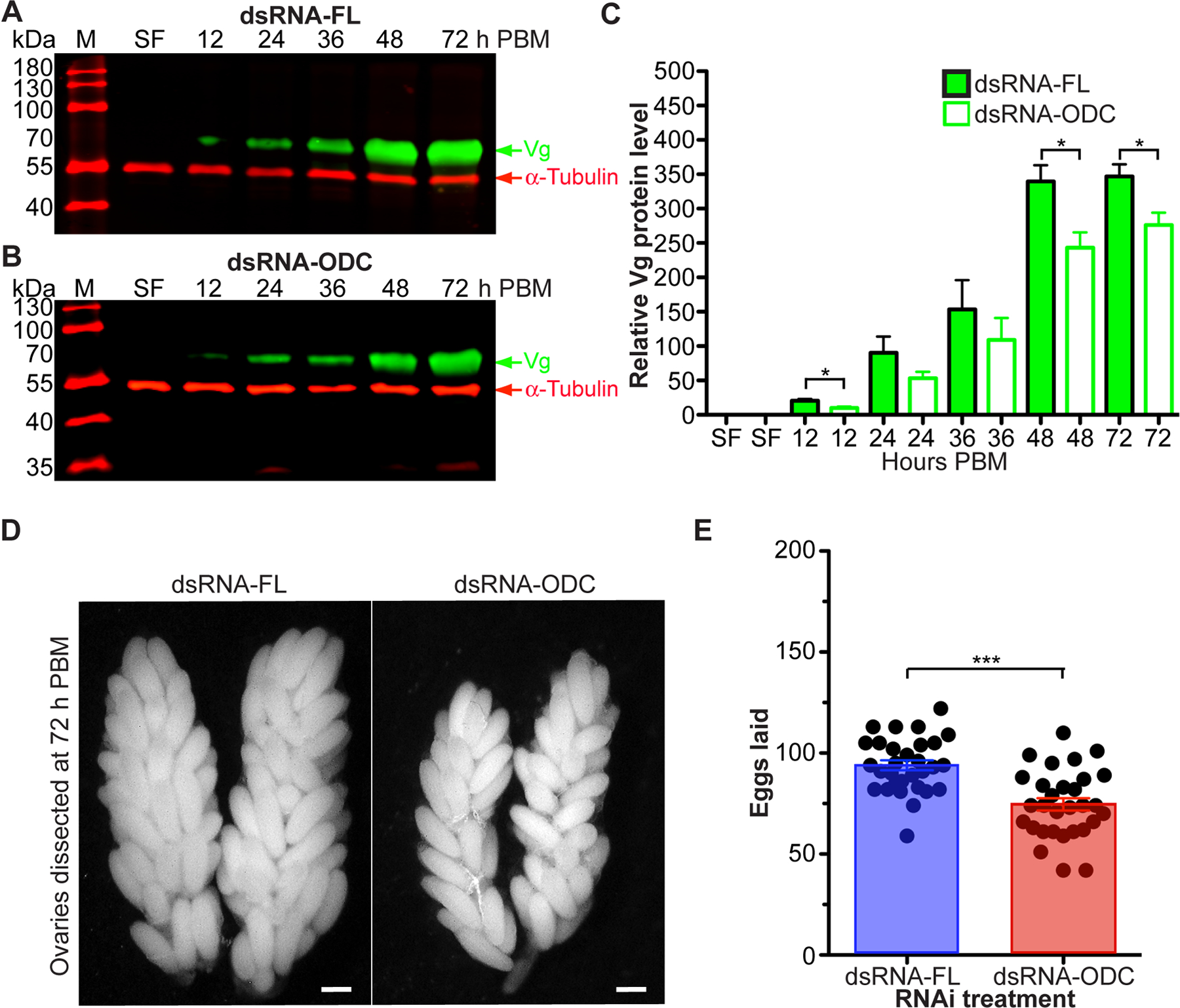
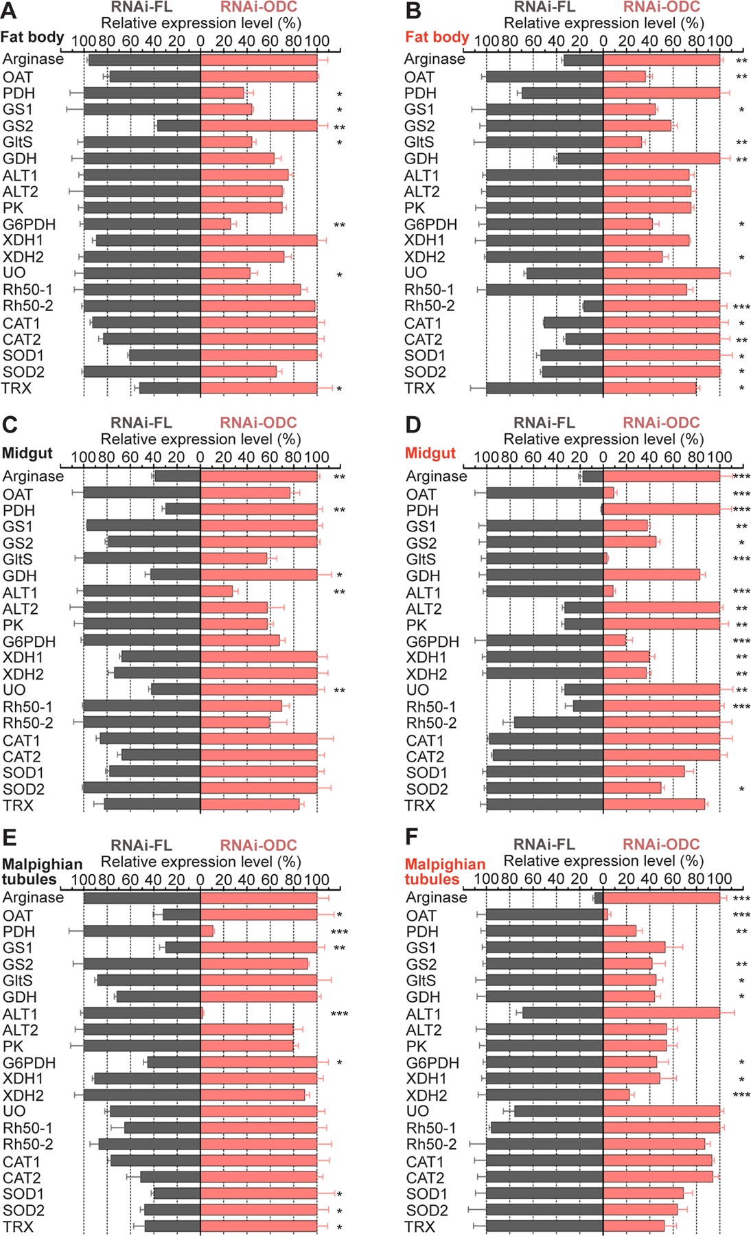
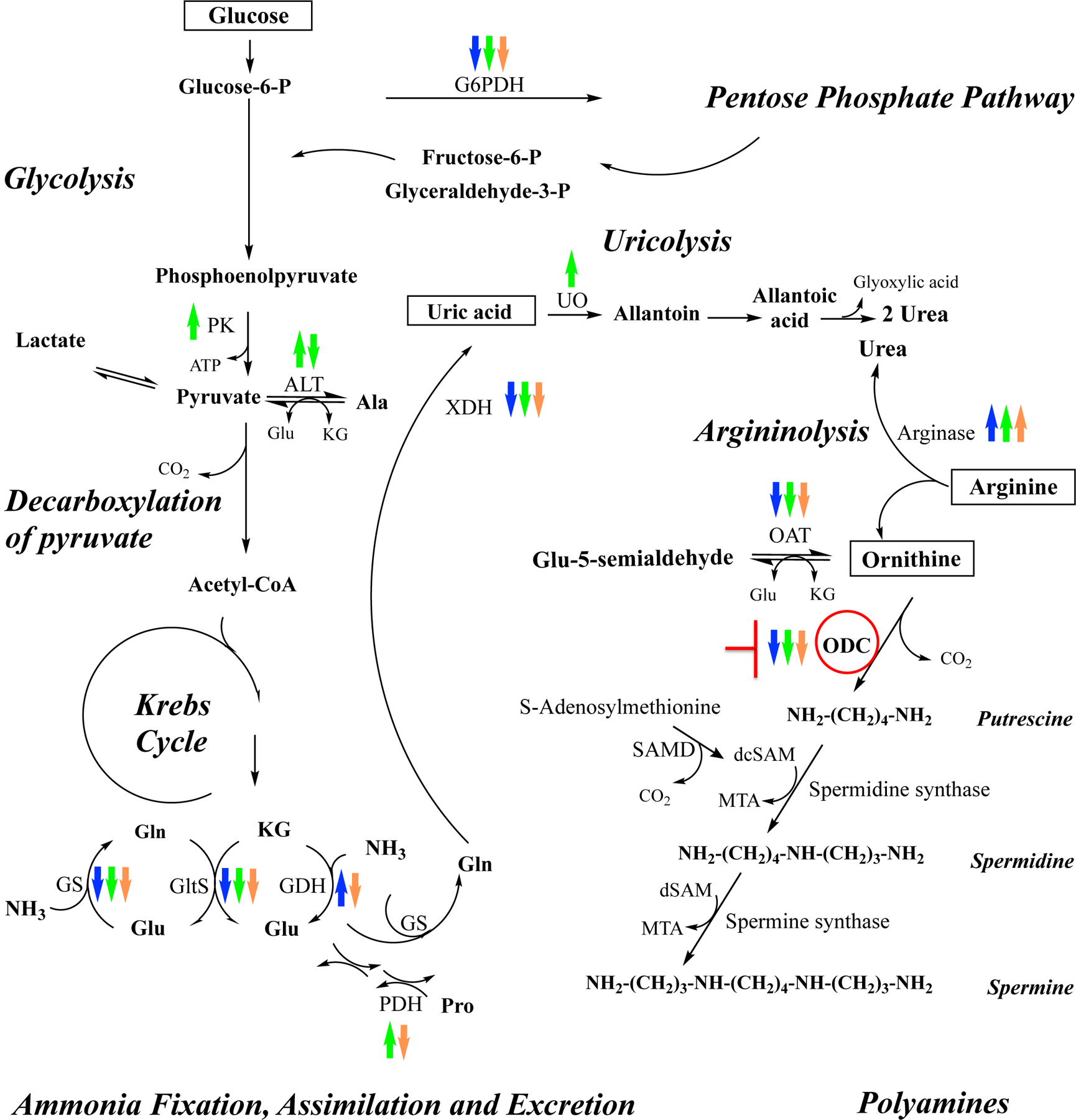
Ornithine decarboxylase (ODC; EC 4.1.1.17) catalyzes the conversion of ornithine to putrescine, the rate-limiting first step for de novo polyamine biosynthesis. Previously, we reported that genetic knockdown of xanthine dehydrogenase 1 (XDH1)-a gene encoding the enzyme involved in the last two steps of uric acid synthesis-causes an increase in ODC transcript levels in fat body of blood-fed Aedes aegypti mosquitoes, suggesting a crosstalk at molecular level between XDH1 and ODC during nitrogen disposal. To further investigate the role of ODC in nitrogen metabolism, we conducted several biochemical and genetic analyses in sugar- and blood-fed A. aegypti females. Distinct ODC gene and protein expression patterns were observed in mosquito tissues dissected during the first gonotrophic cycle. Both pharmacological and RNA interference-mediated knockdown of ODC negatively impacted mosquito survival, disrupted nitrogen waste disposal, delayed oviposition onset, and decreased fecundity in vitellogenic blood-fed females. A lag in the expression of two major digestive serine proteases, a reduction of blood meal digestion in the midgut, and a decrease in vitellogenin yolk protein uptake in ovarian follicles were observed by western blots in ODC-deficient females. Moreover, genetic silencing of ODC showed a broad transcriptional modulation of genes encoding proteins involved in multiple metabolic pathways in mosquito fat body, midgut, and Malpighian tubules prior to and after blood feeding. All together, these data demonstrate that ODC plays an essential role in mosquito metabolism, and that ODC crosstalks with multiple genes and proteins to prevent deadly nitrogen perturbations in A. aegypti females.

Keywords: ammonia metabolism; glucose metabolism; oxidative stress; polyamines; survival.


Conflict of interest statement

CONFLICT OF INTEREST STATEMENT

The authors declare no conflict of interest.

Figures

FIGURE 1
FIGURE 1. Ornithine decarboxylase (ODC) gene and protein expression patterns in Aedes aegypti tissues.
Four-day old sucrose-fed (SF) mosquitoes were fed only on 3% sucrose after adult eclosion. Tissues were dissected from SF and blood-fed mosquitoes at intervals after feeding. Four-day old females were allowed to feed on blood. A-E) ODC mRNA levels obtained by qPCR were normalized to mRNA levels of the ribosomal protein S7. Data are presented as mean ± SEM of 3 independent samples. Each cDNA replicate was prepared from a pool of 10 tissues. N = 300. *P < 0.05, **p < 0.01, ***p < 0.001, ****p < 0.0001 when compared to SF. F-J) ODC protein levels were analyzed using a custom-made polyclonal antibody against Ae. aegypti ODC. For fat body, midgut, and ovary, each lane contains 1 tissue equivalent of the protein extracts. For thorax and Malpighian tubules, each lane contains 0.2 and 2 tissue equivalent of the protein extracts, respectively. Anti-α-tubulin antibody was used as an internal control for protein loading. Western blots are representative of 3 biological replicates. Each protein extract replicate was prepared from a pool of 10 mosquito tissues. N = 270. M = protein markers, PBM = post blood meal.
FIGURE 2
FIGURE 2. Effect of DL-α-difluoromethylornithine (DFMO) on Aedes aegypti survival, excretion and egg production.
Mosquitoes were fed a blood meal (BM) supplemented with 0 or 100 mM DFMO. A) Survival analysis. Mortality was recorded daily throughout the experimental period. Data are expressed as mean percentages ± SEM of 3 independent experiments. In each treatment, 300 females were monitored. N = 600. B) Uric acid concentration in excreta. C) Heme concentration in excreta. Nitrogen waste data (B and C) are expressed as mean ± SEM of 10 individual mosquitoes. In each treatment, 10 individual females were used for the time course analyzed. N = 20. D) Numbers of eggs laid by individual females. Data are expressed as mean ± SEM of 30 individual mosquitoes. In each treatment, 30 individual females were used. N = 60. *P < 0.05, ***p < 0.001, ****p < 0.0001 when compared to control.
FIGURE 3.
FIGURE 3.. Efficiency of ornithine decarboxylase (ODC) knockdown on Aedes aegypti tissues.
Ae. aegypti females were microinjected with dsRNA-firefly luciferase (dsRNA-FL) or dsRNA-ODC. In each treatment, fat body, midgut and Malpighian tubules were dissected from sugar- and blood-fed females at 24 h PBM. A-F) ODC gene expression in individual mosquitoes. Data are expressed as mean ± SEM of 9–12 individual mosquitoes. N = 133. ***P < 0.001 when compared to control (dsRNA-FL). G-I) ODC protein expression. ODC protein levels were analyzed using a custom-made polyclonal antibody against Ae. aegypti ODC. For fat body, each lane contains 0.5 tissue equivalent of the protein extracts. For midgut and Malpighian tubules, each lane contains 2.0 tissue equivalent of the protein extracts. Anti-α-tubulin antibody was used as an internal control for protein loading. Western blots are representative of 3 biological replicates. Each protein extract replicate was prepared from a pool of 5 mosquito tissues. N = 60. M = protein markers, PBM = post blood meal.
FIGURE 4.
FIGURE 4.. Effect of ornithine decarboxylase (ODC) knockdown on Aedes aegypti survival and excretion.
Ae. aegypti females were microinjected with dsRNA-firefly luciferase (dsRNA-FL) or dsRNA-ODC, fed only on 3% sucrose or a blood meal and maintained on 3% sucrose. A) Survival analysis of sugar-fed dsRNA-injected females. Mortality was recorded daily throughout the experiment period. Data are expressed as mean percentages ± SEM of 3 independent experiments. In each treatment, 78 females were monitored. N = 156. B) Survival analysis of blood-fed dsRNA-injected females. Data are expressed as mean percentages ± SEM of 3 independent experiments. C) Uric acid concentration in excreta. D) Heme concentration in excreta. Nitrogen waste data (C and D) are expressed as mean ± SEM of 10 individual mosquitoes. In each treatment, 10 individual females were used for the time course analyzed. N = 20. *P < 0.05, ****p < 0.0001 when compared to control. BM = blood meal, PBM = post blood meal.
FIGURE 5.
FIGURE 5.. Effect of ornithine decarboxylase (ODC) knockdown on Aedes aegypti blood protein digestion.
Ae. aegypti females were microinjected with dsRNA-firefly luciferase (dsRNA-FL) or dsRNA-ODC. Sucrose-fed (SF) mosquitoes were fed only on 3% sucrose. A-D) Western blot analysis of two major midgut serine proteases in response to RNAi treatment using custom-made polyclonal antibodies against Ae. aegypti late trypsin and 5G1. Each lane contains 0.5 tissue equivalent of the midgut protein extracts. Anti-α-tubulin antibody was used as an internal control for protein loading. Western blots are representative of 3 biological replicates. Each protein extract replicate was prepared from a pool of 10 midguts. N = 360. E-F) Densitometry analysis of western blots by ImageJ software. Data are expressed as means ± SEM of 3 biological replicates. *P < 0.05, **p < 0.01, ***p < 0.001 when compared to control. G) Midgut images at 24, 36, 48, 60 and 72 h PBM. Each midgut is presented as anterior to posterior orientation. Images shown are representative of 5 mosquitoes from each time course. N = 50. Each scale bar represents 200 μm. M = protein markers, PBM = post blood meal.
FIGURE 6.
FIGURE 6.. Effect of ornithine decarboxylase (ODC) knockdown on Aedes aegypti reproduction.
Ae. aegypti females were microinjected with dsRNA-firefly luciferase (dsRNA-FL) or dsRNA-ODC. Sucrose-fed (SF) mosquitoes were fed only on 3% sucrose. A-B) Vitellogenin (Vg) protein expression in response to RNAi treatment. Relative abundance of Vg was analyzed using a custom-made polyclonal antibody against Ae. aegypti Vg. Each lane contains 0.5 tissue equivalent of the protein extracts. Anti-α-tubulin antibody was used as an internal control for protein loading. Western blots are representative of 3 biological replicates. Each protein extract replicate was prepared from a pool of 10 ovaries. N = 360. C) Densitometry analysis. Data are expressed as means ± SEM of 3 replicates. D) Ovary images at 72 h PBM. Images shown are representative of 3 mosquitoes from each treatment. N = 6. Each scale bar represents 200 μm. E) Numbers of eggs laid by individual females in the first gonotrophic cycle. Data are expressed as mean ± SEM of 30 individual mosquitoes. In each treatment, 30 individual females were used. N = 60. *P < 0.05, ***p < 0.001 when compared to control. M = protein markers, PBM = post blood meal.
FIGURE 7.
FIGURE 7.. Effect of ornithine decarboxylase (ODC) knockdown on transcripts levels of multiple genes in Aedes aegypti tissues.
Ae. aegypti females were microinjected with dsRNA-firefly luciferase (dsRNA-FL) or dsRNA-ODC. A, C, E) Relative abundance of transcripts in fat body, midgut and Malpighian tubules dissected from sugar-fed mosquitoes. B, D, F) Relative abundance of transcripts in fat body, midgut and Malpighian tubules dissected at 24 h after blood feeding. Transcript levels were normalized to mRNA levels of the ribosomal protein S7. Data are expressed as mean ± SEM of 10 individual tissues. Each cDNA replicate was prepared from 10 individual tissues. N = 120. *P < 0.05, **p < 0.01, ***p < 0.001, ****p < 0.0001 when compared to control. ALT, alanine aminotransferase; CAT, catalase; G6PDH, glucose-6-phosphate dehydrogenase; GDH, glutamate dehydrogenase; GltS, glutamate synthase; GS, glutamine synthetase; OAT, ornithine aminotransferase; ODC, ornithine decarboxylase; PK, pyruvate kinase; PDH, proline dehydrogenase; Rh50, Rhesus 50 glycoprotein; SOD, superoxide dismutase; TRX, thioredoxin; UO, urate oxidase and XDH, xanthine dehydrogenase.
FIGURE 8.
FIGURE 8.. Schematic representation of metabolic interactions between glucose, ammonia, arginine and polyamine pathways in blood-fed Aedes aegypti mosquitoes.
Transcriptional changes of multiple genes in tissues dissected from blood-fed ODC-deficient females at 24 h PBM are shown with up and down arrows, which indicate up-regulation and down-regulation respectively. Blue arrows indicate changes in fat body, green arrows indicate changes in midgut, and orange arrows indicate changes in Malpighian tubules. ALT, alanine aminotransferase; dsSAM: decarboxylated 5-adenosylmethionine; G6PDH, glucose-6-phosphate dehydrogenase; GDH, glutamate dehydrogenase; GltS, glutamate synthase; GS, glutamine synthetase; KG, ketoglutarate; MTA: methylthioadenosine; OAT, ornithine aminotransferase; ODC, ornithine decarboxylase; PK, pyruvate kinase; PDH, proline dehydrogenase; SAMD: S-adenosylmethionine decarboxylase; UO, urate oxidase, and XDH, xanthine dehydrogenase. This scheme is adapted from Scaraffia et al., 2006, 2008 and Horvath et al., 2018 (30, 34, 36). Copyright permission obtained. Copyright (2006) Elsevier. Copyright (2008) National Academy of Sciences, U.S.A. Copyright (2018) John Wiley and Sons.

Similar articles

Cited by

References

    1. Minois N, Carmona-Gutierrez D, Madeo F. Polyamines in aging and disease. Aging (Albany NY) 2011;3:716–732. - PMC - PubMed
    1. Nishimura K, Okudaira H, Ochiai E, et al. Identification of proteins whose synthesis is preferentially enhanced by polyamines at the level of translation in mammalian cells. Int J Biochem Cell Biol 2009;41:2251–2261. - PubMed
    1. Rider JE, Hacker A, Mackintosh CA, et al. Spermine and spermidine mediate protection against oxidative damage caused by hydrogen peroxide. Amino Acids 2007;33:231–240. - PubMed
    1. Murray Stewart T, Dunston TT, Woster PM, Casero RA Jr. Polyamine catabolism and oxidative damage. J Biol Chem 2018;293:18736–18745. - PMC - PubMed
    1. Bae DH, Lane DJR, Jansson PJ, Richardson DR. The old and new biochemistry of polyamines. Biochim Biophys Acta Gen Subj 2018;1862:2053–2068. - PubMed

Publication types