Review
. 2022 Mar 29;145(13):1002-1019.
doi: 10.1161/CIRCULATIONAHA.121.055854. Epub 2022 Mar 28.

Takotsubo Syndrome: Pathophysiology, Emerging Concepts, and Clinical Implications

Affiliations
Review

Takotsubo Syndrome: Pathophysiology, Emerging Concepts, and Clinical Implications

Trisha Singh et al. Circulation. .

Erratum in

Abstract

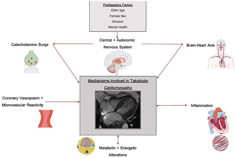
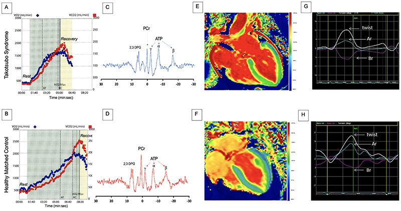
Takotsubo syndrome is a condition characterized by acute transient left ventricular systolic dysfunction, which at presentation can be challenging to distinguish from acute myocardial infarction. Although previously thought to be a benign, self-limiting condition, recent studies have confirmed that patients with takotsubo syndrome have persistent subtle ongoing cardiac dysfunction, and many continue to have limiting symptoms despite restoration of left ventricular ejection fraction. Moreover, these patients have a substantial burden of morbidity and mortality, as well, with high rates of subsequent major adverse cardiovascular events that approach those of patients with acute coronary syndrome. The mechanisms behind this condition remain elusive. Despite substantial research, the medical community continues to have an incomplete understanding of its underlying pathogenesis and pathophysiology. Catecholamine-induced myocardial injury is the most established and well-known theory, but this does not explain all the clinical features and presentations of the condition, and numerous other pathways and abnormalities are emerging. Because of the poor understanding of its underlying pathophysiology, there is a lack of evidence-based interventions to treat the acute episode, to avoid recurrences, and to prevent major adverse cardiovascular events. This highlights the need for further research to gain a better understanding of the underlying pathophysiology to inform appropriate randomized controlled trials of interventions targeting the causative pathways. Only then can evidence-based management strategies be established to improve clinical outcomes of this potentially lethal condition.

Keywords: Takotsubo cardiomyopathy; physiopathology; treatment outcomes.


Conflict of interest statement

Conflicts Of Interest

none

Figures

Figure 1
Figure 1. Takotsubo Syndrome: Anatomical variants
Left ventriculogram demonstrating apical (A, B) ballooning of the left ventricle, similar to the shape of a Japanese octopus trap (C). Basal (D, E), mid-ventricular (F, G) and focal (H,I) variations of Takotsubo syndrome.
Figure 2
Figure 2. Cardiac Magnetic Resonance Imaging and Computed Tomography Findings in Takotsubo Syndrome
Short axis T2 maps (A), T2 polar map (B) and short-axis T1 maps (D), demonstrating elevated T2 and T1 values circumferentially in the mid and apical regions (outside of a coronary territory). Long axis two-chamber view demonstrating transmural fibrotic band pattern typical of takotsubo syndrome 4 months after the index event (C). Long axis four-chamber and short axis views of mid-ventricle demonstrating elevated left ventricular mass in acute phase (E, F) and normalization during convalescence (G, H). Hybrid positive emission tomography with cardiac computed tomography angiography depicting a small left ventricular thrombus in a patient with takotsubo syndrome with no clinically apparent thrombus on conventional imaging. There is subtle hypoattenuation on the computed tomography angiogram (magnified inserts) and increased uptake of an activated platelet and thrombus-specific radiotracer (18F-GP-1; yellow-red) in long axis four-chamber (I, J) and short axis (K, L) views on positron emission tomography.
Figure 3
Figure 3. Diagnostic Criteria and Pathway for Takotsubo Syndrome
OCT, optical coherence tomography, IVUS, intra-vascular ultrasound.
Figure 4
Figure 4. Outcomes in Takotsubo Syndrome
(A), In-hospital complications in patients with takotsubo syndrome (blue) and patients with acute coronary syndrome (red). (B), long-term outcomes in patients with takotsubo cardiomyopathy. (C) Kaplan Meier curves for long-term mortality in patients with takotsubo syndrome compared with patient with acute coronary syndrome. Ghadri et al, 2018 (reference 15). STE-ACS, ST-segment elevation acute coronary syndrome, NSTE-ACS, Non-ST segment elevation acute coronary syndrome.
Figure 5
Figure 5. Mechanisms Involved in Takotsubo Syndrome
Figure 6
Figure 6. Abnormal Exercise Capacity, Energetics and Cardiac Performance in Patients with Takotsubo Syndrome
Cardiopulmonary exercise (treadmill) data from (A) a patient with prior takotsubo syndrome 20 months previously and (B) an age- and sex-matched healthy control subject. The maximal oxygen consumption (VO2, blue dots) achieved by the patient with takotsubo syndrome is markedly reduced, with an earlier anerobic threshold and a shorter duration of exercise when compared to the healthy control subject. Phosporus magnetic resonance spectrum acquisition for a patient with acute takotsubo syndrome (C) and healthy volunteer (D) showing reduced phosphocreatine/adenosine triphosphate ratio. Resonances corresponding to phosphocreatine (PCr), γ, ß, and α adenosine triphosphate (ATP), and 2,3-diphosphoglycerate (2,3 DPG). Manganese-enhanced magnetic resonance imaging in (E) a patient with acute takotsubo syndrome and (F) an age and sex-matched volunteer. The patient demonstrates abnormal calcium activity (green) throughout the mid ventricle and apex with preserved calcium activity in the basal segments (blue). In comparison, the healthy control subject demonstrates normal calcium activity throughout the myocardium (blue). Twist curves in (G) a patient with takotsubo syndrome at 2-year follow up and (H) an age and sex-matched healthy control volunteer. The healthy subject demonstrates the characteristic early systolic twist in a clockwise rotation at the apex (blue trace) and in counter clockwise rotation at the base (purple trace), occurring during isovolumic contraction. This is followed by counter clockwise apical rotation (Ar; blue) and clockwise basal rotation (Br; purple), which results in the net systolic twist during left ventricular ejection (twist, white line). In comparison, the patient with takotsubo syndrome demonstrates incomplete recovery of left ventricular twist, predominantly due to the reduced apical rotation. VO2 (mL/min), volume of oxygen inspired, VCO2 (mL/min), volume of carbon dioxide exhaled, AT, anerobic threshold, t(s), time, VO2 Max, maximal volume of oxygen inspired.

Similar articles

Cited by

References

    1. Sato TH, Uchida T, Dote KMI. Tako-tsubo-like left ventricular dysfunction due to multivessel coronary spasm. Kagakuhyoronsha Publishing Co; Tokyo, Japan: 1990. Clinical Aspect of Myocardial Injury: From Ischemia to Heart Failure.
    1. Engel GL. Sudden and rapid death during psychological stress. Folklore or folk wisdom? Ann Intern Med. 1971;74:771–782. doi: 10.7326/0003-4819-74-5-771. - DOI - PubMed
    1. Rees WD, Lutkins SG. Mortality of bereavement. Br Med J. 1967;4:13–16. doi: 10.1136/bmj.4.5570.13. - DOI - PMC - PubMed
    1. Minhas AS, Hughey AB, Kolias TJ. Nationwide trends in reported incidence of takotsubo cardiomyopathy from 2006 to 2012. Am J Cardiol. 2015;116:1128–1131. doi: 10.1016/j.amjcard.2015.06.042. - DOI - PubMed
    1. Pattisapu VK, Hao H, Liu Y, Nguyen TT, Hoang A, Bairey Merz CN, Cheng S. Sex-and age-based temporal trends in takotsubo syndrome incidence in the united states. J Am Heart Assoc. 2021;10:e019583. doi: 10.1161/JAHA.120.019583. - DOI - PMC - PubMed

Publication types