. 2022 Apr 5;119(14):e2115083119.
doi: 10.1073/pnas.2115083119. Epub 2022 Mar 28.

Genome-wide CRISPR screen reveals CLPTM1L as a lipid scramblase required for efficient glycosylphosphatidylinositol biosynthesis

Affiliations

Genome-wide CRISPR screen reveals CLPTM1L as a lipid scramblase required for efficient glycosylphosphatidylinositol biosynthesis

Yicheng Wang et al. Proc Natl Acad Sci U S A. .

Abstract

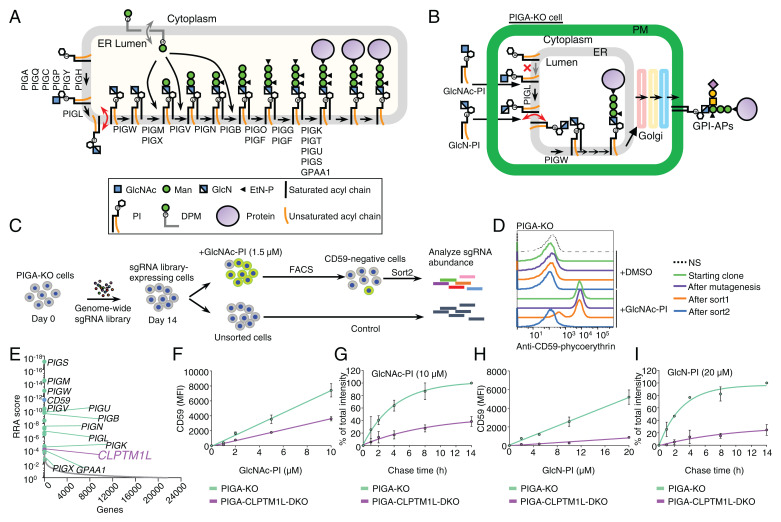
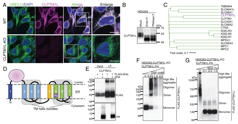
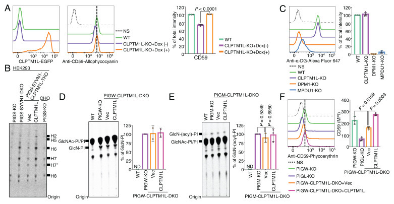
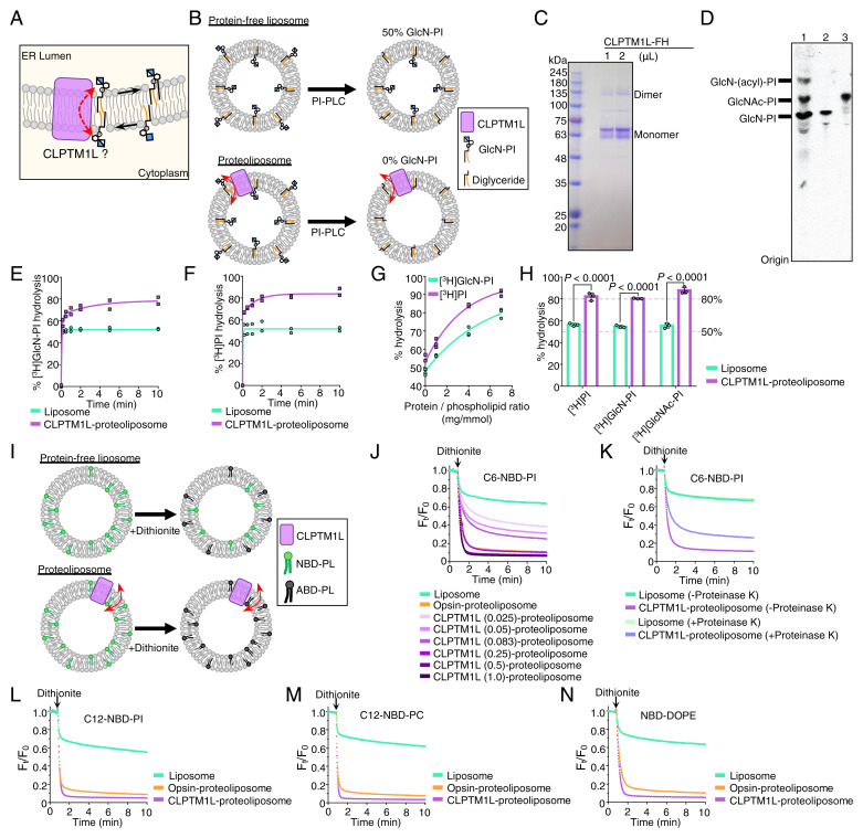
SignificanceScramblases translocate lipids across the lipid bilayer without consumption of ATP, thereby regulating lipid distributions in cellular membranes. Cytosol-to-lumen translocation across the endoplasmic reticulum (ER) membrane is a common process among lipid glycoconjugates involved in posttranslational protein modifications in eukaryotes. These translocations are thought to be mediated by specific ER-resident scramblases, but the identity of these proteins and the underlying molecular mechanisms have been elusive. Here, we show that CLPTM1L, an integral membrane protein with eight putative transmembrane domains, is the major lipid scramblase involved in efficient glycosylphosphatidylinositol biosynthesis in the ER membrane. Our results validate the long-standing hypothesis that lipid scramblases ensure the efficient translocations of lipid glycoconjugates across the ER membrane for protein glycosylation pathways.

Keywords: CLPTM1L; endoplasmic reticulum; glycobiology; glycosylphosphatidylinositol; scramblase.


Conflict of interest statement

The authors declare no competing interest.

Figures

Fig. 1.
Fig. 1.
A genetic screen for components of the GPI biosynthetic pathway identified CLPTM1L. (A) Schematic of the GPI biosynthetic pathway in the ER. The GlcN-PI scrambling step is indicated in red and DPM scrambling is indicated in gray. Genes essential for GPI biosynthesis and GPI anchoring to protein are listed. (B) Schematic of the restoration of GPI biosynthesis in PIGA-KO cells in the presence of chemically synthesized GlcNAc-PI or GlcN-PI. (C) Scheme depicting a FACS-based genome-wide CRISPR screen for genes involved in efficient GlcNAc-PI utilization for GPI biosynthesis using PIGA-KO HEK293 cells. (D) Loss of an ability to restore GPI-AP using exogenous GlcNAc-PI by PIGA-KO cells after the FACS-based genome-wide CRISPR screen. Starting clone, cells after mutagenesis, sort1 cells, and sort2 cells incubated with DMSO or 2 µM GlcNAc-PI overnight were stained with anti-CD59 mAb and analyzed by flow cytometry. NS, cells not stained with first antibody. Representative of two biological repeats. (E) Gene scores in unsorted versus sorted PIGA-KO cells. Known GPI biosynthetic pathway genes are shown in green with some of their names, CD59 is shown in blue, and CLPTM1L is shown in purple. See also Datasets S2 and S3. (F and H) Restoration of CD59 expression in PIGA-KO and CLPTM1L-PIGA-DKO cells in the presence of various concentrations of GlcNAc-PI (F) or GlcN-PI (H) for 24 h. MFI: mean fluorescence intensity. (G and I) Time course of CD59 expression in PIGA-KO and CLPTM1L-PIGA-DKO cells preincubated with GlcNAc-PI (G) or GlcN-PI (I) for 2 h. Data are mean ± SEM in FI of two independent biological repeats.
Fig. 2.
Fig. 2.
CLPTM1L is an ER-resident PQ-loop family protein with eight TM domains. (A) IF detection of endogenous CLPTM1L (magenta) and the ER membrane protein marker UBE2J1 (green) in wild-type (WT) and CLPTM1L-KO HEK293 cells. (Scale bars, 10 μm.) (B) Western blotting of endogenous CLPTM1L. Lysates of HEK293 cells treated with or without EndoH or PNGase F were analyzed by Western blotting. (C) The human PQ-loop family tree. Protein sequences taken from UniProt were aligned by the MAFFT to generate the tree file and illustrated using iTOL (65, 66). Gene names are shown, and CLPTM1L is shown in purple. (D) Topology model of human CLPTM1L in the ER membrane. TM5 is the inversion linker TM helix between two TM helix bundles. The TM domains are indicated by numbers. The PQ motif within TM6 is indicated. (E) Empty vector or CLPTM1L-3HA was coexpressed with CLPTM1L-FLAG-6His (CLPTM1L-FH) in CLPTM1L-KO HEK293 cells. Immunoprecipitates with anti-FLAG antibody resins were separated by SDS/PAGE and analyzed by Western blotting with anti-FLAG and anti-HA antibodies. (F) CLPTM1L-KO HEK293 cells expressing CLPTM1L-FH were incubated with various concentrations of a chemical cross-linker dithiobis (succinimidyl propionate) (DSP). Cell lysates were analyzed by Western blotting. (G) Lysates from CLPTM1L-KO HEK293 cells expressing CLPTM1L-FH were treated with various concentrations of SDS. Proteins were separated by blue-native gel electrophoresis and were analyzed by Western blotting. Experiments in A, B, and EG are representative of two biological repeats.
Fig. 3.
Fig. 3.
CLPTM1L is crucial for efficient utilization of GlcN-PI by the GPI biosynthetic pathway in the ER lumen. (A) Flow cytometry analysis of WT and CLPTM1L-KO HEK293 cells stably expressing CLPTM1L-mEGFP controlled by a doxycycline (Dox)-inducible Tet-On system (Left) stained with anti-CD59 mAb (Center). CLPTM1L-mEGFP expression is induced by 1 µg/mL of Dox. (B) HPTLC analysis of GPI intermediates from cells metabolically labeled with [3H]Man for 1 h. Representative of two biological repeats. (C) Flow cytometry analysis of WT, DPM1-KO, MPDU1-KO, and CLPTM1L-KO HEK293 cells stained with anti–α-dystroglycan (α-DG) mAb. DPM1 and MPDU1 are essential for biosynthesis and utilization of DPM, respectively. (D and E) HPTLC analysis of GlcN-PI (D) or GlcN-(acyl)-PI (E) from cells metabolically labeled with [3H]inositol for 10 h. (F) Flow cytometry analysis of PIGW-KO, PIGL-KO, and PIGW-CLPTM1L-DKO HEK293 cells stably expressing empty vector (Vec) or CLPTM1L stained with anti-CD59 mAb. Data shown in Right panels of A, C, and DF are mean ± SD of three (A, C, E, and F) or two (D) independent biological repeats. In A, P value is from t test (unpaired and two-tailed). In E and F, P values are from one-way ANOVA followed by Dunnett’s test for multiple comparisons.
Fig. 4.
Fig. 4.
CLPTM1L scrambles GlcN-PI, GlcNAc-PI, and various PL in vitro. (A) Schematic of CLPTM1L-mediated ER luminal translocation of GlcN-PI. (B) Schematic of PI-PLC–based scramblase assay. PI-PLC hydrolyzes [3H]GlcN-PI in the outer leaflet of liposomes. (C) SDS/PAGE analysis of purified human CLPTM1L-FLAG-6His. (D) HPTLC analysis of a mixture of early GPI precursors (lane 1), purified [3H]GlcN-PI (lane 2), and purified [3H]GlcNAc-PI. (E and F) Time course of [3H]GlcN-PI (E) or [3H]PI (F) hydrolysis by PI-PLC in CLPTM1L-proteoliposomes using protein to PPR of ∼7 mg/mmol. Protein-free liposomes were used as controls. (G) PI-PLC–mediated hydrolysis of [3H]GlcN-PI or [3H]PI in CLPTM1L-proteoliposomes for 10 min at different PPRs. (H) PI-PLC–mediated hydrolysis of [3H]GlcNAc-PI in CLPTM1L-proteoliposomes (PPR ∼7 mg/mmol) for 10 min. Data are from two (E and F) or three (G and H) independent measurements. In H, P values are from t test (unpaired and two-tailed). (I) Schematic of dithionite-based scramblase assay. NBD-PLs in the outer leaflet of liposomes are bleached to nonfluorescent 7-amino-2,1,3-benzoxadiazol-4-yl (ABD)-PLs by dithionite. (J) Scrambling of acyl-C6-NBD-PI in CLPTM1L-proteoliposomes at different PPRs and bovine opsin-proteoliposomes using PPR of ∼0.8 mg/mmol. The PPR value (mg/mmol) of CLPTM1L-proteoliposome is indicated. Ft/F0 represents the fluorescence intensity at each time point (Ft) divided by F0 at time 0. (K) Scrambling of acyl-C6-NBD-PI in proteinase K or mock-treated CLPTM1L-proteoliposomes using PPR of ∼0.25 mg/mmol. (LN) Substrate specificity of CLPTM1L toward acyl-C12-NBD-PI (L), acyl-C12-NBD-PC (M), and N-NBD-DOPE (N). Scrambling of NBD-PLs in CLPTM1L-proteoliposomes using PPR of ∼1 mg/mmol and opsin-proteoliposomes using PPR of ∼0.8 mg/mmol. Experiments in JN are representative of two independent measurements.
Fig. 5.
Fig. 5.
Other scramblases might contribute to translocation of GlcN-PI in the absence of CLPTM1L. (A) Flow cytometry analysis of various human cultured cell lines with CLPTM1L deficiency. Cells stably expressing lentiCRISPR v2 with CLPTM1L-targeting sgRNAs were stained with anti-CD59 mAb. (B) Flow cytometry analysis of CLPTM1L-KO HEK293 cells stained with anti-CD59 mAb. Overexpression of CLPTM1L-FH, CLPTM1-FH, and mTMEM16K-FH controlled by the Tet-On system was induced by 1 µg/mL Dox. In A and B, data on the right are mean ± SD of three independent biological repeats. In B, P values are from one-way ANOVA followed by Dunnett’s test for multiple comparisons. (C) Scheme depicting a FACS-based genome-wide CRISPR screen for genes involved in GPI biosynthesis using CLPTM1L-KO HEK293 cells. (D) Flow cytometry analysis of CLPTM1L-KO HEK293 cells during the FACS-based genome-wide CRISPR screen. Starting clone, cells after mutagenesis, sort1 cells, and sort2 cells were stained with anti-CD59 and anti-CD55/DAF antibodies. (E) Gene scores in unsorted versus sorted CLPTM1L-KO cells. Known GPI biosynthetic pathway genes are shown in green. CLPTM1 and reported ER scramblase genes, including TMEM41B, VMP1, and TMEM16K/ANO10, are shown in pink. The ranking numbers based on the gene scores are shown in parentheses after gene names. See also Datasets S4 and S5.

Similar articles

Cited by

References

    1. Kinoshita T., Biosynthesis and biology of mammalian GPI-anchored proteins. Open Biol. 10, 190290 (2020). - PMC - PubMed
    1. Takeda J., et al. , Deficiency of the GPI anchor caused by a somatic mutation of the PIG-A gene in paroxysmal nocturnal hemoglobinuria. Cell 73, 703–711 (1993). - PubMed
    1. Bellai-Dussault K., Nguyen T. T. M., Baratang N. V., Jimenez-Cruz D. A., Campeau P. M., Clinical variability in inherited glycosylphosphatidylinositol deficiency disorders. Clin. Genet. 95, 112–121 (2019). - PubMed
    1. Vidugiriene J., Menon A. K., Early lipid intermediates in glycosyl-phosphatidylinositol anchor assembly are synthesized in the ER and located in the cytoplasmic leaflet of the ER membrane bilayer. J. Cell Biol. 121, 987–996 (1993). - PMC - PubMed
    1. Pottekat A., Menon A. K., Subcellular localization and targeting of N-acetylglucosaminyl phosphatidylinositol de-N-acetylase, the second enzyme in the glycosylphosphatidylinositol biosynthetic pathway. J. Biol. Chem. 279, 15743–15751 (2004). - PubMed

MeSH terms

Substances

LinkOut - more resources