

ASSESSMENT OF FLUORESCEIN ANGIOGRAPHY NONPERFUSION IN EYES WITH DIABETIC RETINOPATHY USING ULTRAWIDE FIELD RETINAL IMAGING
- PMID: 35344528
- PMCID: PMC9233028
- DOI: 10.1097/IAE.0000000000003479
ASSESSMENT OF FLUORESCEIN ANGIOGRAPHY NONPERFUSION IN EYES WITH DIABETIC RETINOPATHY USING ULTRAWIDE FIELD RETINAL IMAGING
Abstract
Purpose: Evaluate association of retinal nonperfusion (NP) on ultrawide field (UWF) fluorescein angiography (FA) with diabetic retinopathy (DR) severity and predominantly peripheral lesions (PPL).
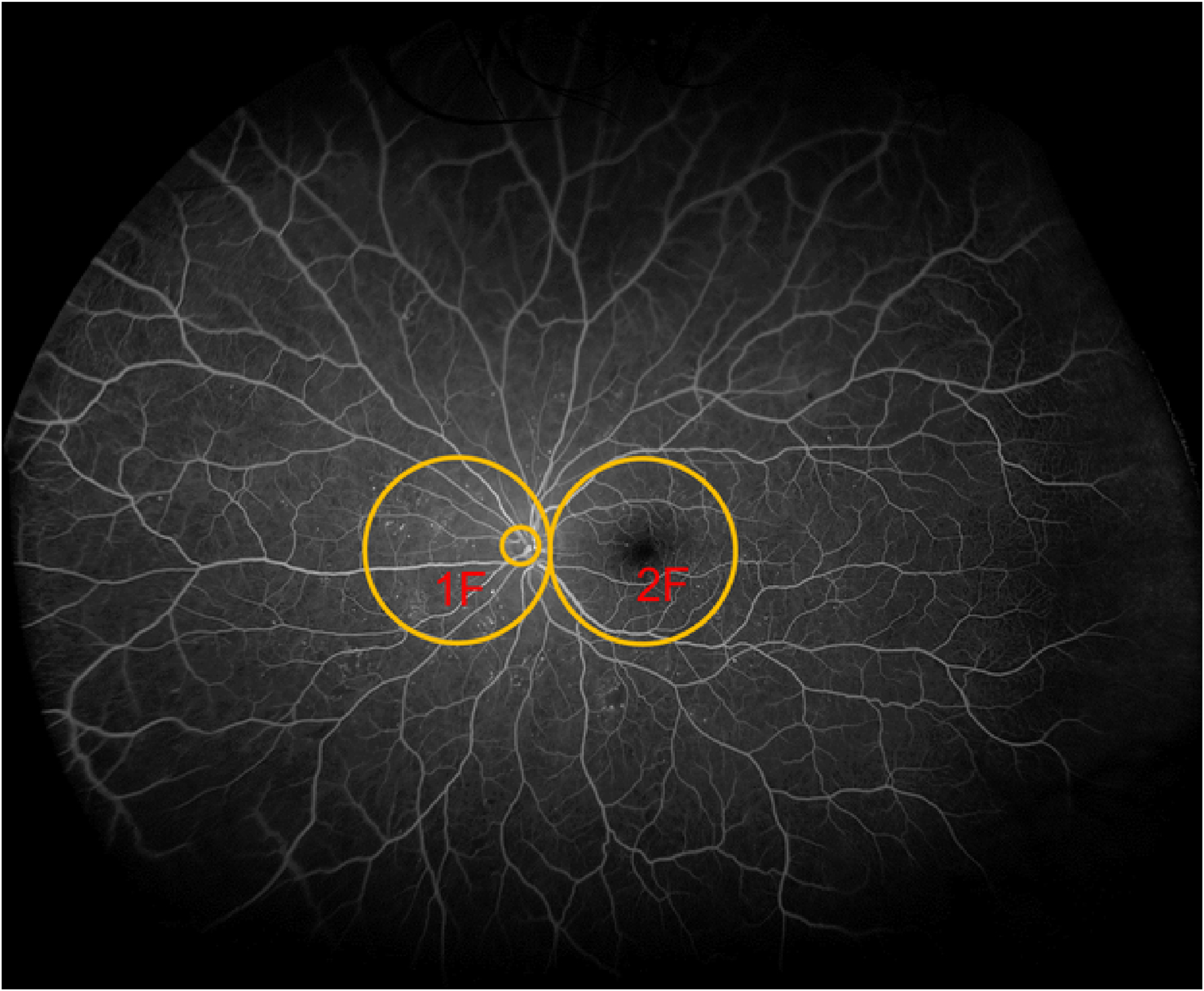
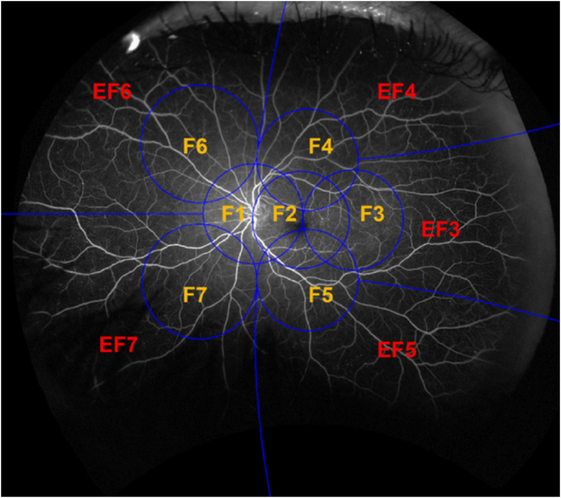
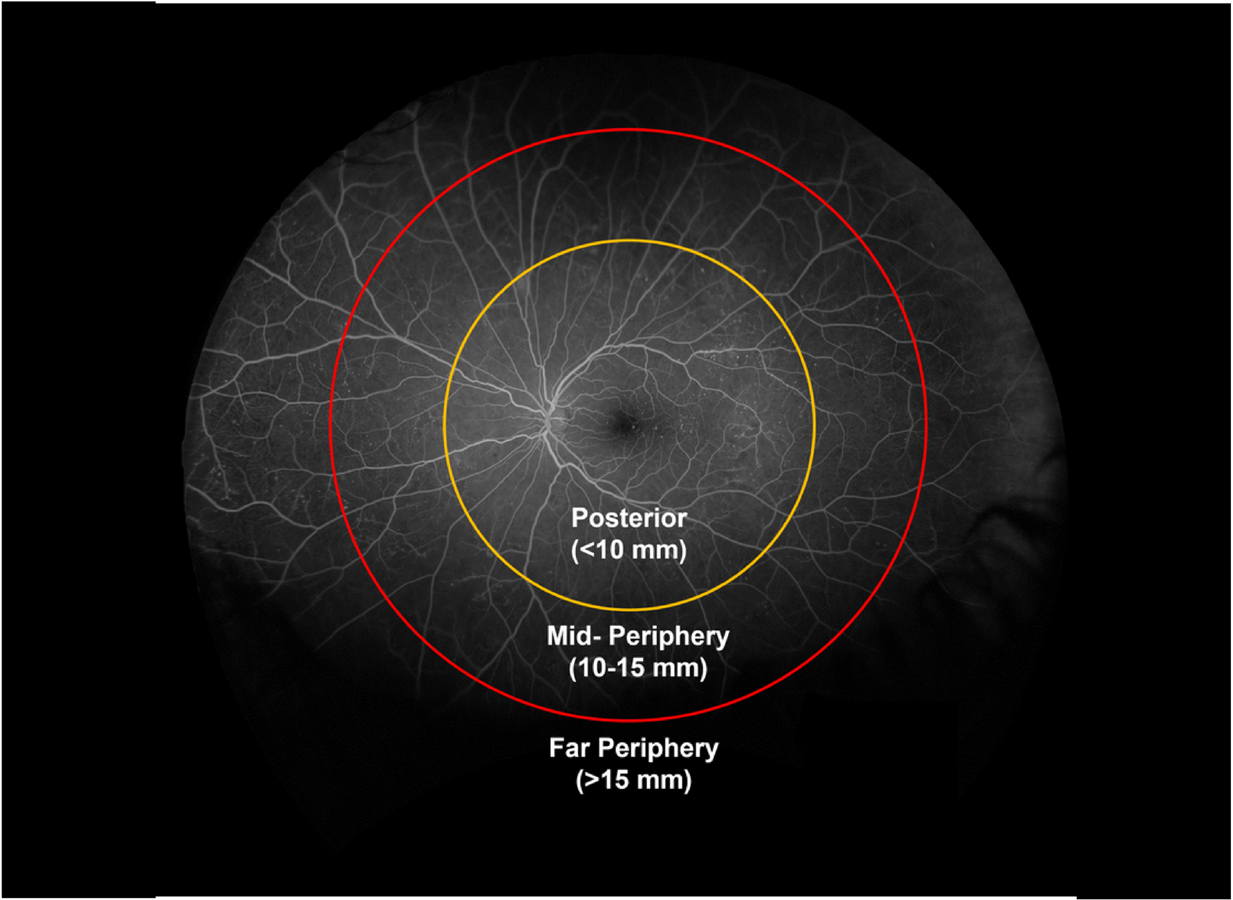
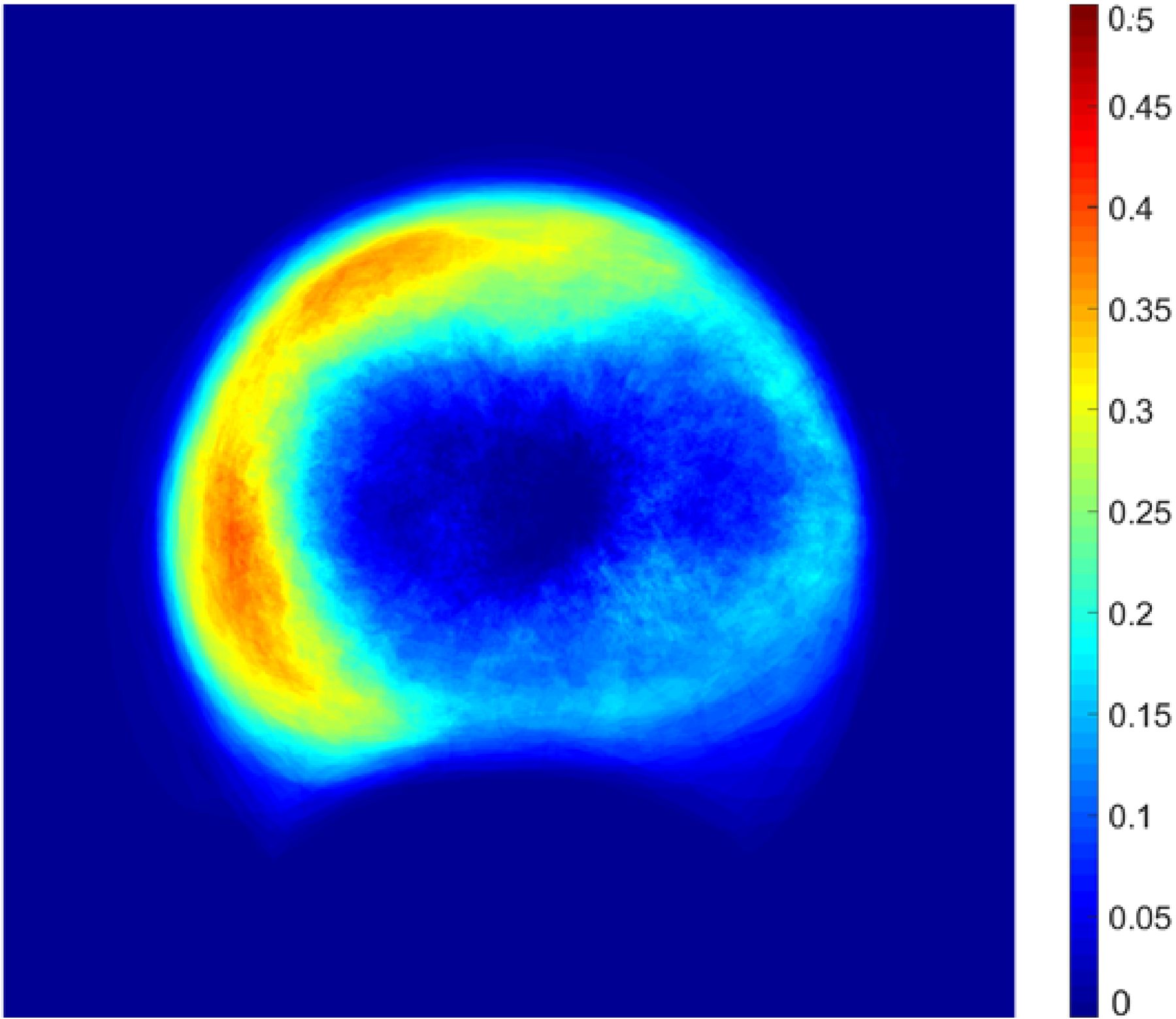
Methods: Multicenter observational study, 652 eyes (361 participants) having nonproliferative DR (NPDR) without center-involved diabetic macular edema in at least one eye. Baseline 200° UWF-color and UWF-FA images were graded by a central reading center for color-PPL and FA-PPL, respectively. UWF-FA was graded for NP index within concentric zones: posterior pole (<10 mm from fovea), midperiphery (10-15 mm), and far periphery (>15 mm).
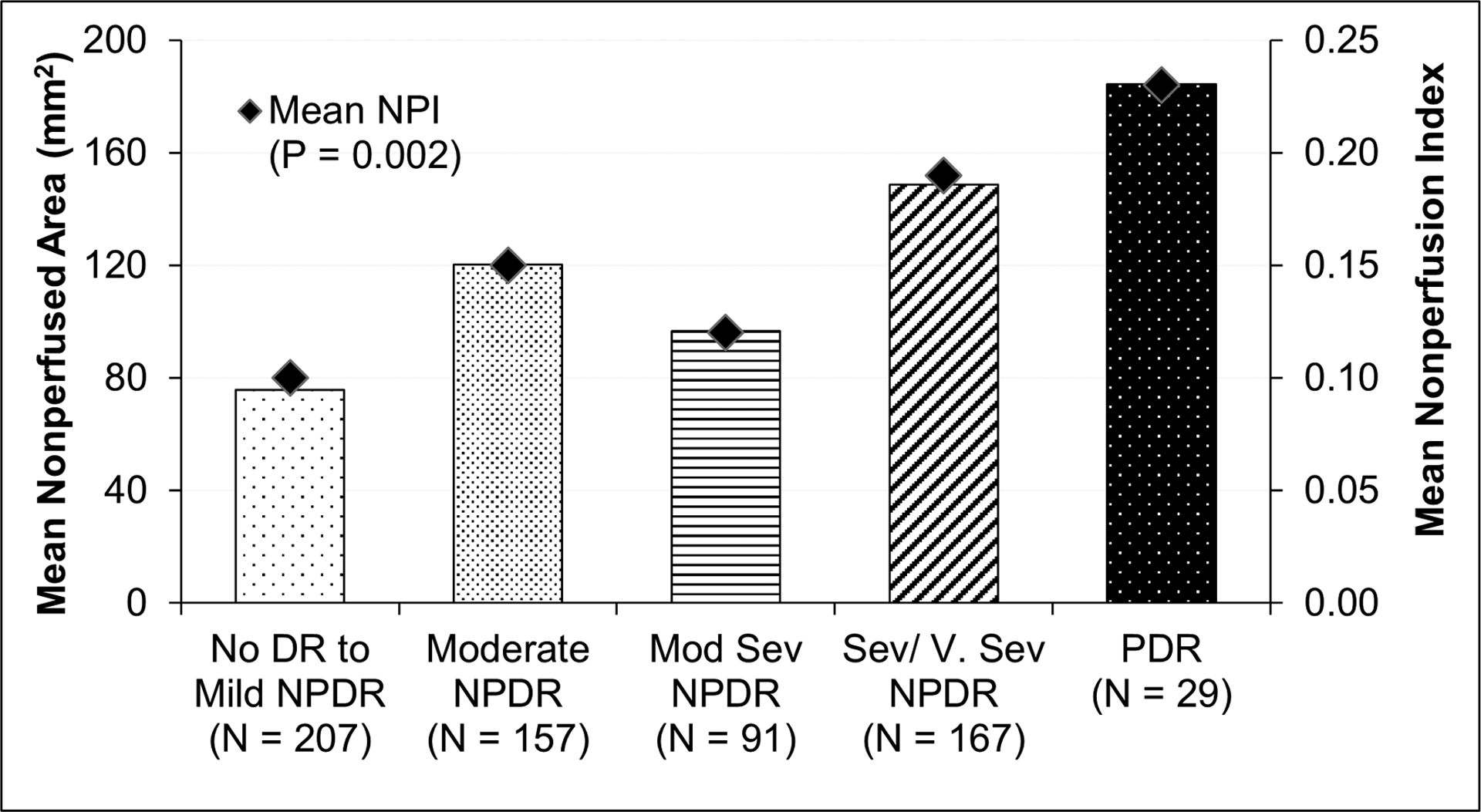
Results: Baseline Early Treatment Diabetic Retinopathy Study DR severity was 31.7% no DR/mild NPDR, 24.1% moderate NPDR, 14.0% moderately severe NPDR, 25.6% severe/very severe NPDR, and 4.6% proliferative DR. Worse DR severity was associated with increased NP index overall (P = 0.002), in the posterior pole (P < 0.001), midperiphery (P < 0.001), and far periphery (P = 0.03). On average, 29.6% of imaged retinal NP was in the posterior pole, 33.7% in midperiphery, and 36.7% in far periphery. Increased NP index was associated with FA-PPL (P < 0.001) but not with color-PPL (P = 0.65).
Conclusion: Approximately, 70% of NP in diabetic eyes is located outside the posterior pole. Increased NP is associated with the presence of FA-PPL, suggesting UWF-FA may better predict future DR worsening than UWF-color alone.
Figures
References
-
- Shimizu K, Kobayashi Y, Muraoka K. Midperipheral fundus involvement in diabetic retinopathy. Ophthalmology. 1981;88(7):601–612. - PubMed
-
- Niki T, Muraoka K, Shimizu K. Distribution of capillary nonperfusion in early-stage diabetic retinopathy. Ophthalmology. 1984;91(12):1431–1439. - PubMed
-
- Silva PS, Dela Cruz AJ, Ledesma MG, et al. Diabetic retinopathy severity and peripheral lesions are associated with nonperfusion on ultrawide field angiography. Ophthalmology. 2015;122(12):2465–2472. - PubMed
-
- Silva PS, Cavallerano JD, Haddad NM, et al. Peripheral lesions identified on ultrawide field imaging predict increased risk of diabetic retinopathy progression over 4 years. Ophthalmology. 2015;122(5):949–956. - PubMed
-
- Silva PS, Cavallerano JD, Sun JK, et al. Peripheral lesions identified by mydriatic ultrawide field imaging: distribution and potential impact on diabetic retinopathy severity. Ophthalmology. 2013;120(12):2587–2595. - PubMed
Publication types
MeSH terms
Grants and funding
LinkOut - more resources
Full Text Sources
Medical
Miscellaneous
