. 2022 Jul;43(7):900-918.
doi: 10.1002/humu.24375. Epub 2022 May 10.

Phenotypic and mutational spectrum of ROR2-related Robinow syndrome

Affiliations

Phenotypic and mutational spectrum of ROR2-related Robinow syndrome

Ariadne R Lima et al. Hum Mutat. 2022 Jul.

Abstract

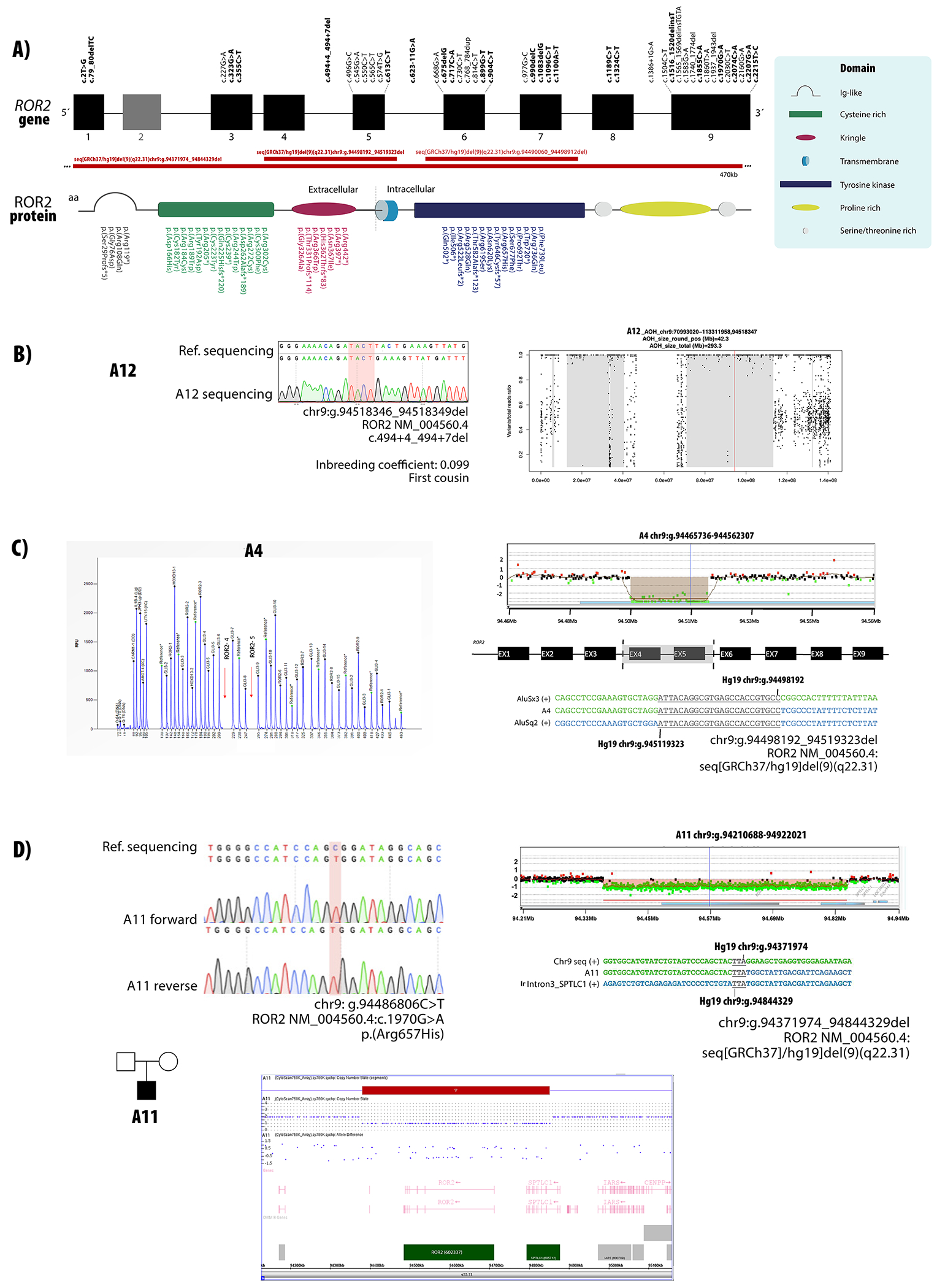
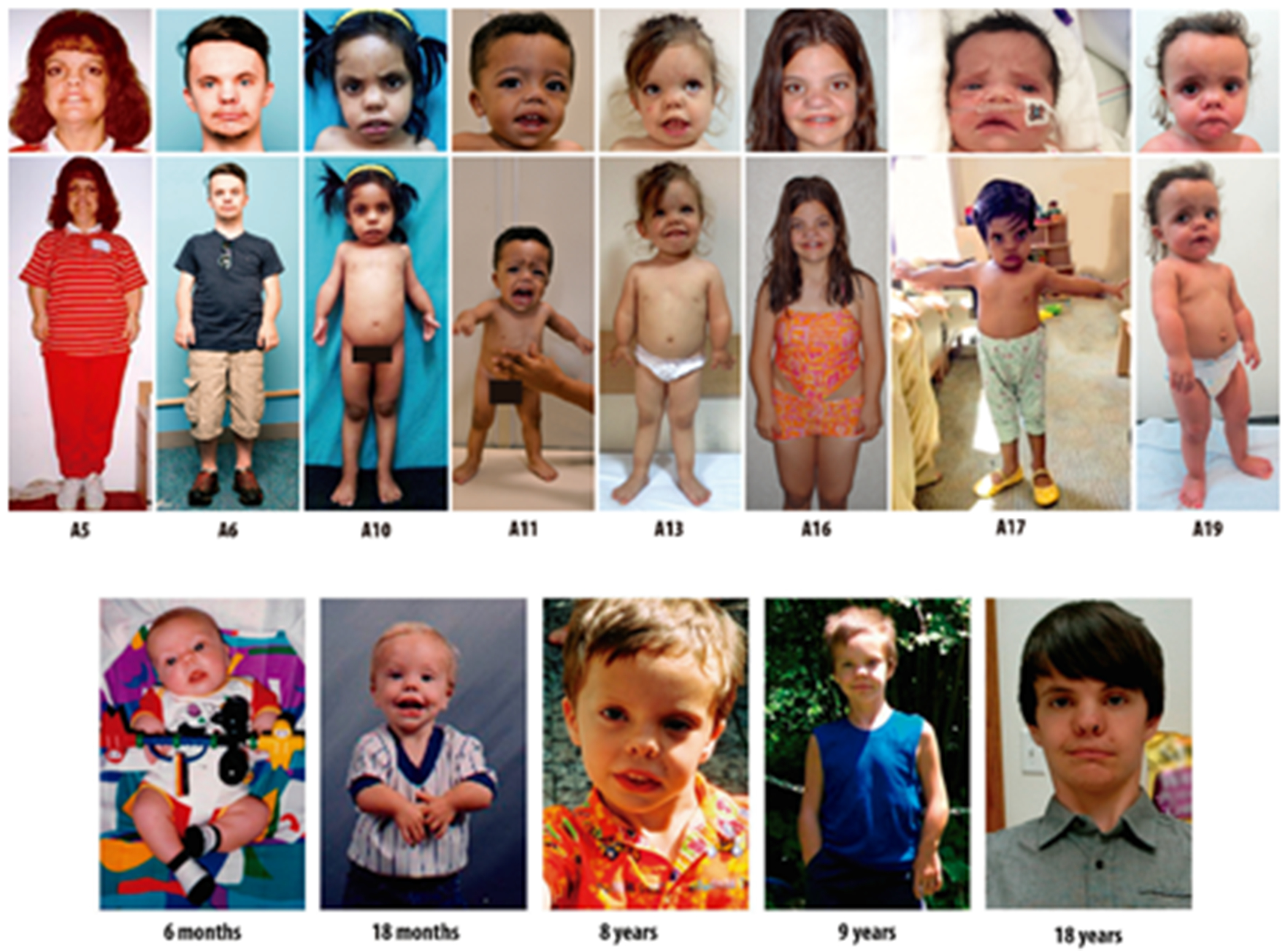
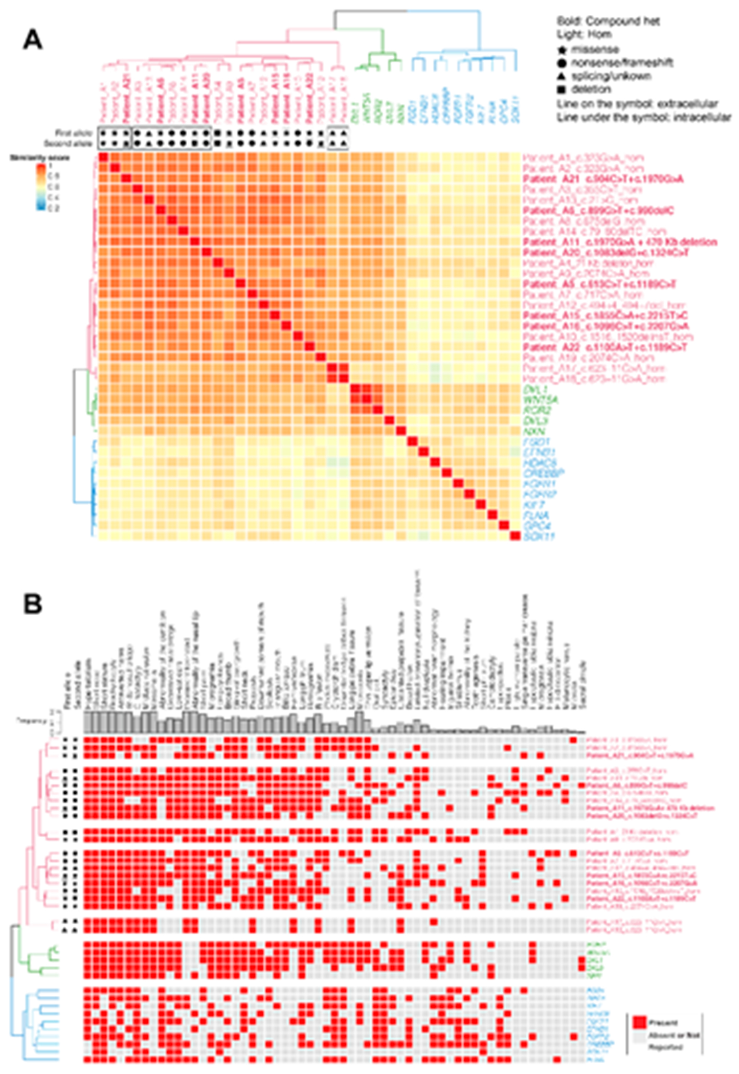
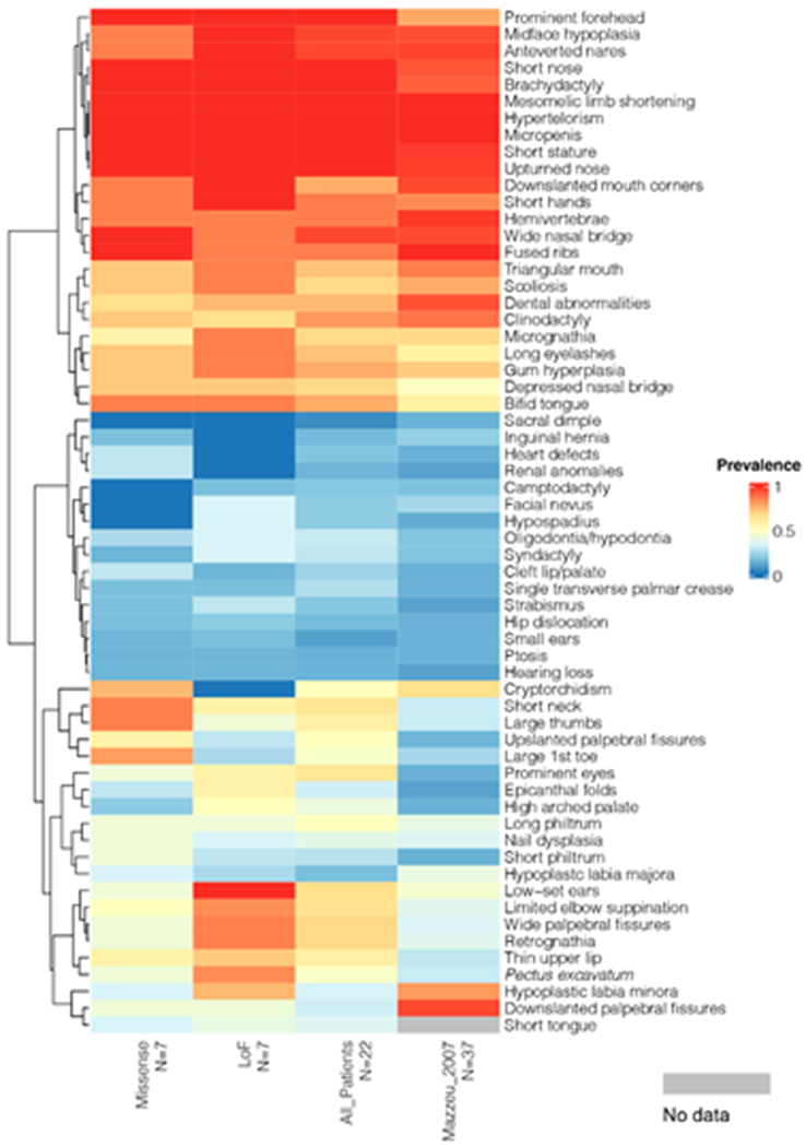
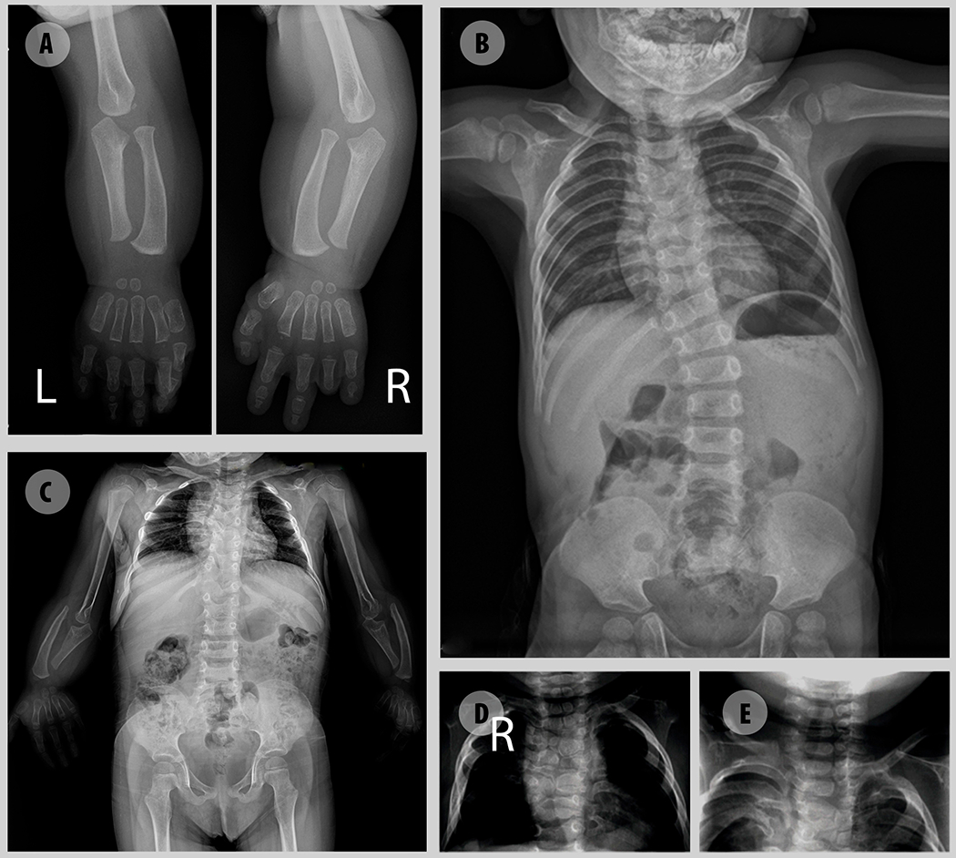
Robinow syndrome is characterized by a triad of craniofacial dysmorphisms, disproportionate-limb short stature, and genital hypoplasia. A significant degree of phenotypic variability seems to correlate with different genes/loci. Disturbances of the noncanonical WNT-pathway have been identified as the main cause of the syndrome. Biallelic variants in ROR2 cause an autosomal recessive form of the syndrome with distinctive skeletal findings. Twenty-two patients with a clinical diagnosis of autosomal recessive Robinow syndrome were screened for variants in ROR2 using multiple molecular approaches. We identified 25 putatively pathogenic ROR2 variants, 16 novel, including single nucleotide variants and exonic deletions. Detailed phenotypic analyses revealed that all subjects presented with a prominent forehead, hypertelorism, short nose, abnormality of the nasal tip, brachydactyly, mesomelic limb shortening, short stature, and genital hypoplasia in male patients. A total of 19 clinical features were present in more than 75% of the subjects, thus pointing to an overall uniformity of the phenotype. Disease-causing variants in ROR2, contribute to a clinically recognizable autosomal recessive trait phenotype with multiple skeletal defects. A comprehensive quantitative clinical evaluation of this cohort delineated the phenotypic spectrum of ROR2-related Robinow syndrome. The identification of exonic deletion variant alleles further supports the contention of a loss-of-function mechanism in the etiology of the syndrome.

Keywords: HPO terms; WNT pathway; chromosome microarray analysis; craniofacial morphology; exonic deletion; next-generation sequencing; quantitative phenotyping cluster heatmap; skeletal dysplasia.


Conflict of interest statement

Conflict of interests:

Baylor College of Medicine (BCM) and Miraca Holdings have formed a joint venture with shared ownership and governance of the Baylor Genetics (BG), which performs clinical microarray analysis and clinical exome sequencing and whole genome sequencing. J.R.L. serves on the Scientific Advisory Board of the BG. J.R.L. has stock ownership in 23andMe, is a paid consultant for Regeneron Genetics Center, and is a coinventor on multiple United States and European patents related to molecular diagnostics for inherited neuropathies, eye diseases, genomic disorders and bacterial genomic fingerprinting. The Department of Molecular and Human Genetics at Baylor College of Medicine derives revenue from molecular genetics and clinical genomics testing offered at BG. The other authors declare no competing financial interests.

Figures

Figure 1.
Figure 1.
ROR2 variants causative of AR-RS. A) Localization of variants described in the present study and in previous reports in relation to their position in the exons and protein domains. Novel variants are highlighted in bold. B) Sanger sequencing chromatogram for patient A12 carrying a homozygous variant in ROR2. AOH plot shows that this variant is within an AOH region of 42.3 Mb. C) Electropherogram of P179 MLPA reaction showing a homozygous deletion of probes corresponding to exons 4 and 5 of ROR2. Customized array CGH plots confirms the 21 kb deletion. Breakpoints determined by Sanger sequencing are represented below. D) Sanger sequencing chromatogram for patient A11 carrying a hemizygous variant in ROR2. Chromosome microarray analysis showing a 470 kb deletion including whole ROR2 and partial SPTLC1 genes. Breakpoints determined by Sanger sequencing are represented below.
Figure 2.
Figure 2.
Facial and whole-body photographs of eight individuals from our cohort showing the spectrum of ROR2-related Robinow syndrome. All individuals exhibit typical dysmorphic features that characterize the syndrome. Patient A6 is shown at different ages to document the evolving facial gestalt.
Figure 3.
Figure 3.
Semantic similarity heatmap and phenotypic annotation grid results between research subjects with biallelic ROR2 variants and significantly similar OMIM annotated known disease gene phenotypes A. *Hierarchical agglomerative clustering (HAC) and visualization of quantitative phenotypic similarity. The dendrogram shown at the top and to the left of the heatmap is based on HAC analysis of the dissimilarity matrix produced from Lin semantic similarity scores and with k set to 3. Unique clusters are represented by different colors, individual probands and significantly similar known disease genes are labeled on top of and to the right of the heatmap. Within the heatmap, dark red indicates a higher similarity while dark blue indicates lower similarity. A key is provided on the left. Bold: subjects who have compound heterozygous variant alleles. Light font type: subjects who have homozygous variant alleles. Star: missense variants. Circle: loss of function (LoF) variants including nonsense variants and frameshifting variant alleles. Triangle: splicing variants or variants with unknown consequence. Rectangle: large exonic deletion (> 50bp) variant alleles. Line on the symbol: variants in the extracellular region. Line under the symbol (i.e. underlined font): variants in the intracellular region. B. Phenotypic annotation grid. Phenotypic annotation grid of phenotypes of all subjects and significantly similar known disease genes. To interpret and understand biology of phenotypes driving semantic similarity in these analyses, HPO terms associated with all subjects and significantly similar known disease genes were annotated and visualized in a gridded array format. Red indicates presence of a phenotype while gray represents absence or not reported. Probands and significantly similar known disease genes are labeled to the right (italicized gene symbols) and are ordered by HAC. The frequency of each phenotype in probands from this cohort is shown on top of the grid.
Figure 4.
Figure 4.
Semantic similarity heatmap between ROR2 subjects and significantly similar OMIM annotated known disease phenotypes (p<0.005). The dendrogram shown at the top and to the left of the heatmap is based on HAC analysis of the dissimilarity matrix produced from Lin semantic similarity scores and with k set to 3. Unique clusters are represented by different colors, individual probands and significantly similar known diseases are labeled on top of and to the right of the heatmap. Within the heatmap, dark red indicates a higher similarity while dark blue indicates lower similarity. A key is provided on the left. Bold: subjects who have compound heterozygous variant alleles. Light font type: subjects who have homozygous variant alleles. Star: missense variants. Circle: loss of function (LoF) variants including nonsense variants and frameshifting variant alleles. Triangle: splicing variants or variants with unknown consequence. Rectangle: large exonic deletion (> 50bp) variant alleles. Line on the symbol: variants in the extracellular region. Line under the symbol (i.e. underlined font): variants in the intracellular region.
Figure 5.
Figure 5.
Phenotypic analysis of subjects with bi-allelic missense variants and LoF variants. Prevalence (0-1.0) of phenotypes in subjects with bi-allelic missense variants (A1, A2, A9, A15, A16, A19, A21), bi-allelic LoF variants (A3, A5, A7, A10, A14, A20), all subjects (N=22), and subjects published in Mazzeu et al. 2007 (N=37) is displayed by heatmap. Probands with other mutation types were not included in this analysis because of their limited numbers (N<3). Within the heatmap, red indicates a higher prevalence while blue indicates lower prevalence; light grey indicates these specific data are not available. The phenotypes are ordered by dendrogram shown on the left based on hierarchical agglomerative clustering (HAC) analysis. A prevalence key is provided on the right.
Figure 6.
Figure 6.
Radiographic images illustrative of major skeletal defects in AR-RS. A – Upper limb showing mesomelia, brachydactyly with pronounced shortening of distal phalanges and absence of medial and distal phalanges of 4th finger. B –Thoracic scoliosis due to multiple hemivertebra C- Multiple hemivertebra, butterfly vertebrae, rib fusions and mesomelia with malformation of the olecranon and coronoid process. Absence of the humero-radial joint. D, E – Hemivertebra.

Similar articles

Cited by

References

    1. Abu-Ghname A, Trost J, Davis MJ, Sutton VR, Zhang C, Guillen DE, Carvalho C, & Maricevich RS (2021). Extremity anomalies associated with Robinow syndrome. American journal of medical genetics. Part A, 185(12), 3584–3592. 10.1002/ajmg.a.61884 - DOI - PubMed
    1. Adzhubei IA, Schmidt S, Peshkin L, Ramensky VE, Gerasimova A, Bork P, Kondrashov AS, & Sunyaev SR (2010). A method and server for predicting damaging missense mutations. Nature methods, 7(4), 248–249. 10.1038/nmeth0410-248 - DOI - PMC - PubMed
    1. Afzal AR, Rajab A, Fenske CD, Oldridge M, Elanko N, Ternes-Pereira E, Tüysüz B, Murday VA, Patton MA, Wilkie AO, & Jeffery S (2000). Recessive Robinow syndrome, allelic to dominant brachydactyly type B, is caused by mutation of ROR2. Nature genetics, 25(4), 419–422. 10.1038/78107 - DOI - PubMed
    1. Aglan M, Amr K, Ismail S, Ashour A, Otaify GA, Mehrez MA, Aboul-Ezz EH, El-Ruby M, Mazen I, Abdel-Hamid MS, & Temtamy SA (2015). Clinical and molecular characterization of seven Egyptian families with autosomal recessive robinow syndrome: Identification of four novel ROR2 gene mutations. American journal of medical genetics. Part A, 167A(12), 3054–3061. 10.1002/ajmg.a.37287 - DOI - PubMed
    1. Ali BR, Jeffery S, Patel N, Tinworth LE, Meguid N, Patton MA, & Afzal AR (2007). Novel Robinow syndrome causing mutations in the proximal region of the frizzled-like domain of ROR2 are retained in the endoplasmic reticulum. Human genetics, 122(3-4), 389–395. 10.1007/s00439-007-0409-0 - DOI - PubMed

Publication types

MeSH terms

Substances

Supplementary concepts