. 2022 Aug;88(2):880-889.
doi: 10.1002/mrm.29236. Epub 2022 Mar 28.

Optimization of undersampling parameters for 3D intracranial compressed sensing MR angiography at 7 T

Affiliations

Optimization of undersampling parameters for 3D intracranial compressed sensing MR angiography at 7 T

Matthijs H S de Buck et al. Magn Reson Med. 2022 Aug.

Abstract

Purpose: 3D time-of-flight MRA can accurately visualize the intracranial vasculature but is limited by long acquisition times. Compressed sensing reconstruction can be used to substantially accelerate acquisitions. The quality of those reconstructions depends on the undersampling patterns used. In this work, we optimize sets of undersampling parameters for various acceleration factors of Cartesian 3D time-of-flight MRA.

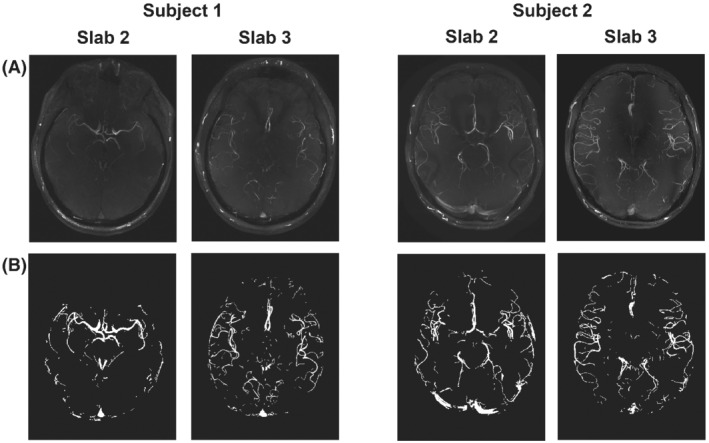
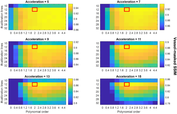
Methods: Fully sampled datasets, acquired at 7 Tesla, were retrospectively undersampled using variable-density Poisson disk sampling with various autocalibration region sizes, polynomial orders, and acceleration factors. The accuracy of reconstructions from the different undersampled datasets was assessed using the vessel-masked structural similarity index. Identified optimal undersampling parameters were then evaluated in additional prospectively undersampled datasets. Compressed sensing reconstruction parameters were chosen based on a preliminary reconstruction parameter optimization.

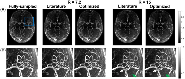
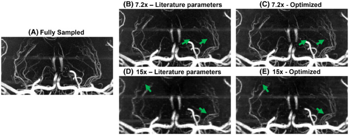
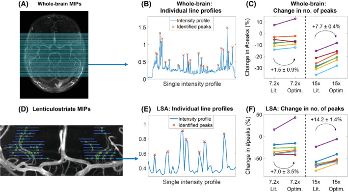
Results: For all acceleration factors, using a fully sampled calibration area of 12 × 12 k-space lines and a polynomial order of 2 resulted in the highest image quality. The importance of parameter optimization of the sampling was found to increase for higher acceleration factors. The results were consistent across resolutions and regions of interest with vessels of varying sizes and tortuosity. The number of visible small vessels increased by 7.0% and 14.2% when compared to standard parameters for acceleration factors of 7.2 and 15, respectively.

Conclusion: The image quality of compressed sensing time-of-flight MRA can be improved by appropriate choice of undersampling parameters. The optimized sets of parameters are independent of the acceleration factor and enable a larger number of vessels to be visualized.

Keywords: MR angiography; compressed sensing; lenticulostriate arteries; time-of-flight MRA; ultra-high field; undersampling.


Conflict of interest statement

Matthijs de Buck receives studentship support from Siemens Healthineers. Peter Jezzard is the Editor‐in‐Chief of Magnetic Resonance in Medicine. In line with COPE guidelines, he wishes to recuse himself from all involvement in the review process of this paper, which was handled by an associate editor. He and the other authors have no access to the identity of the reviewers.

Figures

FIGURE 1
FIGURE 1
The 4 slabs used to test retrospective undersampling strategies (from Cohort 1). (A) MIPs of the fully sampled reconstruction for each slab. (B) The corresponding vessel masks, which were used for computing the vessel‐masked SSIM. MIP, maximum intensity projection; SSIM, structural similarity index
FIGURE 2
FIGURE 2
Average SSIMs for various sets of undersampling parameters. Each datapoint represents the mean vessel‐masked SSIM values of the 4 slabs shown in Figure 1. The results for each of the 4 slabs are separately shown in Supporting Information Figures S3‐S6. The scaling of the SSIM values in each individual figure runs from the maximum value (for the given acceleration factor) to 90% of that maximum value to maximize the visibility of the relative image quality for each acceleration factor. Red boxes indicate the proposed optimized undersampling parameters
FIGURE 3
FIGURE 3
Comparison of axial MIPs from optimized (calib = 12) and literature‐based (calib = 32) undersampling schemes for R = 7.2 and R = 15. (A) Reconstructed images from fully sampled data and the different prospectively undersampled acquisitions. (B) Closeup of the region marked with a blue square in (A) for all acquisitions. Green arrows indicate examples of improved vessel visibility when using optimized undersampling parameters; white arrows indicate improved vessel sharpness. The windowing was reduced for (B) to improve the visibility of small vessels. calib, calibration region size; R, undersampling factor
FIGURE 4
FIGURE 4
Comparison of coronal MIPs of the LSAs from optimized and literature‐based undersampling schemes. Images shown for (A) fully sampled data; (B,C) data for R = 7.2 using literature‐based (B) and optimized (C) prospectively undersampled acquisitions; and (D,E) data for R = 15 using literature‐based (D) and optimized (E) prospectively undersampled acquisitions. LSA, lenticulostriate arteries
FIGURE 5
FIGURE 5
Quantification of the change in the number of detected peaks. (A–C) Whole‐brain MIPs. (A) The 100 lines along which peaks in the intensity profiles were identified on all datasets (as shown in (B) for line 25). (C) The change in the identified number of peaks for optimized and literature‐based undersampled acquisitions, relative to fully sampled acquisitions. Each color indicates a subject from Cohort 2. (D–F): The same as (A–C) for MIPs of lenticulostriate ROIs. Line locations were drawn manually at 5‐pixel intervals. Lit., literature‐based; Optim., optimized; ROI, region of interest

References

    1. Degnan AJ, Gallagher G, Teng Z, Lu J, Liu Q, Gillard JH. MR angiography and imaging for the evaluation of middle cerebral artery atherosclerotic disease. Am J Neuroradiol. 2012;33:1427‐1435. - PMC - PubMed
    1. Farahmand M, Farahangiz S, Yadollahi M. Diagnostic accuracy of magnetic resonance angiography for detection of intracranial aneurysms in patients with acute subarachnoid hemorrhage; a comparison to digital subtraction angiography. Bull Emerg Trauma. 2013;1:147‐151. - PMC - PubMed
    1. Kapsalaki EZ, Rountas CD, Fountas KN. The role of 3 Tesla MRA in the detection of intracranial aneurysms. Int J Vasc Med. 2012;2012:792834. - PMC - PubMed
    1. Hendrikse J, Zwanenburg JJ, Visser F, Takahara T, Luijten P. Noninvasive depiction of the lenticulostriate arteries with time‐of‐flight MR angiography at 7.0 T. Cerebrovasc Dis. 2008;26:624‐629. - PubMed
    1. Greenberg SM. Small vessels, big problems. N Engl J Med. 2006;354:1451‐1453. - PubMed

Publication types

MeSH terms