. 2022 Apr 14;185(8):1389-1401.e18.
doi: 10.1016/j.cell.2022.03.009. Epub 2022 Mar 10.

Broad neutralization of SARS-CoV-2 variants by an inhalable bispecific single-domain antibody

Affiliations

Broad neutralization of SARS-CoV-2 variants by an inhalable bispecific single-domain antibody

Cheng Li et al. Cell. .

Abstract

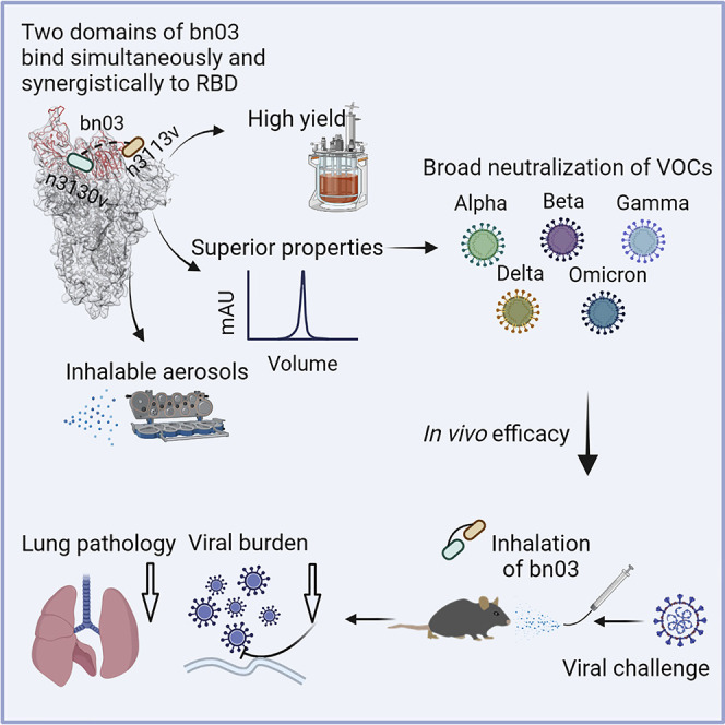
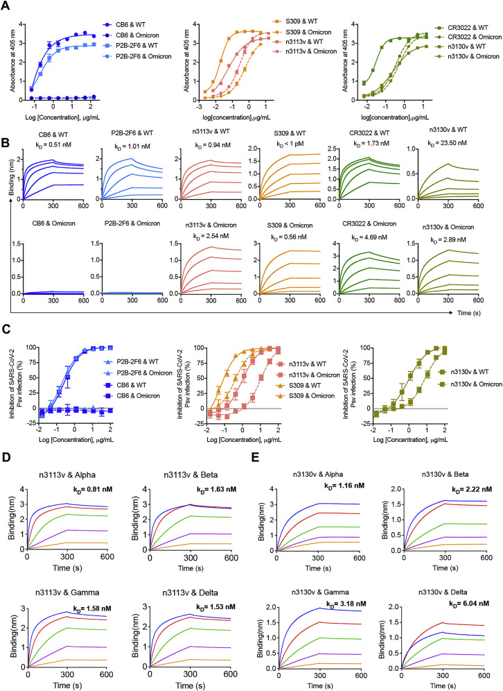
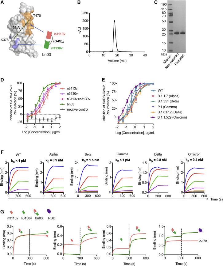
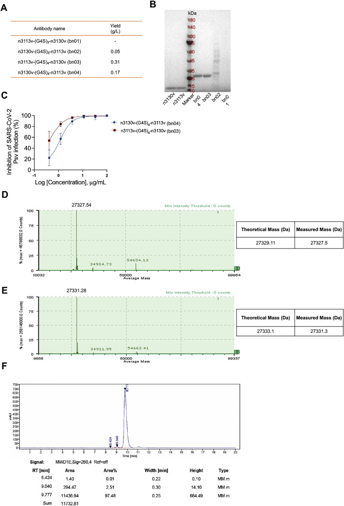
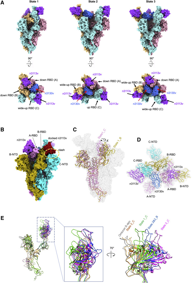
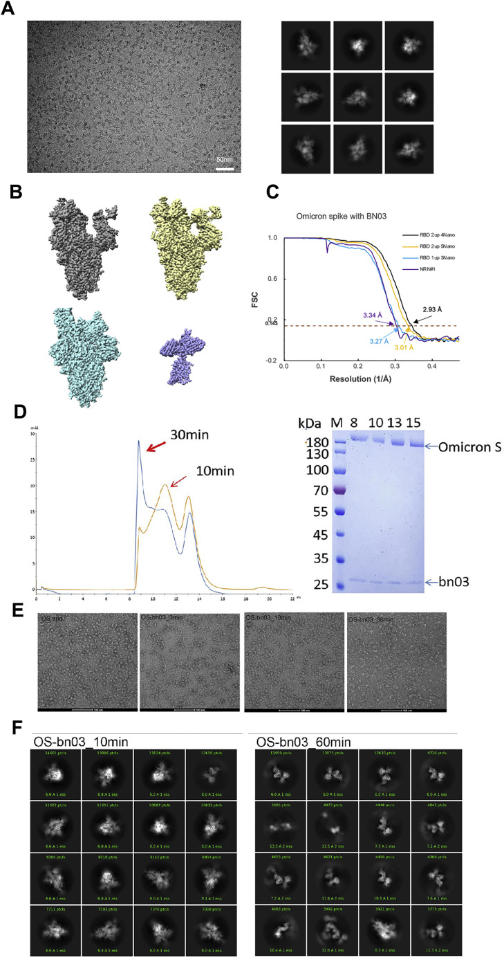
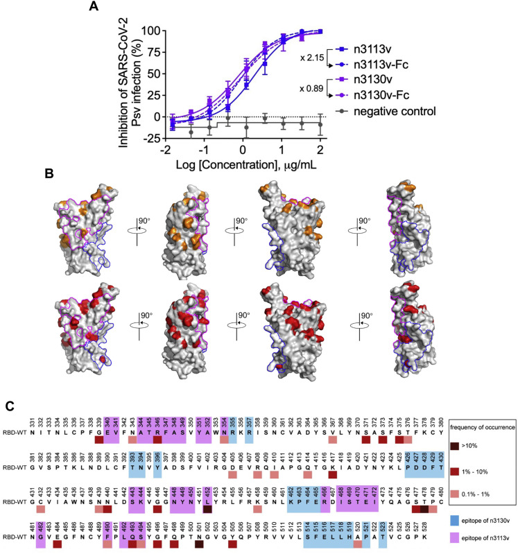
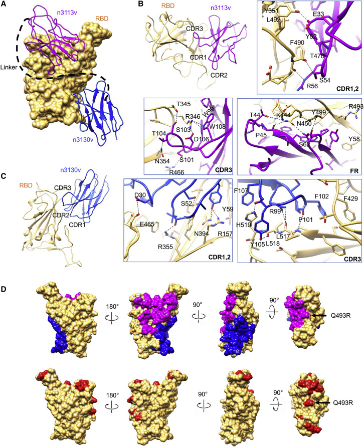
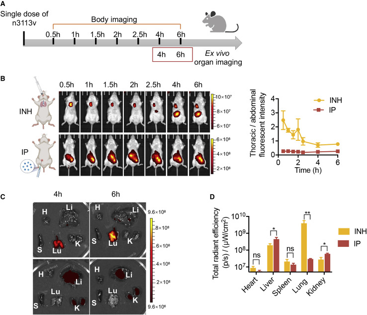
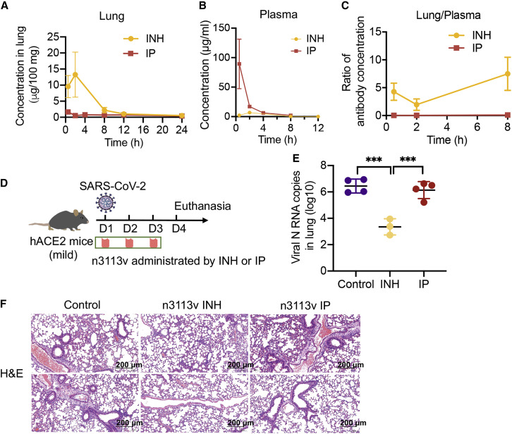
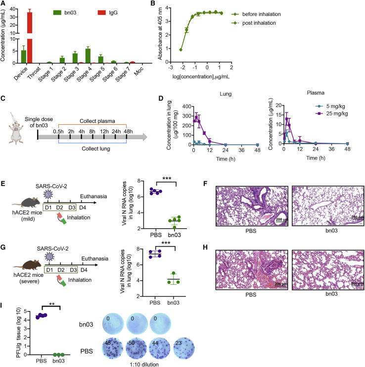
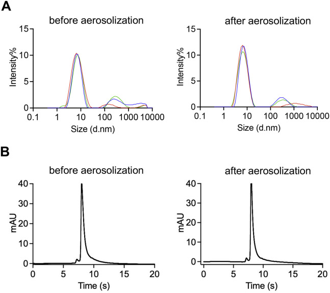
The effectiveness of SARS-CoV-2 vaccines and therapeutic antibodies have been limited by the continuous emergence of viral variants and by the restricted diffusion of antibodies from circulation into the sites of respiratory virus infection. Here, we report the identification of two highly conserved regions on the Omicron variant receptor-binding domain recognized by broadly neutralizing antibodies. Furthermore, we generated a bispecific single-domain antibody that was able to simultaneously and synergistically bind these two regions on a single Omicron variant receptor-binding domain as revealed by cryo-EM structures. We demonstrated that this bispecific antibody can be effectively delivered to lung via inhalation administration and exhibits exquisite neutralization breadth and therapeutic efficacy in mouse models of SARS-CoV-2 infections. Importantly, this study also deciphered an uncommon and highly conserved cryptic epitope within the spike trimeric interface that may have implications for the design of broadly protective SARS-CoV-2 vaccines and therapeutics.

Keywords: SARS-CoV-2; bispecific antibody; broad neutralization; inhalation; single-domain antibody.


Conflict of interest statement

Declaration of interests C.L., Y.W., and T.Y. are listed as inventors on two patent applications related to this work.

Figures

None
Graphical abstract
Figure 1
Figure 1
Non-RBM antibodies confer resistance against the SARS-CoV-2 Omicron variant (A) Epitope clustering of RBD-targeting antibodies on the spike protein and RBD. The Omicron S was shown as surface, with RBD colored in gray; mutations are highlighted in red. The three classes of epitope are circled in S and highlighted in RBD as blue, green, and yellow, respectively. (B) The amino acid mutations in the RBD region of the Omicron variant are shown as colored boxes. The key mutations found in binding sites of three classes of antibody, such as RBM-binding site, lateral surface, and cryptic site, are displayed. (C) Binding and neutralization of vaccinee plasma against SARS-CoV-2 WT and Omicron variant. Values denote the geometric mean titer ± SD. Three independent experiments were performed in triplicate. (D) Illustration of RBD mAbs, RBM mAbs, and non-RBM mAbs in plasma, as measured by BLI (left). Correlation coefficient between plasma neutralization ID50 against the Omicron variant with RBM mAbs or non-RBM mAbs (right), analyzed by Spearman’s rank correlation test. (E) The binding affinity and neutralization of six antibodies. Values indicate the fold change relative to WT. See also Figures S1, S2, and S3.
Figure S1
Figure S1
Illustration of mutations on spike of Omicron variant and epitopes for RBD-targeting antibodies, related to Figure 1 (A) The mutations in Omicron variant are shown on spike. The monomeric spike is shown as gray cartoon. The mutations are depicted as spheres in indicated colors. (B) Epitopes for ACE2 and three classes of RBD-targeting antibodies. The residues involved in interaction with antibodies were calculated by ePISA (Proteins, Interfaces, Structures, and Assemblies) and COOT (Crystallographic Object-Oriented Toolkit) and are depicted in colored squares.
Figure S2
Figure S2
The Omicron variant reduces the binding activity and neutralizing potency of boosted vaccinee plasma, related to Figure 1 (A) Binding curves for WT or Omicron RBD by individual plasma, as determined by ELISA. (B) Neutralization for WT or Omicron pseudoviruses by individual plasma. (C) The RBD mAbs, RBM mAbs, and non-RBM mAbs in boosted vaccinee plasma were measured by competition assay using BLI. (D) Summary of the ED50, ID50 of boosted vaccinee plasma against WT or Omicron variant. The RBD mAbs, RBM mAbs, and non-RBM mAbs in plasma denoted by binding signal.
Figure S3
Figure S3
Binding affinity and neutralization of distinct antibody clusters to WT and Omicron variant, related to Figure 1 (A) Binding capacity of antibodies to WT and Omicron RBD, as measured by ELISA. (B) Binding kinetics of antibodies to WT and Omicron RBD, as measured by BLI. (C) Neutralization for WT or Omicron pseudoviruses by antibodies. (D and E) Single-domain antibody n3113v (D) and n3130v (E) exhibited broadly binding capacities against VOCs with high affinity, as measured by BLI.
Figure 2
Figure 2
The design of bispecific single-domain antibody bn03 (A) The bispecific single-domain antibody bn03 contains n3130v and n3113v linked with a linker (GGGGS)4. n3113v and n3130v are colored in pink and green, respectively. The RBD is depicted as gray surface with the epitopes of n3113v and CR3022 (targeting the same epitopes with n3130v) highlighted in orange and blue, respectively. K378 and T470 that are involved in the recognition of CR3022 and n3113, respectively, are shown as sticks. The distance between Cα of K378 and T470 was measured and is labeled in dashed green lines. (B) Size exclusion chromatography profile of the bispecific antibody bn03. (C) Reducing and non-reducing SDS-PAGE analysis of bn03. (D) Neutralization of SARS-CoV-2 pseudoviruses by a panel of single-domain antibodies, including n3113v, n3130v, cocktail of n3113v and n3130v, and bn03. Three independent experiments were performed in triplicate. (E) Neutralizing potency of bn03 against pseudoviruses of WT and five VOCs. Three independent experiments were performed in triplicate. (F) Binding affinity of bn03 to RBDs of WT and five VOCs. The KD values are shown. (G) The bispecific single-domain antibody bn03 simultaneously binds two distinct epitopes on the RBD. The immobilized RBD was incubated with bn03, n3113v (orange), or n3130v (green) until saturation and then incubated with second antibody or the RBD. The binding curves were monitored. See also Figure S4.
Figure S4
Figure S4
The properties and neutralization of bispecific single-domain antibodies containing n3113v and n3130v connected with different linkers, related to Figure 2 (A) Four types of bispecific single-domain antibodies (bn01-04) were expressed by E. coli and purified by Ni-NTA resin. The yield was shown. “-” represents no detectable. (B) SDS-PAGE analysis of n3113v, n3130v and bispecific single-domain antibodies (bn01-04). (C) Neutralizing activity of bn03 and bn04 against SARS-CoV-2 WT pseudovirus. (D) Mass spectrometry analysis of intact bn03 using ESI. (E) Mass spectrometry analysis of reduced bn03 using ESI. (F) HPLC analysis of bn03.
Figure 3
Figure 3
Cryo-EM structures of Omicron S trimer in complex with bispecific single-domain antibody bn03 (A) Bispecific single-domain antibody bn03 binds to Omicron S trimers in 3 states. Two perpendicular views of Omicron S-bn03 are depicted as surface, with n3113v in magenta, n3130v in blue, and the trimeric spike in cyan, pink, and yellow. (B) Superposition of RBD-bound n3113v on B-RBD of state 1 structure indicates a clash. A-RBD-bound n3113v is shown as magenta surface. The docked n3113v is shown in red surface. (C) Alignment of down-state spike from molecule C (pink) and molecule B (yellow) of state 1 structure. The down-state spike is shown as cartoon. Protomer A and C in Omicron S-bn03 structure are depicted as gray surface. (D) Quaternary binding of n3130 and RBDs. Top view of the state 1 structure shown in cartoon representation. (E) Comparison of the up-RBDs of bn03-bound Omicron S complexes with the up-RBD of Omicron S-apo (gray). The n3113v-bound state-2 C-RBD is colored orange. The n3113v-n3130v-bound state-1-C, state 2&3-B, and state 3-C are colored green, blue, and magenta, respectively. See also Figure S5.
Figure S5
Figure S5
Cryo-EM data collection and processing of bn03 bound SARS-CoV-2 Omicron S, related to Figure 3 (A) Representative electron micrograph and 2D classification results of XG014 bound SARS-CoV-2 S. (B) The reconstruction map of the complex structures at three states and one local refinement map. (C) Gold-standard Fourier shell correlation curves for each structure. The 0.143 cut-off is indicated by a horizontal dashed line. (D) Purification of bn03-Oimcron S complex. The gel-filtration curved showed that incubation of bn03 with Omicron-S-induced aggregation of S protein. (E) Negative stain images of bn03-Omicron S complex, showing that incubating with bn03 for 30 min leads to Omicron S trimer disassembly. The disassembled monomers tend to aggregate. (F) The 2D classification result of cryo-EM data collected using 10-min incubation complex and 60-min incubation complex.
Figure S6
Figure S6
Neutralizing activity of n3113v, n3113v-Fc, n3130v, and n3130v-Fc against SARS-CoV-2 WT pseudovirus, and illustration of epitopes of single-domain antibodies on RBD, related to Figure 4 (A) Fold change in IC50 values reflects the increase or decrease of neutralizing activity post fusing to Fc region. (B) The RBD was represented as gray surface. Mutations found in VOCs (Alpha, Beta, Gamma, Delta, and Omicron) were highlighted in orange in the upper row. Mutations in RBD with high frequency (occurred in over 0.1% of the total reported sequences) from public database (https://ngdc.cncb.ac.cn/ncov/variation/spike) were indicated in red in the lower row. The epitope of n3130v and n3113v was circled in blue and magenta lines, respectively. (C) Reported mutations in RBD from public database (https://ngdc.cncb.ac.cn/ncov/variation/spike) were marked by squares in different color. Residues involved in the binding to n3130v and n3113v were highlighted in blue magenta, respectively.
Figure 4
Figure 4
Two conserved epitopes recognized by bn03 (A) Close-up view of the interactions between bn03 and Omicron RBD. The Omicron RBD is displayed in yellow surface. n3113v and n3130v are shown as cartoon colored in magenta and blue, respectively. (B and C) The interaction of n3113v (B) and n3130v (C) with Omicron RBD. The residues involved in interactions are represented as sticks. Polar interactions are indicated as dotted lines. (D) The epitopes of n3113v and n3130v on Omicron RBD. Omicron RBD is shown as yellow surface. Four degrees of surface representation of Omicron RBD with epitopes of n3113v and n3130v highlighted in magenta and blue, respectively, are shown. The mutations in Omicron are colored in red. See also Figure S6.
Figure 5
Figure 5
Effective delivery of single-domain antibody to lung via inhalation (A) Schematic diagram of n3113v biodistribution in mice by inhalation and intraperitoneal injection. (B) Bio-imaging of mice body at different time points (left) and fluorescence intensity ratio of thoracic cavity against abdomen (right, n = 3). (C) Mice were sacrificed at 4 and 6 h post antibody administration and dissected organs were imaged (n = 3). Lu, lung; K, kidney; H, heart; S, spleen; Li, liver. (D) Quantification of organ tissue fluorescence signal.
Figure 6
Figure 6
Inhalation of single-domain antibody exhibits effective therapeutic effects (A–C) Concentration of n3113v in lung (A), plasma (B), and the ratio of n3113v in lung relative to plasma (C), n = 2. (D) Experimental design of therapeutic evaluation of n3113v by inhalation and intraperitoneal injection in authentic SARS-CoV-2-infected hACE2 transgenic mice. (E and F) Lung viral load (E) and lung histology (F) in infected mice with indicated treatments.
Figure 7
Figure 7
Inhalation of bn03 effectively treats SARS-CoV-2 infection in mice (A) The aerosol performances of bn03 and IgG (S309) by NGI. The concentration of aerosolized particles in different stages is quantified. Three independent experiments were performed in triplicate. (B) Comparison of the binding ability of bn03 to RBD before or after inhalation using the high-pressure microsprayer. Three independent experiments were performed in triplicate. (C) Experimental design for evaluation of bn03 in mice by inhalation. BALB/c mice received bn03 via inhalation at a dose of 5 or 25 mg/kg, and blood and lung samples were collected at indicated time points. (D) Antibody concentration of bn03 in lung (left, n = 2) and plasma (right, n = 2). (E–H) Therapeutic efficacy of bn03 via inhalation in a hACE2 transgenic mouse model with mild (E) or severe symptoms (G) after authentic SARS-CoV-2 infection. Lung viral load (E and G) and lung histology (F and H) in mice with indicated treatments. (I) Determination of focus-forming units (FFUs) with lung homogenate in 1:10 dilutions. Lung homogenate of each mouse was serially diluted 10-fold and incubated with Vero E6 cells. The FFUs of each well were counted. The total FFUs of lung tissue were determined and represented (n = 3–4). The right panel shows representative FFUs. Ordinary one-way ANOVA was used in the statistical analysis. Statistical significance is represented as ∗∗∗p < 0.001, ∗∗p < 0.01, p < 0.05; ns, no significance. See also Figure S7.
Figure S7
Figure S7
The properties of bn03 were determined by DLS and HPLC before and after aerosolization by microsprayer aerosolizer, related to Figure 7 (A) Properties of bn03 determined by DLS and (B) properties of bn03 determined by HPLC.

Similar articles

Cited by

References

    1. Bangaru S., Lang S., Schotsaert M., Vanderven H.A., Zhu X., Kose N., Bombardi R., Finn J.A., Kent S.J., Gilchuk P., et al. A site of vulnerability on the influenza virus hemagglutinin head domain trimer interface. Cell. 2019;177:1136–1152.e18. - PMC - PubMed
    1. Baum A., Fulton B.O., Wloga E., Copin R., Pascal K.E., Russo V., Giordano S., Lanza K., Negron N., Ni M., et al. Antibody cocktail to SARS-CoV-2 spike protein prevents rapid mutational escape seen with individual antibodies. Science. 2020;369:1014–1018. - PMC - PubMed
    1. Bianco F., Salomone F., Milesi I., Murgia X., Bonelli S., Pasini E., Dellacà R., Ventura M.L., Pillow J. Aerosol drug delivery to spontaneously-breathing preterm neonates: lessons learned. Respir. Res. 2021;22:71. - PMC - PubMed
    1. Cameroni E., Bowen J.E., Rosen L.E., Saliba C., Zepeda S.K., Culap K., Pinto D., VanBlargan L.A., De Marco A., di Iulio J., et al. Broadly neutralizing antibodies overcome SARS-CoV-2 Omicron antigenic shift. Nature. 2022;602:664–670. - PMC - PubMed
    1. Cao Y., Wang J., Jian F., Xiao T., Song W., Yisimayi A., Huang W., Li Q., Wang P., An R., et al. Omicron escapes the majority of existing SARS-CoV-2 neutralizing antibodies. Nature. 2022;602:657–663. - PMC - PubMed

Publication types

Supplementary concepts