. 2022 May;605(7909):340-348.
doi: 10.1038/s41586-022-04661-w. Epub 2022 Mar 28.

A TMPRSS2 inhibitor acts as a pan-SARS-CoV-2 prophylactic and therapeutic

Affiliations

A TMPRSS2 inhibitor acts as a pan-SARS-CoV-2 prophylactic and therapeutic

Tirosh Shapira et al. Nature. 2022 May.

Abstract

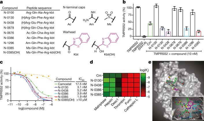
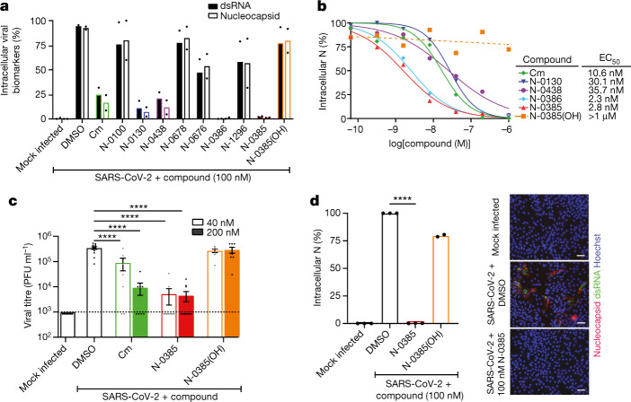
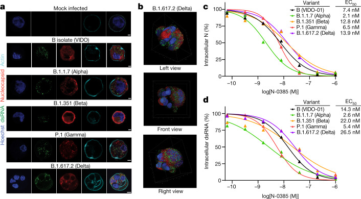
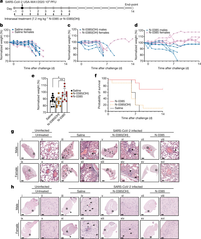
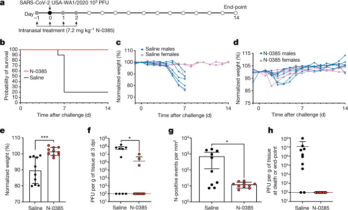
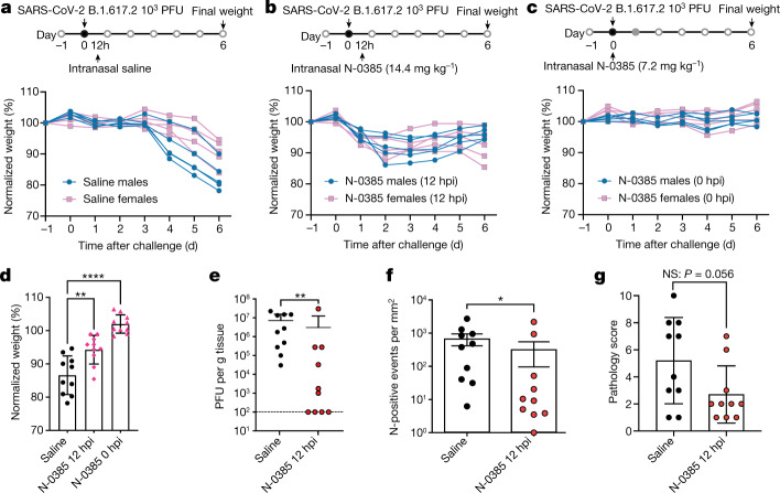
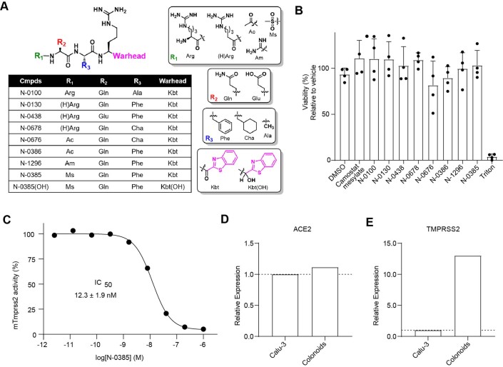
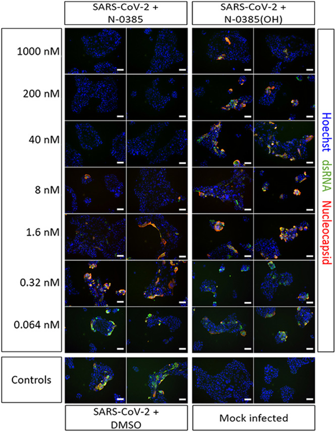
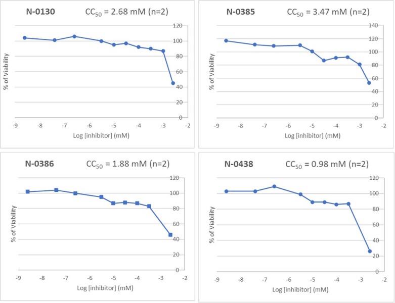
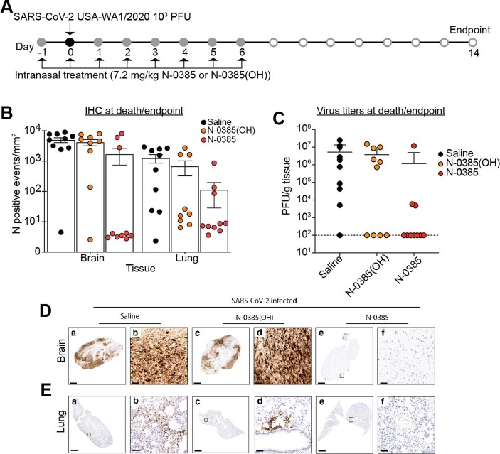
The COVID-19 pandemic caused by the SARS-CoV-2 virus remains a global public health crisis. Although widespread vaccination campaigns are underway, their efficacy is reduced owing to emerging variants of concern1,2. Development of host-directed therapeutics and prophylactics could limit such resistance and offer urgently needed protection against variants of concern3,4. Attractive pharmacological targets to impede viral entry include type-II transmembrane serine proteases (TTSPs) such as TMPRSS2; these proteases cleave the viral spike protein to expose the fusion peptide for cell entry, and thus have an essential role in the virus lifecycle5,6. Here we identify and characterize a small-molecule compound, N-0385, which exhibits low nanomolar potency and a selectivity index of higher than 106 in inhibiting SARS-CoV-2 infection in human lung cells and in donor-derived colonoids7. In Calu-3 cells it inhibits the entry of the SARS-CoV-2 variants of concern B.1.1.7 (Alpha), B.1.351 (Beta), P.1 (Gamma) and B.1.617.2 (Delta). Notably, in the K18-human ACE2 transgenic mouse model of severe COVID-19, we found that N-0385 affords a high level of prophylactic and therapeutic benefit after multiple administrations or even after a single administration. Together, our findings show that TTSP-mediated proteolytic maturation of the spike protein is critical for SARS-CoV-2 infection in vivo, and suggest that N-0385 provides an effective early treatment option against COVID-19 and emerging SARS-CoV-2 variants of concern.


Conflict of interest statement

P.-L.B. and R.L. are inventors on patent applications (US9365853B2 and US10988505B2) that cover matriptase and other type II transmembrane serine proteases inhibitors for treating and preventing viral infections, respiratory disorders, inflammatory disorders, pain disorders, tissue disorders, hyperproliferative disorders, and disorders associated with iron overload. The remaining authors declare that they have no competing interests.

Figures

Fig. 1
Fig. 1. Ketobenzothiazole-based peptidomimetics are potent TMPRSS2 inhibitors.
a, Peptidomimetic compounds used in this study along with their respective sequences. The structures of N-terminal caps, the ketobenzothiazole warhead and the alcohol ketobenzothiazole are shown on the right. (H)Arg, desamino arginine; kbt, ketobenzothiazol. b, Vero E6 cells were transfected with an empty vector (mock), wild-type TMPRSS2 or the inactive mutant TMPRSS2(S441A) for 24 h. The indicated compounds (10 nM) were added concomitantly with a fluorogenic substrate on cells for an additional 24 h before fluorescence reading. Relative TMPRSS2 activity was measured using the mock-subtracted fluorescence and is reported as the percentage of residual activity relative to the vehicle-treated cells (0.01% DMSO). Data are presented as mean ± s.d. (n = 3 independent experiments). c, Dose–response curves were generated for the indicated compounds using the assay described in b, and IC50 values were determined using nonlinear regression analysis. Representative IC50 curves are shown, with the mean value of independent experiments (n = 3 for N-0130, N-0386 and N-0385(OH); n = 4 for Cm and N-0385; n = 5 for N-0438). d, Specificity of selected compounds toward other serine proteases. Data are the mean of log(Ki); n = 3 independent experiments (except cathepsin L versus N-0385, n = 4) and are shown as a heat map. e, Main image, docking of N-0385 (green; warhead in purple) in the binding pocket of TMPRSS2 (homology model). Residues of the catalytic triad are shown in cyan. Inset, interaction of N-0385 with TMPRSS2 residues. N-0385 forms a covalent bond with the catalytic triad residue Ser441. Source data
Fig. 2
Fig. 2. Peptidomimetics active against TMPRSS2 are potent low nanomolar inhibitors of SARS-CoV-2 in a human lung epithelial cell line and in human colonoids.
a, Calu-3 cells were pretreated with 100 nM of the indicated compounds followed by SARS-CoV-2 (VIDO) infection (multiplicity of infection (MOI) = 2)). Intracellular infection levels were evaluated by high-content screening of cell nuclei, dsRNA and nucleocapsid and then quantified relative to DMSO-treated cells (n = 2 independent experiments). b, Dose–response curves were generated for the lead antiviral peptidomimetic compounds in Calu-3 cells using nucleocapsid (N) staining of cells that were pretreated with the indicated compounds before infection (Cm, n = 5; N-0130, n = 5; N-0438, n = 3; N-0386, n = 4; N-0385, n = 8; N-0385(OH), n = 5). c, Plaque assays were performed using two of the experimental conditions evaluated in the dose–response analysis (40 nM and 200 nM) to determine the viral titres (amount of infectious virus) produced in cells that were pretreated with the indicated compounds before infection (n = 3 independent experiments); dotted line represents limit of detection. d, Colonoids were pretreated with 100 nM of the indicated compounds and infected with SARS-CoV-2 (MOI ≈ 1). Intracellular infection was relatively quantified using N staining. (N-0385, n = 3; N-0385(OH), n = 2). Representative fluorescent images of colonoids subjected to the indicated treatments are shown (Hoechst in blue, nucleocapsid in red and dsRNA in green). Scale bars, 50 μm. One-way ANOVA with Bonferroni correction was used to determine significance in c, d; **** indicates modified P < 0.0001. Error bars, s.e.m. Source data
Fig. 3
Fig. 3. N-0385 is a nanomolar inhibitor of four SARS-CoV-2 VOCs.
a, Representative fluorescent images of SARS-CoV-2-infected Calu-3 cells. Calu-3 cells infected with the indicated SARS-CoV-2 variants and mock infected are shown. Scale bars, 5 μm. b, Representative images from a 3D volume rendering of Delta-infected cells. In a, b, Hoechst is shown in blue, nucleocapsid (N) in red, dsRNA in green and actin in cyan; images were captured with a Leica TCS SP8 3× STED microscope. c, d, Dose–response curves were generated for N-0385 in Calu-3 cells using N staining (c) and dsRNA staining (d) of N-0385 pretreated cells infected with the indicated VOCs (n = 4 independent experiments). Source data
Fig. 4
Fig. 4. N-0385 reduces morbidity and mortality in the K18-hACE2 mouse model of SARS-CoV-2.
a, Mice (n = 10 per treatment group) were treated daily on days −1 to +6 relative to infection. Surviving mice were euthanized at the study end-point. bd, Weight change of mice treated with saline (control) (b), N-0385(OH) (c) or N-0385 (d). e, Weight loss difference at 6 dpi; *P = 0.0193, **P = 0.0083. Data are mean ± s.d., with two-tailed Student’s t-tests used to determine significance. f, Probability of survival. g, Representative haematoxylin and eosin (H&E) staining of lung histopathology (one slice analysed per mouse) at death or study end-point in untreated mice (i, ii, ix, x) or mice treated with saline (iii, iv, day 7; xi, xii, day 6), N-0385(OH) (v, vi, xiii, xiv, day 6) or N-0385 (vii, viii, xv, xvi, day 14). Uninfected mice tissues (i, ii, ix, x) were normal. Challenged mice (iii–viii, xi–xvi) developed perivascular infiltrates of inflammatory cells (arrowheads). Severe inflammatory changes including alveolar fibrin and oedema (asterisks) were found only in the saline group (iii, iv, xi, xii). Perivascular inflammatory cell infiltrates (arrowheads) were more widespread in saline (iii, xi) and control N-0385(OH) (v, xiii) compared to N-0385 mice (vii, xv). Surviving N-0385 mice (vii, viii, xv, xvi) had smaller and fewer perivascular inflammatory infiltrates (arrowheads) and occasional type II pneumocyte hyperplasia (red arrows). h, Representative H&E images of brain histopathology (one slice analysed per mouse) in untreated mice (i, ii, ix, x) or mice treated with saline (iii, iv, day 7; xi, xii, day 8), N-0385(OH) (v, vi, day 6; xiii, xiv, day 7) or N-0385 (vii, viii, xv, xvi, day 14). Saline-treated mice (i, ii, ix, x) developed perivascular cuffs of inflammatory cells (asterisks), necrotic neurons (arrows), gliosis and meningeal infiltrates (arrowheads). Brain lesions were reduced in N-0385(OH) mice (v, vi, xiii, xiv) and absent in surviving N-0385 mice (vii, viii, xv, xvi). The magnified areas were selected to best represent the presence of inflammatory cells and pathological changes. Scale bars in g, h: i, iii, v, vii, ix, xi, xiii, xv, 1 mm; ii, iv, vi, viii, x, xii, xiv, xvi, 50 µm. Source data
Fig. 5
Fig. 5. N-0385 reduces viral burden and weight loss and completely prevents mortality in a K18-hACE2 mouse model of SARS-CoV-2 after an early four-day treatment regimen.
a, K18-hACE2 mice were treated once daily on day −1 to day 2 relative to SARS-CoV-2 infection; surviving mice were euthanized on day 14. b, Probability of survival. c, Weight change of saline control mice. d, Weight change of N-0385-treated mice. e, Differences in weight loss between treatment groups from c, d at 6 dpi) (n = 10 mice (5 males and 5 females) per group); ***P = 0.0004. Two-tailed unpaired t-test was used to determine significance. Data are mean ± s.d. f, Virus titres (PFU per g of tissue) from lungs of infected mice at 3 dpi (n = 10 mice per group). Titres were significantly lower for the N-0385 group compared to the saline control group; *P = 0.0290. Two-tailed Student’s t-test was used to determine significance. Data are mean ± s.d. Plaque assays were performed twice per sample from each mouse and the average was used to determine the PFU per g. g, Numbers of events per mm2 that were positive for SARS-CoV-2 nucleocapsid (N) by IHC staining in lung tissue at 3 dpi; the reduction in positive cells was significantly greater for the N-0385 treatment versus the saline control; *P = 0.0433. Two-tailed Mann–Whitney test was used to determine significance. Data are mean ± s.d. One complete lung section per mouse was analysed (n = 10 mice per treatment group). h, Virus titres (PFU per g of tissue) from the lungs of infected mice (n = 10 mice per group) at the time of death or the study end-point. Statistical analysis was not performed as samples are from different time points. Data are mean ± s.d. (logarithmic scale precludes negative values being shown). Plaque assays were performed twice per sample from each mouse and the average was used to determine the PFU per g. Source data
Fig. 6
Fig. 6. A single dose of N-0385 reduces weight loss and viral burden in K18-hACE2 mice infected with the SARS-CoV-2 B.1.617.2 (Delta) VOC.
a, Weight change in K18-hACE2 mice treated once with saline at 12 hpi. b, Weight change in mice treated once with N-0385 at 12 hpi (14.4 mg kg−1). c, Weight change in mice treated once with N-0385 at 0 hpi (7.2 mg kg−1). d, Differences in weight loss across three treatment groups (ac) at 6 dpi (n = 10 mice per treatment group); **P = 0.0012; ****P ≤ 0.0001. One-way ANOVA using Dunnett’s multiple comparisons test was used to assess significance. Data are mean ± s.d. e, Virus titres (PFU per g of tissue) from the lungs of infected mice 3 dpi. Titres were significantly lower for the N-0385 group compared to the saline control group; **P = 0.0081. Two-tailed Mann Whitney test was used to determine significance. Data are mean ± s.d., n = 10 mice per group. Plaque assays were performed twice per sample from each mouse and the average was used to determine the PFU per g. f, Numbers of events per mm2 that were positive for SARS-CoV-2 nucleocapsid (N) by IHC staining in lung tissue at 3 dpi; the reduction in positive cells was significantly greater for the N-0385 treatment versus the saline control; *P = 0.0355. Two-tailed Mann–Whitney test was used to determine significance. Data are mean ± s.d. One whole lung section was analysed per mouse (n = 10 mice per group). g, Total lung pathology scores of infected mice at 3 dpi as assessed on IHC were improved by approximately 1.9-fold (or 46%) and approach statistical significance; P = 0.053. Two-tailed Mann–Whitney test was used to determine significance Data are mean ± s.d. One complete lung section per mouse was analysed (n = 10 mice per group). Source data
Extended Data Fig. 1
Extended Data Fig. 1. Characterization of peptidomimetic inhibitors.
(A) Backbone structure of peptidomimetic compounds used in this study along with the groups used in the N-terminal R1 position, R2, R3 and the C-terminal warhead (ketobenzothiazole; kbt) and alcohol warhead (Kbt(OH)). The P1 position is Arginine in all compounds. (B) Cytotoxicity of compounds in Vero E6 cells. Cellular viability was evaluated after 24 h exposure to 10 μM of the indicated compounds (n = 4 independent experiments performed in duplicate). Results are background corrected and presented as the mean viability (%) ± standard deviation (SD) compared to one replicate of vehicle treated cells (DMSO 0.01%) in each experiment. Triton X-100 0.01% was used as a toxicity control. (C) N-0385 inhibition of mouse TMPRSS2 (mTMPRSS2). Vero E6 cells were transfected with either an empty vector or mTMPRSS2 for 24 h. N-0385 was added concomitantly with a fluorogenic substrate on cells for an additional 24 h before fluorescence reading. Relative activity was measured using the mock-subtracted fluorescence and reported as the percentage of residual activity. Dose–response curves were generated and IC50 values were determined using nonlinear regression analysis. One representative IC50 curve is shown. The IC50 value shown represents the mean ± SD from n = 3 independent experiments. (D, E) Real-time PCR analysis for relative expression of (D) ACE2 and (E) TMPRSS2 expression in Calu-3 cells and colonoids. Relative expression levels normalized using 3 housekeeping genes (YWHAZ, PUM1, MRPL19) are shown for one sample (RNA extract) performed in technical triplicates Source data
Extended Data Fig. 2
Extended Data Fig. 2. N-0385 inhibits SARS-CoV-2 infection in a dose-dependent manner.
Representative images (CellInsight CX7 High Content Screening platform) are shown from Calu-3 cells treated with the indicated doses of N-0385 and N-0385(OH) for 3 h prior to infection with SARS-CoV-2 (VIDO) for two days. Cells are stained for Hoechst 33342 (blue), dsRNA (green), nucleocapsid (red). Each image represents one of nine fields of view from a single well of a 96-well plate. Each independent experiment was performed n = 8 for N-0385 and n = 5 N-0385(OH), with 3 wells of each condition analysed per experiment. Scale bars = 50 μm.
Extended Data Fig. 3
Extended Data Fig. 3. Dose–response CC50 curves for N-130, N-0385, N-0386 and N-0438 in Calu-3 lung epithelial cells.
n = 2 independent experiments were performed for each compound Source data
Extended Data Fig. 4
Extended Data Fig. 4. Representative fluorescent images of SARS-CoV-2-infected colonoids.
(A) Colonoids infected with SARS-CoV-2 (VIDO) + 0.1% DMSO are shown. Scale bar: 20 μm. (B) Mock, SARS-CoV-2 infected, and SARS-CoV-2 + 100 nM N-0385 treated colonoids are shown. Images in (B) represent Hoechst, dsRNA, Nucleocapsid and composite images presented in Fig. 2d. Scale bars are 50 μm. For (A) and (B) Hoechst is shown in blue, nucleocapsid in red and dsRNA in green. Images captured with EVOS M7000 Imaging System. The images are representative of n = 3 independent experiments.
Extended Data Fig. 5
Extended Data Fig. 5. SARS-CoV-2 in the lungs of mice treated with N-0385 as demonstrated by IHC and plaque assay.
(A) Mice were treated daily on days −1 to +6 relative to challenge, with surviving mice terminated on Day 14 (same mice as Fig. 4). (B) Number of cells/mm2 positive for SARS-CoV-2 nucleocapsid (N) at time of death or end-point by IHC staining. One whole lung slice evaluated per mouse. Data presented are mean ± SD (C) Virus titers (plaque-forming unit (PFU)/g of tissue) from the lungs of infected mice at time of death or end-point. Plaque assays were performed twice using a sample from each mouse and the average used to determine PFU/g. Data presented are mean ± SD (D) Representative sections of SARS-CoV-2 N staining in the brains of SARS-CoV-2 infected mice at time of death or end-point. Mice treated with saline (a, b: day 8) often had positive immunoreactivity in neurons throughout the brain. Immunoreactivity for SARS-CoV-2 was rare to absent in mice that survived to the study end-point (c, d, e, f: day 14). (E) Representative sections of SARS-CoV-2 nucleocapsid in the lung of SARS-CoV-2-infected mice at time of death or end-point. Mice treated with saline (a, b: day 7) had immunoreactivity against SARS-CoV-2 throughout the lung. A similar pattern of patchy infection was present in mice treated with N-0385(OH) (c, d: day 6) but was not present in all mice. Immunoreactivity for SARS-CoV-2 was rare to absent in N-0385-treated mice that survived to the study end-point (e, f: day 14). Scale bar for D, E: a, c, e = 1 mm; b, d, f = 50 μm. For each experiment, 10 mice (5 males; 5 females) were analysed per treatment group. For histopathology and IHC analyses, representative images were selected based on the prevalent trend for a given treatment group, showing images representative of the mean pathological score Source data

Comment in

References

    1. Planas, D. et al. Considerable escape of SARS-CoV-2 Omicron to antibody neutralization. Nature602, 671–675 (2021). - PubMed
    1. Tao, K. et al. The biological and clinical significance of emerging SARS-CoV-2 variants. Nat. Rev. Genet.22, 757–773 (2021). - PMC - PubMed
    1. Shang, J. et al. Cell entry mechanisms of SARS-CoV-2. Proc. Natl Acad. Sci. USA117, 11727–11734 (2020). - PMC - PubMed
    1. Hoffmann, M. et al. SARS-CoV-2 cell entry depends on ACE2 and TMPRSS2 and is blocked by a clinically proven protease inhibitor. Cell181, 271–280 (2020). - PMC - PubMed
    1. Wong, J. P. & Damania, B. SARS-CoV-2 dependence on host pathways. Science371, 884–885 (2021). - PubMed

MeSH terms

Substances

Supplementary concepts