. 2022 Mar 22:17:1343-1360.
doi: 10.2147/IJN.S343479. eCollection 2022.

Development and Preclinical Evaluation of a Near-Infrared Fluorescence Probe Based on Tailored Hepatitis B Core Particles for Imaging-Guided Surgery in Breast Cancer

Affiliations

Development and Preclinical Evaluation of a Near-Infrared Fluorescence Probe Based on Tailored Hepatitis B Core Particles for Imaging-Guided Surgery in Breast Cancer

Rui-Qin Yang et al. Int J Nanomedicine. .

Abstract

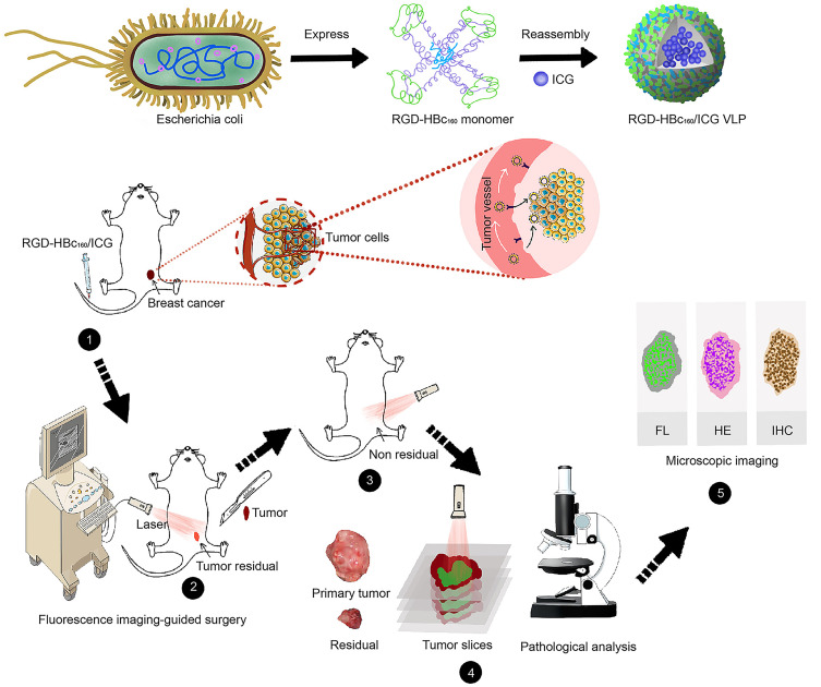
Purpose: Tumor-free surgical margin is crucial but challenging in breast-conserving surgery (BCS). Fluorescence imaging is a promising strategy for surgical navigation that can reliably assist the surgeon with visualization Of the tumor in real-time. Notably, finding an optimized fluorescent probe has been a challenging research topic. Herein, we developed a novel near-infrared (NIR) fluorescent probe based on tailored Hepatitis B Core virus-like protein (HBc VLP) and presented the preclinical imaging-guided surgery.

Methods: The RGD-HBc160 VLP was synthesized by genetic engineering followed encapsulation of ICG via disassembly-reassembly. The applicability of the probe was tested for cell and tissue binding capacities through cell-based plate assays, xenograft mice model, and MMTV-PyVT mammary tumor transgenic mice. Subsequently, the efficacy of RGD-HBc160/ICG-guided surgery was evaluated in an infiltrative tumor-bearing mouse model. The protein-induced body's immune response was further assessed.

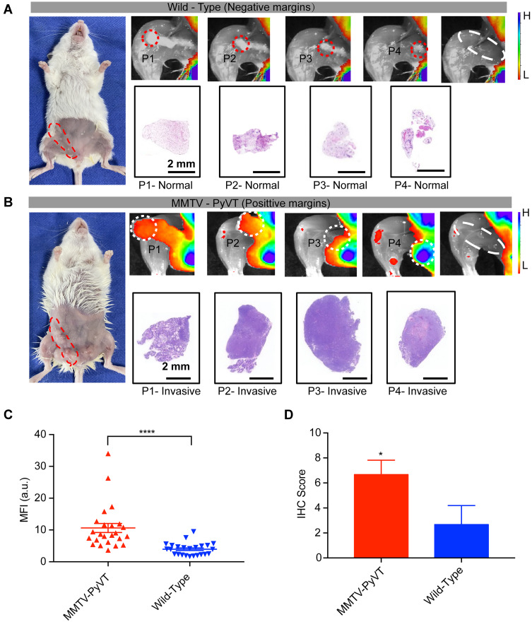
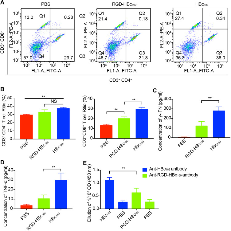
Results: The prepared RGD-HBc160/ICG showed outstanding integrin αvβ3 targeting ability in vitro and in vivo. After intravenous administration of probe, the fluorescence guidance facilitated more complete tumor resection and improved overall survival Of the infiltrative tumor-bearing mice. The probe also showed the excellent capability to differentiate between benign and malignant breast tissues in the mammary tumor transgenic mice. Interestingly, the ingenious tailoring of HBc VLP could not only endow its tumor-targeting ability towards integrin αvβ3 but also significantly reduce the humoral and cellular immune response.

Conclusion: The RGD-HBc160/ICG holds promise as an effective tool to delineate tumor margin. These results have translational potential to achieve margin-negative resection and improve the stratification of patients for a potentially curative.

Keywords: Hepatitis B core virus-like particles; NIR fluorescence imaging; breast Cancer; imaging-guided surgery; tumor margin.


Conflict of interest statement

The authors report no conflicts of interest in this work.

Figures

None
Graphical abstract
Figure 1
Figure 1
Characterization and cellular uptake of RGD-HBc160/ICG. (A) Transmission electron microscopy images of RGD-HBc160/ICG. (B) Diameter distribution of RGD-HBc160/ICG. Representation of absorption spectra (C), and fluorescence emission spectra (D), of free ICG and RGD-HBc160/ICG. (E) Representation of MFI at fluorescence microscopy in MDA-MB-231 and MCF-10A cells following incubation with different RGD-HBc160/ICG concentrations (left), and comparison of MFI (right) at the selected concentration. (F) Representative fluorescence microscopy of MDA-MB-231 and MCF-10A treated with RGD-HBc160/ICG. Representative fluorescence microscopy images from MDA-MB-231 cells after treated with RGD-HBc160/ICG, HBc183/ICG, and ICG (G), and comparison of MFI (H). (I) Comparison of MFI in MDA-MB-231 cells after incubation with RGD-HBc160/ICG and blocking with c(RGDfK) peptides. **P<0.01, ***P<0.001.
Figure 2
Figure 2
Validation of the target specificity of RGD-HBc160/ICG in subcutaneous breast cancer tumor models. (A) Representative quantification of TBR in MDA-MAB-231 tumor mice as a function of time after intravenous injection with different doses of RGD-HBc160/ICG and comparison of TBR at 36 h time points (B). (C) Representative quantification of TBR in MDA-MAB-231 tumor mice as a function of time after intravenous injection with RGD-HBc160/ICG, HBc183/ICG, ICG, and the Fluorescence images of mice (D). (E) Comparison of specific binding with RGD-HBc160/ICG compared with HBc183/ICG and the free ICG at maximum TBR (right). (F) Fluorescence images from organs and tumors excised at 36 h after injection of RGD-HBc160/ICG, and semi-quantitative analysis of total fluorescence intensity from the samples (G). *P<0.05, **P<0.01.
Figure 3
Figure 3
Fluorescent imaging-guided surgical resections of infiltrative MDA-MB-231-luc tumors. (A) The procedure of tumor resection in represented mice guided by white light (WL) plus fluorescence imaging (FL) (blue arrow, residual fluorescence signal in the surgery bed). (B) Ex-vivo fluorescence imaging of excised tissue (red arrow, primary tumor excised under white light only; blue arrow, residual tumor tissue; green arrow, negative surgical margins). (C) Fluorescence image, upper, and H/E stain, lower, for residual tumor tissue and negative margins. (D) Representative preoperative and postoperative bioluminescence images following white light surgery only (n = 10) and white light plus fluorescence-guided surgery (n = 10) with Kaplan-Meier survival analysis of two groups (E).
Figure 4
Figure 4
Fluorescence imaging-guided surgical resection in a mammary tumor transgenic model. (A) Simulated negative surgical margins in fluorescence images of combined fourth and fifth mammary glands from an 8-week-old wild-type female mouse. The tissue to be resected is highlighted with a dashed circle. The second row shows representative histological staining of resected portions (P1-4). (B) Simulated positive surgical margins in fluorescence images of combined fourth and fifth mammary glands from an 8-week-old MMTV-PyVT female mouse following the same excision sequence as above. (C) Representative mean fluorescence intensity of tissues with positive and negative surgical margins pooled from the 6 mice. (D) Representation of immunohistochemistry scores for αv-stained breast tissues from MMTV-PyVT and wild-type mice. *P<0.05, ****P<0.0001.
Figure 5
Figure 5
Distribution of the fluorescent probe in the transgenic mouse mammary gland. (A) Schematic illustration of the performance evaluation (left) and the receiver operating characteristic (ROC) curve of fluorescence imaging to differentiate between normal tissues and cancer (right). (B) Microscopic bio-distribution of RGD-HBc160/ICG in breast tissues based on micro-segmentation analyses (left). The upper row shows a representative example of the fluorescence ROI per tissue type based on H/E staining. The lower row shows the corresponding H/E staining image. MFI of all per-tissue type ROIs are shown in the right panel. (C) Ex vivo fluorescent imaging of breast tissue showing corresponding fluorescence intensity (upper), H/E sections (middle), and immunohistochemical stain of αv expression (lower).
Figure 6
Figure 6
RGD-HBc160 induced immune responses in BALB/c mice. (A) Flow cytometry determination of Ths (CD3+ CD4+) and CTLs (CD3+ CD8+) in the spleen tissue from each group, (B) Quantitative analysis of the means between the different groups. Comparison of the level of serum γ-IFN(C) and TNF-α (D) from RGD-HBc160-immunized, HBc183-immunized, and non-immunized mice. (E) Comparison of the corresponding serum antibody levels from each group on 7 days after the second immunization. **P<0.01.

References

    1. Sung H, Ferlay J, Siegel RL, et al. Global cancer statistics 2020: GLOBOCAN estimates of incidence and mortality worldwide for 36 cancers in 185 countries. CA Cancer J Clin. 2021;71(3):209–249. doi:10.3322/caac.21660 - DOI - PubMed
    1. Fisher B, Anderson S, Bryant J, et al. Twenty-year follow-up of a randomized trial comparing total mastectomy, lumpectomy, and lumpectomy plus irradiation for the treatment of invasive breast cancer. N Engl J Med. 2002;347(16):1233–1241. doi:10.1056/NEJMoa022152 - DOI - PubMed
    1. Waljee JF, Hu ES, Newman LA, Alderman AK. Predictors of re-excision among women undergoing breast-conserving surgery for cancer. Ann Surg Oncol. 2008;15(5):1297–1303. doi:10.1245/s10434-007-9777-x - DOI - PubMed
    1. Biglia N, Maggiorotto F, Liberale V, et al. Clinical-pathologic features, long term-outcome and surgical treatment in a large series of patients with invasive lobular carcinoma (ILC) and invasive ductal carcinoma (IDC). Eur J Surg Oncol. 2013;39:455–460. - PubMed
    1. Tipirneni KE, Warram JM, Moore LS, et al. Oncologic procedures amenable to fluorescence-guided surgery. Ann Surg. 2017;266(1):36–47. doi:10.1097/SLA.0000000000002127 - DOI - PMC - PubMed

Substances