. 2022 Mar 8:13:829862.
doi: 10.3389/fphar.2022.829862. eCollection 2022.

CYT387, a JAK-Specific Inhibitor Impedes Osteoclast Activity and Oophorectomy-Induced Osteoporosis via Modulating RANKL and ROS Signaling Pathways

Affiliations

CYT387, a JAK-Specific Inhibitor Impedes Osteoclast Activity and Oophorectomy-Induced Osteoporosis via Modulating RANKL and ROS Signaling Pathways

Jing Li et al. Front Pharmacol. .

Abstract

Osteoclasts are of hematopoietic lineage and have the ability to degrade mineralized bone tissues. Abnormalities in osteoclastic activity under certain pathological conditions are common in bone diseases such as osteoporosis, osteosclerosis, and arthritis. Although many kinds of drugs are currently used to treat osteoporosis, they have obvious adverse reactions and limitations. CYT387 is a new small-molecule Janus kinase (JAK) inhibitor involved in hematopoiesis, immune modulation, fertility, lactation, and embryonic development. However, it has remained unclear whether CYT387 functionally impacts osteoclast formation. Our study demonstrated through osteoclast formation assay in vitro, that the use of CYT387 is a potential drug candidate for treating osteoclast-associated bone disease. The effects of CYT387 on osteoclast formation, bone resorption, NFATc1 activation, and especially intracellular ROS levels were investigated in vitro. Further, we examined the preclinical prospects of CYT387 using an oophorectomy (OVX) mouse model of osteoporosis with its anti-osteoclast activity in vivo. On the whole, this study shows that CYT387 holds promise for treating osteoclast-related bone illnesses including osteoporosis.

Keywords: CYT387; ERK; ROS; osteoclast; osteoporosis.


Conflict of interest statement

The authors declare that the research was conducted in the absence of any commercial or financial relationships that could be construed as a potential conflict of interest.

Figures
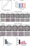
FIGURE 1
FIGURE 1
CYT387 suppresses RANKL-induced osteoclastogenesis in vitro. (A) The chemical structure and molecular formula of CYT387. (B) After being treated with different concentrations of CYT387 for 48 h, cell proliferation was detected by a CCK-8 assay (n = 3 per group). (C) Representative images of TRAcP staining showed that CYT387 inhibited osteoclast formation in a dose-dependent manner after 7 days of 50 ng/ml RANKL stimulation. (D) Representative images of TRAcP staining showed time-dependent inhibition of osteoclast formation by CYT387 at 1 µM. (E) Quantification of TRAcP-positive cells per well. The data were expressed as mean ± SD (n = 3 per group). (F) Quantification of TRAcP-positive cells per well when treated with CYT387 in different periods (n = 3 per group). The data are shown as the means ± SD. *p < 0.05, **p < 0.01 and ***p < 0.001. Scale bar = 2000 μm. BMMs, bone marrow macrophages; CCK-8, cell counting kit-8; RANKL, receptor activator of the nuclear factor-κB ligand; TRAcP, tartrate-resistant acid phosphatase.
FIGURE 2
FIGURE 2
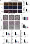
CYT387 impairs the bone resorption function of mature osteoclasts in vitro and inhibits the mRNA expression of osteoclast marker genes. (A) The representative images of podosome belts in osteoclasts treated with different concentrations of CYT387 were observed by confocal microscopy. Scale bar = 1,000 μm. (B) The mean F-actin ring area was quantified (n = 3 per group). (C) The number of osteoclasts per field was quantified. (D) The same number of mature osteoclasts stimulated by 50 ng/ml RANKL were seeded with bone slices, and then the cells were treated with different concentrations of CYT387 to obtain representative bone resorption pit images. The number and area of bone pits were used to evaluate the bone resorption capacity of osteoclasts. Scale bar = 200 μm. (E,F) The number of osteoclasts and the total area of bone resorption were evaluated by ImageJ 1.53 software. (G) qRT-PCR showed that mRNA expression levels of osteoclast-specific genes, including c-Fos, Ctsk, Dcstamp, Mmp9, Nfatc1 and Acp5, were detected in the absence or presence of different concentrations of CYT387. *p < 0.05, **p < 0.01 and ***p < 0.001.
FIGURE 3
FIGURE 3
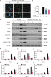
CYT387 inhibits RANKL-induced NFATc1 activation. (A) Immunofluorescence images of NFATc1 nuclear translocation following RANKL stimulation without or with (0.5 and 1 μM) CYT387 treatment (Magnification = ×20). Scale bar = 50 μm. (B) Transcriptional activity of NFATc1 promoter was detected by the luciferase reporter gene. (C) After treatment with CYT387 (1 μM) for 0, 1, 3, and 5 days after RANKL (50 ng/ml) stimulation, western blot was used to detect the expression of RANKL downstream signaling protein. (D–I) Quantification of the ratios of band intensity of NFATc1, c-Fos, CTSK, ATP6V0D2, MMP-9 and Integrin αV/β3 relative to β-actin. All experimental data are expressed as mean ± SD. *p < 0.05, **p < 0.01 and ***p < 0.001.
FIGURE 4
FIGURE 4
CYT387 negatively regulates osteoclast formation and bone resorption by inhibiting MAPK signaling. (A) In the early stage of osteoclast formation, CYT387 inhibited ERK degradation but did not inhibit the phosphorylation of JNK and P38 MAPKs. Total cell protein was extracted with BMM pretreated with 1 μM CYT387 for 1 h, and then stimulated with 50 ng/ml RANKL for a specific time. Protein expression and phosphorylation status of ERK, JNK, and P38 were assessed using specific antibodies. (B) CYT387 did not significantly inhibit IκBα degradation and P65 phosphorylation. Protein expression and phosphorylation status of P65 and IκBα were detected using specific antibodies. (C) The ratio of phosphorylated p-ERK, p-JNK, p-P38, p-P65 to the corresponding total protein band strength and the ratio of IκBα to β-actin were quantitatively determined. (D) Representative images of intracellular Ca2+ were obtained 48 h after RANKL (50 ng/ml) stimulation with or without CYT387 (0.5, 1 μM) using the Fluo-4 assay kit. (E) The fluorescence intensity of intracellular Ca2+ was measured. Scale bar = 300 μm. All experimental data are expressed as mean ± SD. *p < 0.05, **p < 0.01 and ***p < 0.001.
FIGURE 5
FIGURE 5
CYT387 reduces RANKL-induced ROS production during osteoclastogenesis. (A) RANKL-induced osteoclast formation was treated with different concentrations of CYT387 (0, 0.5, 1 μM), and the representative images produced by ROS were detected by DCFHDA. Scale bar = 300 µm. (B) Quantification of the number of ROS-positive cells per field (n = 3 per group). (C) qRT-PCR was used to detect the expression of antioxidant enzymes Nfe2l2, Keap1, Homx-1, Cat and Gsr in BMMs treated by RANKL and CYT387 for 48 h. (D) Representative Western Blot images of the effects of CYT387 on the expression of antioxidant enzymes, including HO-1, GSR, CAT, SOD2 and GCLC. (E) Quantification of the ratios of band intensity of HO-1, GSR, CAT, SOD2 and GCLC relative to β-actin (n = 3 per group). All experimental data are presented as mean ± SD. *p < 0.01, **p < 0.01 and ***p < 0.001.
FIGURE 6
FIGURE 6
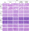
CYT387 has no significant toxic effects on heart, liver, spleen and kidney. Representative images of hematoxylin-eosin staining of the heart, liver, spleen and kidney. 10× multiple scale bar = 100 μm. 40× multiple scale bar = 20 μm.
FIGURE 7
FIGURE 7
CYT387 treatment can partially reverse bone destruction in ovariectomized mice. (A) 3D reconstruction of representative Micro CT images of the proximal tibia in each group. (B) The following bone structure parameters were evaluated by quantitative analysis: bone volume to total volume ratio (BV/TV), trabecular thickness (Tb.Th), trabecular number (TB.N), and trabecular separation (Tb.Sp) (n = 6 per group). All experimental data are expressed as mean ± SD. *p < 0.05, **p < 0.01 and ***p < 0.001.
FIGURE 8
FIGURE 8
CYT387 ameliorates osteoclast activity in a mouse model of osteoporosis induced by ovariectomy. (A) Representative images of histological analysis of femur stained with HE. (B) Bone trabecular percentage (BS/TS) was quantitatively analyzed (n = 6 per group). (C) Representative images of histological analysis of femur stained with TRAcP. The red arrows indicate TRAcP-positive osteoclasts. (D) TRAcP-positive cell area per trabecular surface (TRAcP+ Oc.S/BS) was quantitatively analyzed (n = 6 per group). (E) CTSK-positive cells were marked as brownish yellow. (F) The level of CTSK was quantitatively measured in the tibia (n = 6 per group). All experimental data are expressed as mean ± SD. *p < 0.05, **p < 0.01 and ***p < 0.001. OVX, ovariectomized.
FIGURE 9
FIGURE 9
CYT387 does not affect osteoblast differentiation in vitro or in vivo. (A,B) Representative images of Alkaline Phosphatase staining and quantification of BMSCs (n = 3 per group). (C) qRT-PCR showed that mRNA expression levels of osteoblast-specific genes, including Col1a1, OPG and Runx2, were detected in the absence or presence of different concentrations of CYT387. (D,E) The expression and quantification of OCN in proximal tibia were detected by immunohistochemical staining (n = 6 per group). All experimental data are expressed as mean ± SD. *p < 0.05, **p < 0.01 and ***p < 0.001.
FIGURE 10
FIGURE 10
A proposed scheme for the inhibition of CYT387 in osteoclastogenesis. Our results demonstrate for the first time that CYT387 inhibits the level of ROS and RANKL-induced signaling pathways, accompanied by increased expression of antioxidant enzymes, thereby inhibiting the formation and function of osteoclasts. NFATc1, nuclear factor of activated T cells 1; c-Fos, Proto-oncogene C-Fos; Ctsk, cathepsin K; Dcstamp, dendritic cell-specific transmembrane protein; Mmp9, matrix metallopeptidase 9; Acp5, tartrate resistant acid phosphatase; RANKL, receptor activator of nuclear factor-κB (NF-κB) ligand; NF-κB, nuclear factor-κB; MAPKs, mitogen-activated protein kinases; ROS, reactive oxygen species.

References

    1. Abubaker K., Luwor R. B., Escalona R., McNally O., Quinn M. A., Thompson E. W., et al. (2014). Targeted Disruption of the JAK2/STAT3 Pathway in Combination with Systemic Administration of Paclitaxel Inhibits the Priming of Ovarian Cancer Stem Cells Leading to a Reduced Tumor Burden. Front. Oncol. 4, 75. 10.3389/fonc.2014.00075 - DOI - PMC - PubMed
    1. Bakirezer S. D., Yaltirik C. K., Kaya A. H., Yilmaz S. G., Ozdogan S., Billur D., et al. (2019). The Evaluation of Glutathione Reductase and Malondialdehyde Levels in Patients with Lumbar Disc Degeneration Disease. In Vivo 33, 811–814. 10.21873/invivo.11543 - DOI - PMC - PubMed
    1. Bigarella C. L., Liang R., Ghaffari S. (2014). Stem Cells and the Impact of ROS Signaling. Development 141, 4206–4218. 10.1242/dev.107086 - DOI - PMC - PubMed
    1. Bouchemal K., Bories C., Loiseau P. M. (2017). Strategies for Prevention and Treatment of Trichomonas Vaginalis Infections. Clin. Microbiol. Rev. 30, 811–825. 10.1128/CMR.00109-16 - DOI - PMC - PubMed
    1. Campos-Ordonez T., Gonzalez-Perez O. (2015). Cyclohexane, a Potential Drug of Abuse with Pernicious Effects on the Brain. Front. Pharmacol. 6, 291. 10.3389/fphar.2015.00291 - DOI - PMC - PubMed