. 2022 Mar 26;22(1):137.
doi: 10.1186/s12876-022-02208-x.

RIP3 knockdown inhibits necroptosis of human intestinal epithelial cells via TLR4/MyD88/NF-κB signaling and ameliorates murine colitis

Affiliations

RIP3 knockdown inhibits necroptosis of human intestinal epithelial cells via TLR4/MyD88/NF-κB signaling and ameliorates murine colitis

Chaoqin Duan et al. BMC Gastroenterol. .

Abstract

Background: Ulcerative colitis (UC) is a common inflammatory bowel disease, during which cell necroptosis plays key roles in driving inflammation initiation and aggravation. Previous studies reported Receptor Interacting Protein Kinase 3 (RIP3)-mediated necroptosis in multiple diseases, and RIP3 protein in Paneth cells significantly enriched in the intestines of both humans and mice. Therefore, we hypothesized targeting RIP3 to inhibit necroptosis may depress UC.

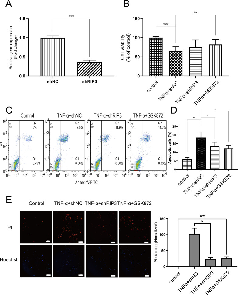
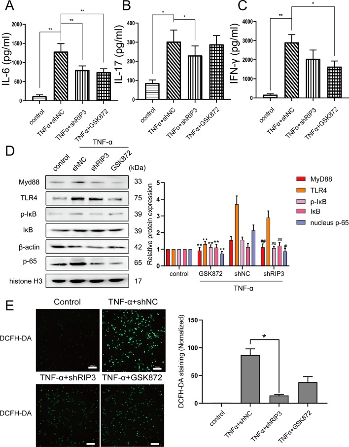
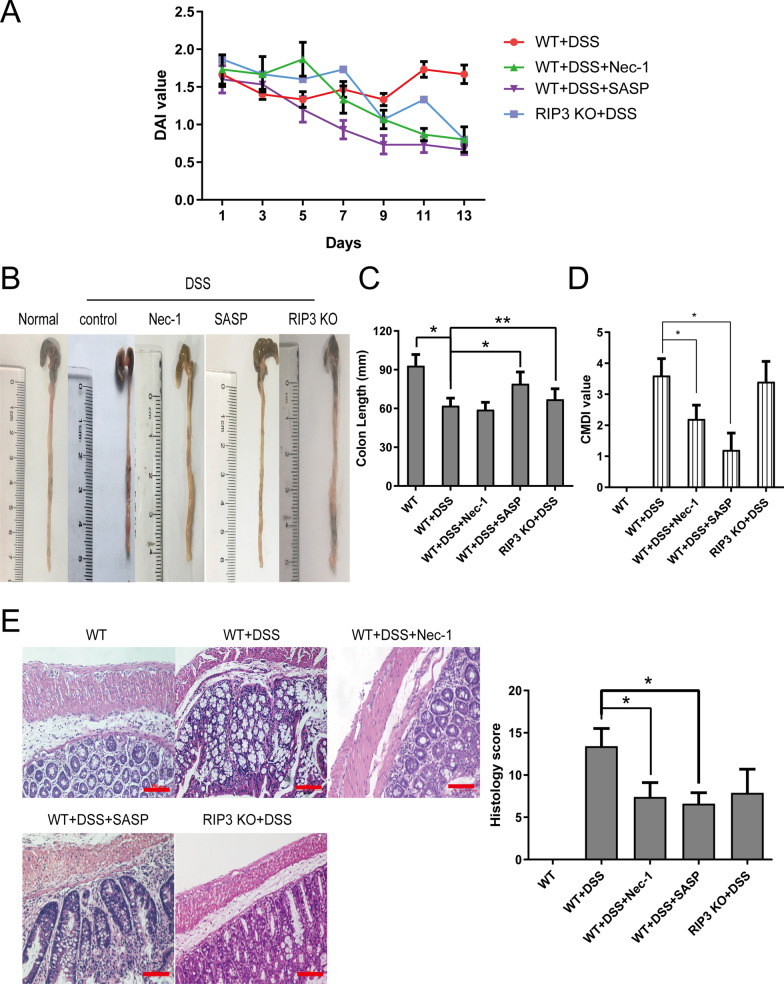
Methods: We classified clinical UC samples according to the modified Truelove & Witts criterion. The expression of RIP3 was measured by western blot and immunohistochemistry. Cell proliferation and apoptosis were analyzed by MTT assay and flow cytometry. ROS production and the secretion of inflammatory cytokines were measured by DCFH-DA probe and ELISA assay. TLR4/MyD88/NF-κB signaling pathway was analyzed by western blot. We established experimental colitis model in RIP3 knockout and wild-type mice and disease activity index (DAI) score was calculated. The expression and distribution of tight junction protein were analyzed by immunofluorescence. The ratio of CD4+Foxp3+ T cells in the spleen was detected by flow cytometry. Oxidative damage of mouse colon was assessed by detecting the levels of SOD, MDA and MPO. Data were analyzed by one-way ANOVA or student's t test.

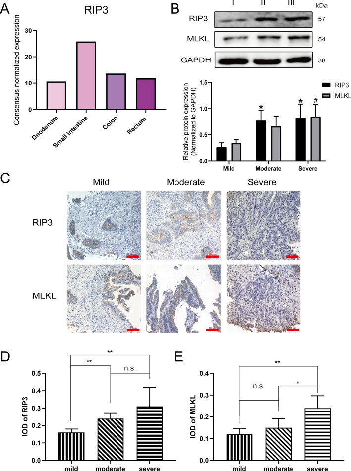
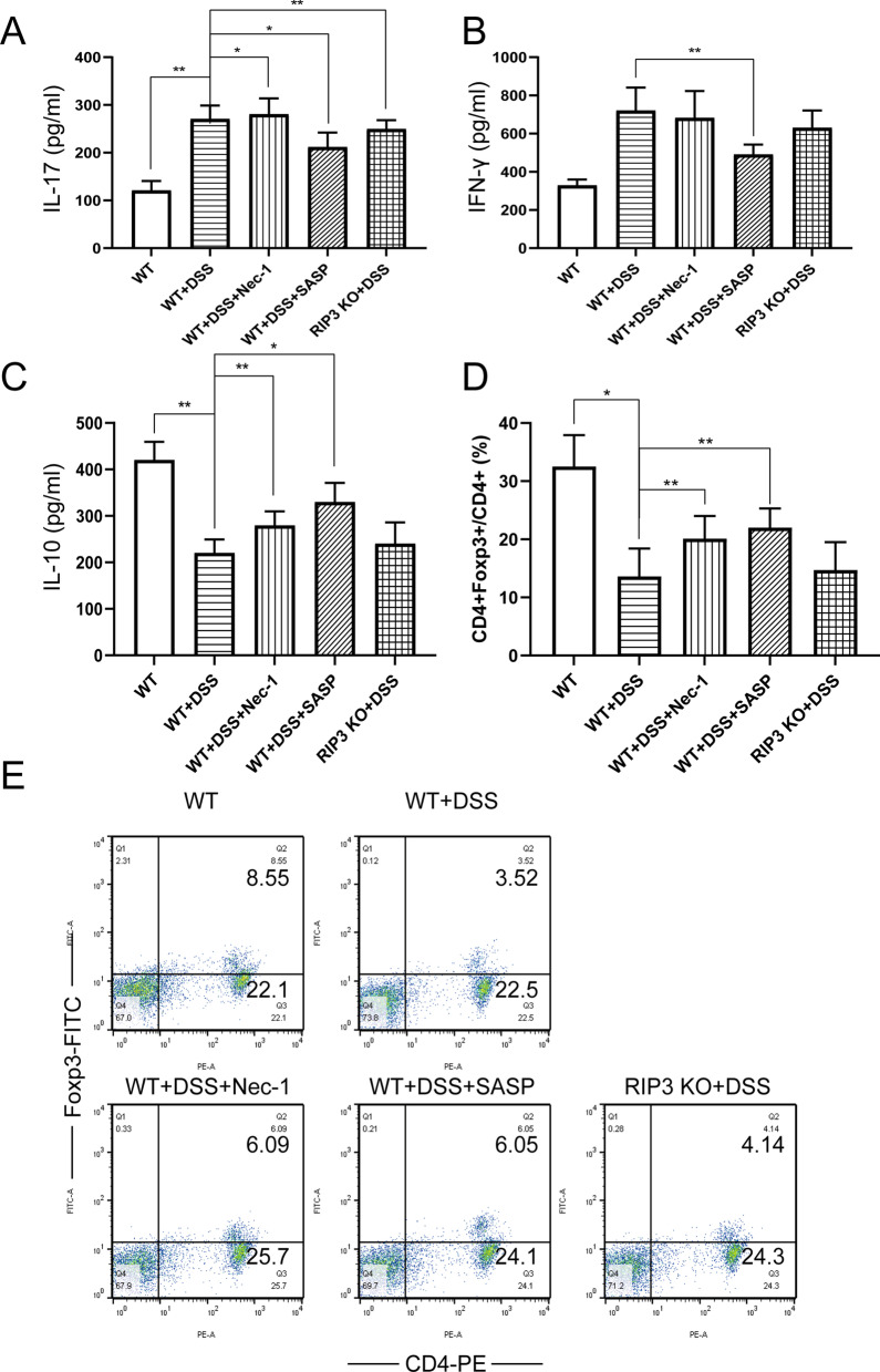
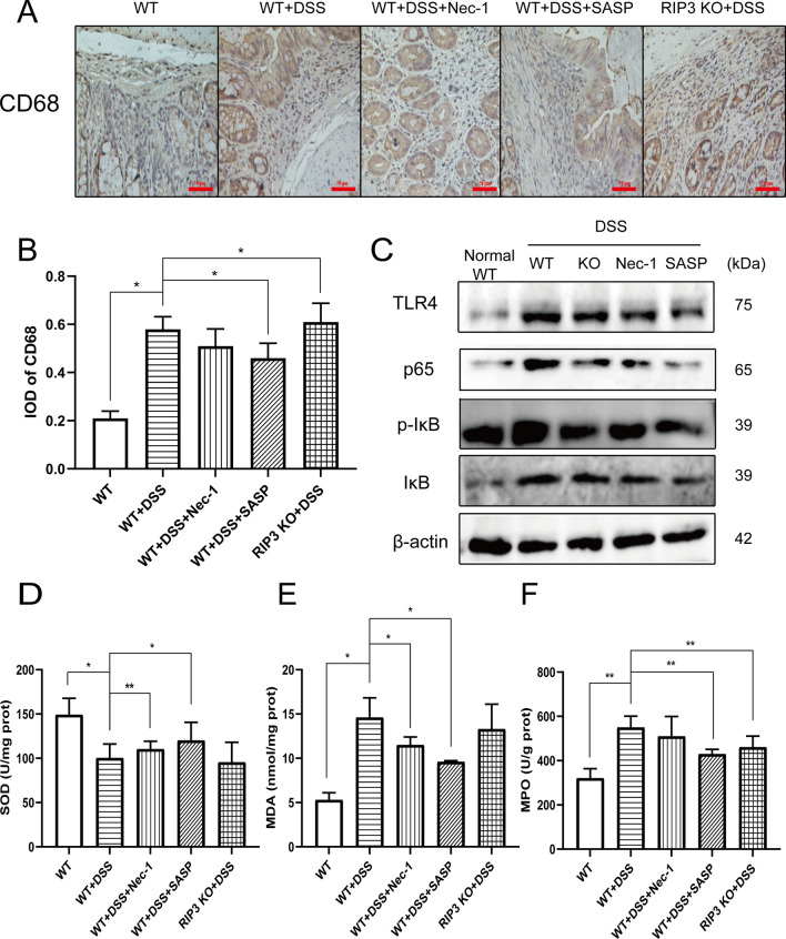
Results: The expression of RIP3 in human colon is positively associated with the severity of UC. RIP3 inhibitor GSK872 or RIP3 knockdown reverses the inhibitory effect of TNF-α on proliferation and the promoting effect of TNF-α on apoptosis and necrosis in human intestinal epithelial cells. In addition, RIP3 deficiency inhibits the secretion of inflammatory cytokines (IL-16, IL-17 and IFN-γ) and ROS production induced by TNF-α. In vivo, RIP3 inhibitor Nec-1 effectively improves DSS-induced colitis in mice. In mechanism, RIP3 depression could upregulate the proportion of CD4+Foxp3+ immunosuppressive Treg cells in the spleen while suppressed TLR4/MyD88/NF-κB signaling pathway and ROS generation, and all these anti-inflammation factors together suppress the secretion of inflammatory cytokines and necroptosis of intestinal epithelial cells.

Conclusions: This study preliminarily explored the regulating mechanism of RIP3 on UC, and Nec-1 may be a promising drug to alleviate the inflammation and necroptosis of the colon in UC patients.

Keywords: Nec-1; Necroptosis; Receptor interacting protein kinase 3; TLR4-MyD88; Ulcerative colitis.


Conflict of interest statement

The authors declared no conflicts of interest with respect to the research, authorship and publication of this article.

Figures

Fig. 1
Fig. 1
The expression of RIP3 in the colon is positively proportional to the severity of UC. A The Human Protein Atlas (https://www.proteinatlas.org/search/RIPK3) analyzed RNA expression of RIP3 in more than 60 human tissues and organs. Interestingly, among more than 60 human tissues and organs, RIP3 has the most RNA expression in small intestines. We have shown the RNA expression of RIP3 in Duodenum, Small intestine, Colon, and Rectum. B Western blot detected the expression levels of RIP3 and MLKL in the colon tissues from patients with mild (I), moderate (II) and severe (III) UC based on the modified Truelove & Witts severity classification. The semi-quantitative analysis of western blotting via Image J software. RIP3: *P < 0.05, ~ versus Mild; MLKL: #P < 0.05, ~ versus Mild. C Immunohistochemistry detected the expression and distribution of RIP3 and MLKL in the colon tissues from patients with mild, moderate and severe UC. DE The mean optical density value (IOD) of the target protein in each field was analyzed by Image-Pro Plus 6.0 software. Scale bar: 100 μm. The data were presented as mean ± SD. The asterisk indicates a significant difference (*P < 0.05, **P < 0.01) between groups. Abbreviations: n.s., no significance
Fig. 2
Fig. 2
Knockdown of RIP3 expression inhibits the pro-apoptotic effect of TNF-α on intestinal epithelial cells. A RT-qPCR demonstrated the knockdown efficiency of shRIP3 plasmid. B MTT assay detected the effect of shRIP3 knockdown plasmid and RIP3 inhibitor GSK872 (2 μM) on cell viability after TNF-α (10 ng/ml, 12 h) treatment. C Annexin V-FITC and PI double staining detected the effect of shRIP3 knockdown plasmid and RIP3 inhibitor GSK872 on cell apoptosis. D Semi-quantitative analysis of cell apoptosis (The sum of the percentages of the first and fourth quadrants). E PI staining detected cell necrosis and the results were observed under fluorescence microscope with a 20 × objective. Blue color: Hochest 33342 stains the nucleus; Green color: PI Dye. Scale bar: 200 μm. The semi-quantitative analysis of immunofluoresence via Image J software (right panel)
Fig. 3
Fig. 3
Knockdown of RIP3 expression inhibits inflammatory signaling pathways in vitro. AC ELISA assay detected the effect of RIP3 knockdown plasmid and RIP3 inhibitor GSK872 on the secretion of inflammatory factors IL-16, IL-17 and IFN-γ after TNF-α (10 ng/ml, 12 h) treatment. D Western blot detected the effect of shRIP3 knockdown plasmid and RIP3 inhibitor GSK872 on the expression of key molecules in the TLR4/MyD88/NF-κB signaling pathway. The semi-quantitative analysis of western blotting via Image J software (right panel). E After cultured with TNF-α (10 ng/ml, 12 h), shRIP3 plasmid, or RIP3 inhibitor GSK872, cells were incubated with DAFH-DA probe and the results were observed under fluorescence microscope with a 10 × objective. Scale bar: 200 μm. The semi-quantitative analysis of immunofluoresence via Image J software (right panel)
Fig. 4
Fig. 4
Nec-1, a RIP3 inhibitor, treatment could relieve intestinal pathology and histopathology in DSS-induced colitis mice. AD Balb/c wild-type and RIP3 knockout mice freely drank 3% DSS solution for 7 days. On the 8th day, Nec-1 (5 mg/kg), SASP (50 mg/kg) and placebo (1% DMSO) were injected intraperitoneally for 14 consecutive days. A DAI, C the length of mouse colon and D CMDI scores were measured, and B the entire colonic intestine from the anus to the end of the cecum was isolated and photographed. E H&E staining results were imaged under a 20 × objective. Scale bar: 100 μm. The histology scores are shown on the right panel. The data are presented as mean ± SD. The asterisk indicates a significant difference (*P < 0.05) between groups. Abbreviations: DSS, dextran sulfate sodium; SASP, sulfasalazine; DAI, disease activity index; CMDI, colon gross morphological damage index
Fig. 5
Fig. 5
Nec-1 treatment could relieve DSS-induced intestinal inflammation in vivo. AC ELISA detected the secretion of IL-17, IFN-γ and IL-10 cytokines in mouse colon tissue. DE Flow cytometry analyzed the proportion of CD4+ Foxp3+ T cells in CD4+ T cells of mouse spleen
Fig. 6
Fig. 6
Knockdown of RIP3 expression inhibits inflammatory signaling pathways in vivo. A Immunohistochemistry detected the expression and distribution of CD68 in the mouse colon. Scale bar: 50 μm. B The mean optical density value (IOD) of CD68 protein in each field was analyzed by Image-Pro Plus 6.0 software. C Western blot detected the effect of RIP3 knockout or knockdown in vivo on key molecules of TLR4/NF-κB signaling pathway. DF The levels of D SOD, E MDA and F MPO in the mouse colon collected after 7 days’ administration

Similar articles

Cited by

References

    1. Boal Carvalho P, Cotter J. Mucosal healing in ulcerative colitis: a comprehensive review. Drugs. 2017;77(2):159–173. - PubMed
    1. Sun W, Wu X, Gao H, Yu J, Zhao W, Lu JJ, Wang J, Du G, Chen X. Cytosolic calcium mediates RIP1/RIP3 complex-dependent necroptosis through JNK activation and mitochondrial ROS production in human colon cancer cells. Free Radical Biol Med. 2017;108:433–444. - PubMed
    1. Yang Z, Wang Y, Zhang Y, He X, Zhong CQ, Ni H, Chen X, Liang Y, Wu J, Zhao S, et al. RIP3 targets pyruvate dehydrogenase complex to increase aerobic respiration in TNF-induced necroptosis. Nat Cell Biol. 2018;20(2):186–197. - PubMed
    1. Vandenabeele P, Declercq W, Van Herreweghe F, Vanden Berghe T. The role of the kinases RIP1 and RIP3 in TNF-induced necrosis. Sci Signal. 2010;3(115):re4. - PubMed
    1. Wang G, Xu B, Shi F, Du M, Li Y, Yu T, Chen L. Protective effect of methane-rich saline on acetic acid-induced ulcerative colitis via blocking the TLR4/NF-κB/MAPK pathway and promoting IL-10/JAK1/STAT3-mediated anti-inflammatory response. Oxid Med Cell Longev. 2019;2019:7850324. - PMC - PubMed

Substances