. 2022 Mar 26;18(1):118.
doi: 10.1186/s12917-022-03218-3.

Retrospective analysis of radiographic signs in feline pleural effusions to predict disease aetiology

Affiliations

Retrospective analysis of radiographic signs in feline pleural effusions to predict disease aetiology

Lily Hung et al. BMC Vet Res. .

Abstract

Background: The objectives of the study were to determine the prevalence of underlying conditions causing pleural effusion in cats and to calculate the positive predictive values, negative predictive values, sensitivity and specificity of radiographic signs to predict aetiology of the pleural fluid.

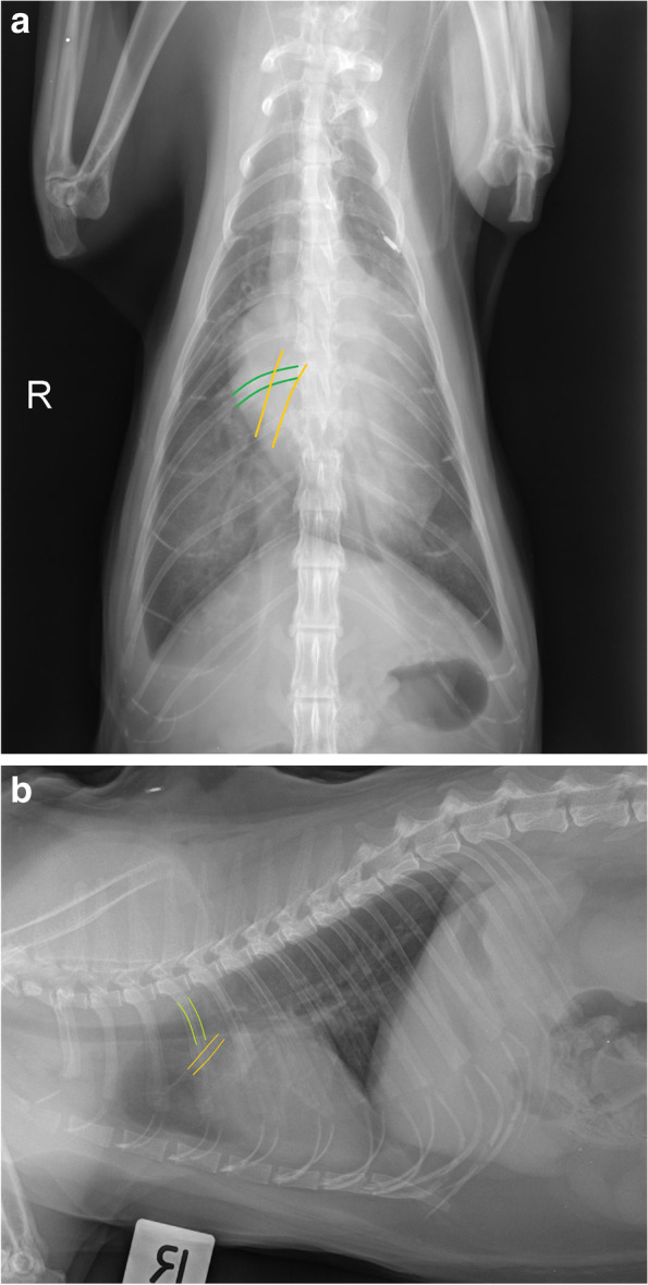
Methods: Data from 148 cats with pleural effusion and diagnosed with known aetiologies were retrospectively analysed. Sixty one cats had thoracic radiographs evaluated by consensus through pre-defined radiographic signs by two radiologists blinded to the diagnoses.

Results: Congestive heart failure (53.4%) was the most common diagnosis, followed by neoplasia (20.3%), pyothorax (10.8%), idiopathic chylous effusion (5.4%), feline infectious peritonitis (1.4%) and "other" or cats with multiple diagnoses (total 8.8%). Cats with an enlarged cardiac silhouette had a high positive predictive value of congestive heart failure (90%). Mediastinal masses (100%)and pulmonary masses (100%) were highly predictive of neoplastic disease. Pulmonary nodules (50%) were poorly predictive of neoplastic disease. The remainder of the radiographic variables were not informative predictors of underlying disease.

Conclusions: In our sample of cats, congestive heart failure was the most common cause of pleural effusion. Radiographically enlarged cardiac silhouette and presence of a mediastinal mass may be useful predictors of aetiology, however there are limitations to the use of radiography alone as a diagnostic tool.

Keywords: Cardiomegaly; Congestive heart failure; Feline pleural effusion; Idiopathic chylothorax; Mediastinal mass; Pleural effusion; Positive and negative predictive values; Pyothorax; Radiographic parameters; Thorax radiography.


Conflict of interest statement

The authors declare that they have no competing interests.

Figures

Fig. 1
Fig. 1
A patient with a large volume of pleural effusion with complete effacement of cardiac silhouette, indistinct pulmonary vasculature and marked difficulty assessing pleural margins and mediastinum
Fig. 2
Fig. 2
Cardiac silhouette size was measured using two methods. The first method was the vertebral heart score. The base apex length and craniocaudal length were transposed onto the vertebral column and recorded as the corresponding number of vertebrae measured from the cranial edge of T4 vertebral body. VHS more than 8 was considered enlarged and likely in heart failure. The base apex length was measured from the ventral wall of the carina to apex (orange solid line). The craniocaudal length was measured perpendicular to the base-apex length, at the widest width of the cardiac silhouette (green solid line). The second method used the base apex length and compared this against the length of sternebrae 2 to 4. If the length extended beyond three sternebrae, the cardiac silhouette was considered enlarged (dashed orange line). This patient has VHS of 9 and an elongated base-apex length suggestive of congestive heart failure
Fig. 3
Fig. 3
VD projection. Caudal lobar vein (depicted by orange solid line) and veins at where they intersect with the 9th rib (depicted by green solid line) were considered enlarged if they were more than 1:1 ratio. This cat had bilateral enlarged caudal pulmonary veins and arteries with a scant volume of pleural effusion. b Lateral projection. Cranial lobar veins (depicted by orange solid line) were considered enlarged if it was more than 0.7 times the proximal third of the 4th rib (depicted by green solid line). This cat had enlarged cranial lobar veins and arteries, severely enlarged globoid cardiac silhouette with a scant volume of pleural effusion
Fig. 4
Fig. 4
The mediastinum has a large soft tissue mass with rounded caudal and lateral margins causing severe deviation of the mediastinal structures (trachea, oesophagus) and lung lobes with complete effacement of the cardiac silhouette. This patient had mediastinal lymphoma
Fig. 5
Fig. 5
Rounded and contracted pleural margins secondary to chronic fibrosis is often described in cats with pyothorax. This cat represents the classic chronic chylothorax pleural margin abnormalities; however, this was not a predictive sign of chylothorax in our cohort

References

    1. Beatty JA, Barrs VR. Pleural effusion in the cat. J Feline Med Surg. 2010;12(11):693–707. doi: 10.1016/j.jfms.2010.07.013. - DOI - PMC - PubMed
    1. Ruiz MD, Vessières F, Ragetly GR, Hernandez JL. Characterization of and factors associated with causes of pleural effusion in cats. J Am Vet Med Assoc. 2018;253(2):181–187. doi: 10.2460/javma.253.2.181. - DOI - PubMed
    1. König A, Hartmann K, Mueller RS, Wess G, Schulz BS. Retrospective analysis of pleural effusion in cats. J Feline Med Surg. 2019;21(12):1102–1110. doi: 10.1177/1098612X18816489. - DOI - PMC - PubMed
    1. Suter P. Thoracic radiography: a text atlas of thoracic diseases of the dog and cat. Switzerland: Wettswil; 1984. Pleural abnormalities; pp. 648–764.
    1. Mandell D. Respiratory distress in cats. In: King LG. Respiratory disease in dogs and cats. Vol. 50, Saunders. W.B. Saunders; 2004. 12–17.