. 2022 Mar 28;41(1):111.
doi: 10.1186/s13046-022-02339-9.

PABPC1-induced stabilization of IFI27 mRNA promotes angiogenesis and malignant progression in esophageal squamous cell carcinoma through exosomal miRNA-21-5p

Affiliations

PABPC1-induced stabilization of IFI27 mRNA promotes angiogenesis and malignant progression in esophageal squamous cell carcinoma through exosomal miRNA-21-5p

Ying Zhang et al. J Exp Clin Cancer Res. .

Abstract

Background: Emerging evidence has demonstrated that RNA-binding protein dysregulation is involved in esophageal squamous cell carcinoma (ESCC) progression. However, the role of poly (A) binding protein cytoplasmic 1 (PABPC1) in ESCC is unclear. We therefore aimed to explore the functions and potential mechanisms of PABPC1 in ESCC progression.

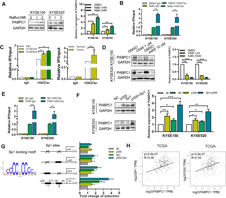
Methods: PABPC1 expression was characterized using immunohistochemistry and qRT-PCR in ESCC tissues and cell lines. Chromatin immunoprecipitation (ChIP) and luciferase reporter assays were used to detect histone acetylation in the promoter region of PABPC1. A series of in vitro and in vivo assays were further applied to elucidate the functions and underlying molecular mechanisms of PABPC1 in ESCC angiogenesis and malignant procession.

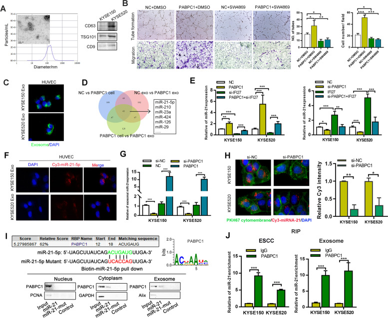
Results: PABPC1 expression was upregulated in ESCC tissues compared with in normal esophageal epithelial tissues. Elevated PABPC1 expression was correlated with tumor cell differentiation and poor prognosis in patients. Sp1 and p300 cooperated to increase the level of H2K37ac in the PABPC1 promoter. Functionally, PABPC1 overexpression enhanced esophageal squamous cell proliferation and invasion by activating the IFN/IFI27 signaling pathway. PABPC1 interacted with eIF4G to increase the stability of IFI27 mRNA by competing with RNA exosomes in ESCC. Furthermore, PABPC1/IFI27 could increase miR-21-5p expression to enable exosomal delivery of miR-21-5p to human umbilical vein endothelial cells to increase angiogenesis via inhibiting CXCL10.

Conclusion: PABPC1 plays a critical role in ESCC malignant progression by interacting with eIF4G to regulate IFI27 mRNA stability and promote angiogenesis via exosomal miR-21-5p/CXCL10. Taken together, our results suggest that PABPC1 is a promising therapeutic target for ESCC.

Keywords: ESCC; IFI27; PABPC1; eIF4G; miR-21-5p.


Conflict of interest statement

The authors declare no conflicts of interest.

Figures

Fig. 1
Fig. 1
Expression of PABPC1 in ESCC tissues with its clinical significance. A Different expression levels of RNA-binding proteins, between normal and ESCC tissues, based on TCGA (https://cancergenome.nih.gov/). B Expression of PABPC1 in different types of cancer in the GEPIA database (http://gepia.cancer-pku.cn/). C Expression of PABPC1 in 24 pairs of ESCC samples (T) and corresponding non-tumor tissues (N) was detected by western blot analysis. D PABPC1 mRNA expression was detected by qRT-PCR in ESCC samples and non-tumor tissues (n = 48) and in TCGA. E Immunohistochemical staining of PABPC1 in primary ESCC samples and non-tumor tissues (n = 190) (scale bars: 200 µm, 50 µm). F Immunohistochemical staining of PABPC1 and CD34 in primary ESCC samples (n = 190) (scale bars: 200 µm). G Expression of PABPC1 in the immortalized esophageal cell line NE1 and ESCC cell lines (KYSE150, KYSE520, KYSE510, KYSE410, KYSE450, KYSE180, 81 T, and TE1) was detected by qRT-PCR and western blotting. H Expression of PABPC1 in ESCC cell lines KYSE150 and KYSE520 was detected by immunofluorescence (scale bars: 20 µm). I Expression of PABPC1 was higher in poorly differentiated than well differentiated tissues. J Comparison of overall survival of ESCC patients with high and low PABPC1 protein expression was determined by Kaplan–Meier survival analysis. Data represent the mean ± SD of 3 separate experiments. *p < 0.05, **p < 0.01, ***p < 0.001 by Student’s t test
Fig. 2
Fig. 2
P300/Sp1 upregulates PABPC1 expression by modulating PABPC1 promoter H3K27 acetylation. A Expression of PABPC1 was detected by western blotting and qRT-PCR following treatment of KYSE150 and KYSE520 cells with different doses of NaBu. B ChIP assay demonstrating that H3K27 acetylation occurred in the PABPC1 promoter in ESCC cell lines following treatment with NaBu (2 mM). C ChIP assay demonstrating that H3K27 acetylation occurred in the PABPC1 promoter in the immortalized esophageal cell line NE1 and KYSE150 and KYSE520 ESCC cell lines. ChIP assay demonstrating that H3K27 acetylation occurred in the PABPC1 promoter in normal and ESCC tissues (n = 3). D Expression of PABPC1 was detected by western blotting and qRT-PCR following treatment of KYSE150 and KYSE520 cells with C646 or A485. E ChIP assay demonstrating that H3K27 acetylation occurred in the promoter of PABPC1 in ESCC cell lines following transfection of p300. F Expression of PABPC1 was detected after co-transfection of Sp1 and p300 plasmids and detection by western blot and qRT-PCR. G HEK293T cells were co-transfected with different combinations of wild-type (wt) and Sp1-site-mutated reporter constructs (mt 1, mt 2, and mt 3), p300 and control. The relative luciferase activity was analyzed. H Correlation of PABPC1 and Sp1 or p300 in TCGA was analyzed. Data represent the mean ± SD of 3 separate experiments. *p < 0.05, **p < 0.01, ***p < 0.001 by Student’s t test
Fig. 3
Fig. 3
PABPC1 promotes cell proliferation, migration, and invasion in vivo and in vitro. A Confirmation of PABPC1 plasmid and siRNA transfection by western blot and qRT-PCR. B Cell invasion and migration was detected by transwell assay in PABPC1-overexpressing or knockdown ESCC cell lines (scale bar: 200 µm). C Cell migration was detected by wound healing assay in PABPC1-overexpressing or knockdown ESCC cell lines (scale bar: 200 µm). D Capillary tube formation assay of HUVECs co-cultured with PABPC1-overexpressing or knockdown ESCC cells. E Effects of PABPC1 overexpression on ESCC progression in subcutaneous xenograft mouse models. Tumor growth volume and weight of xenografts derived from control or PABPC1 cells are shown. F Representative photographs of immunohistochemical staining of tumor tissues from mice inoculated with PABPC1-overexpressing and control cells (scale bar: 200 µm). G Representative images of mass and H&E-stained sections from metastatic nodules in the lung (scale bar: 200 µm). Data represent the mean ± SD of 3 separate determinations. *p < 0.05, **p < 0.01, ***p < 0.001 by Student’s t test
Fig. 4
Fig. 4
PABPC1 mediates IFI27 to promote ESCC malignancy. A Scatter plot of the top 20 KEGG pathway enrichment of DEGs after PABPC1 transfection. The enrichment factor is the ratio of the DEG number to the background number in a corresponding pathway. The size of the dots represents the number of genes, and the color of the dots represents the range of the q-value. B Expression of the indicated protein was detected in PABPC1-overexpressing or knockdown ESCC cell lines. C The top DEGs are listed in the heatmaps. D IFI27 mRNA expression levels were determined by qRT-PCR in ESCC cells overexpressing PABPC1 or with PABPC1 knockdown. E IFI27 protein expression levels were determined by western blotting in ESCC cells overexpressing PABPC1 or with PABPC1 knockdown. F IFI27 and PABPC1 protein expression was detected by immunofluorescence (scale bar: 20 µm). G Transwell assays were performed to determine cell invasion by the indicated transfected KYSE150 cells (scale bars: 200 µm). H Capillary tube formation assay of HUVECs treated with exosomes from PABPC1-overexpressing or PABPC1 knockdown ESCC cells. I Tumor volume and weight of xenografts derived from the indicated KYSE150 cell group (one of the mice in the shIFI27 group had two masses). J Expression of PABPC1 and IFI27 was detected by immunohistochemistry in a cohort of ESCC patients (scale bars: 200 µm, 100 µm). Expression of PABPC1 was positively correlated with IFI27. K Kaplan–Meier analysis indicating the patients with both high expression of PABPC1 and IFI27 have the worst survival, and patients with weak expression of both PABPC1 and IFI27 have better survival. Data represent the mean ± SD of 3 separate determinations. *p < 0.05, **p < 0.01, ***p < 0.001 by Student’s t test
Fig. 5
Fig. 5
PABPC1 interacts with eIF4G to prevent IFI27 mRNA degradation. A PABPC1-overexpressing and control cells were treated with the transcription inhibitor actinomycin D (5 mg/mL). IFI27 mRNA levels were quantified using qRT-PCR. B 5-Ethynyluridine (EU) labeling assay to determine IFI27 mRNA stability following PABPC1 overexpression or knockdown. C RNA immunoprecipitation (RIP) assay, using PABPC1 antibody, demonstrating enrichment of IFI27 mRNA compared to the negative control IgG (upper panel). RNA pull-down was performed using biotin-labeled IFI27 RNA (lower panel). D Flag-tagged RRM1-4 and MLLE were transfected into HEK293T cells, then RIP and RNA pull-down were performed to detect the binding of IFI27 mRNA and PABPC1, respectively. E IFI27 mRNA expression levels were determined in ESCC cells following si-eIF4G transfection after actinomycin D (5 mg/mL) treatment. F The co-localization of PABPC1 and eIF4G was detected by immunofluorescence (Scale bar: 20 µm). G Co-binding of flag-tagged wildtype PABPC1, PABPC1M161A/D165K and HA-tagged eIF4G was detected by co-immunoprecipitation in HEK293T cell. H Expression of IFI27 mRNA in KYSE150 and KYSE520 cells was detected after the indicated transfection and treatment with actinomycin D (5 mg/mL). I QRT-PCR analysis demonstrating IFI27 mRNA levels following knockdown of EXOSC2 cells with two separate siRNAs in KYSE150 and KYSE520 cells. J QRT-PCR and western blotting were performed to detect the expression of IFI27 in the indicated transfected ESCC cells. K ESCC cells transfected with the indicated siRNAs were treated with actinomycin D (5 mg/mL), and IFI27 mRNA stability was assessed. L 5-EU labeling assay to determine IFI27 mRNA stability under PABPC1 or EXOSC2 knockdown conditions. Data represent the mean ± SD of 3 separate determinations. *p < 0.05, **p < 0.01, ***p < 0.001 by Student’s t test
Fig. 6
Fig. 6
PABPC1/IFI27 regulate angiogenesis through exosome-transferred miR-21-5p regulation. A Representative images of exosomes by electron microscopy, NanoSight and western blotting (scale bars: 200 nm). B Transwell invasion and capillary tube formation assay of HUVECs treated with the indicated treatment and transfection (Scale bar: 50 µm). C HUVEC cells were incubated with PKH67-labeled exosomes from ESCCs for 24 h, and the green exosome signal was detected by confocal microscopy (scale bar, 20 µm).D MiRNA microarray was performed using PABPC1 or control transfected KYSE150 cells and cell-derived exosomes. E Expression of miR-21-5p was detected after the indicated transfection of KYSE150 and KYSE520 cells. F ESCCs transiently transfected with Cy3-tagged miR-21-5p were co-cultured with HUVEC cells for 48 h. Fluorescence microscopy was used to detect the red fluorescent signals in HUVECs (scale bar, 20 µm). G Expression of miR-21-5p was detected in ESCC-derived exosomes after the indicated transfection. H HUVECs were co-cultured with ESCCs concurrently transfected with Cy3-miR-21-5p and specific siRNAs targeting PABPC1 for 48 h. Fluorescence microscopy was used to detect fluorescent signals in HUVEC cells (scale bar, 10 µm). I Western blot analysis of PABPC1 expression in samples derived from miRNA pulldowns performed with nuclear, cytoplasmic, or exosomal ESCC lysates and the indicated biotinylated wildtype or mutated miR-21-5p; biotinylated poly(G) was used as a negative control. J. RIP assays with anti-PABPC1 antibody (or IgG as control) were performed on the cell or exosomal lysates from ESCCs. MiR-21-5p levels in immunoprecipitated samples were determined by qRT-PCR and were reported as percentages in respect to the input sample (% input). Data represent the mean ± SD of 3 separate determinations. *p < 0.05, **p < 0.01, ***p < 0.001 by Student’s t test
Fig. 7
Fig. 7
Exosomes enhance tumor angiogenesis by suppressing CXCL10 in endothelial cells. A RNA-seq was performed to detect the downstream pathways and differentially-expressed genes of HUVECs co-cultured with PABPC1-overexpressing ESCC cells. B The differentially-expressed genes are shown, and expression of selected genes was detected using qRT-PCR. C Detection of CXCL10 by western blotting. D Detection of CXCL10 by ELISA. E Predicted miR-21-5p target sequences in the 3’UTRs of CXCL10 mRNA. Relative CXCL10 reporter activities were detected in 293 T cells co-transfected with miR-21-5p and luciferase reporters. F Expression of miR-21-5p was detected after miR-21-5p transfection. G Transwell invasion and capillary tube formation assay of HUVECs treated with the indicated transfection (scale bar: 50 µm). H Proposed model illustrating the modulatory role of PABPC1 in promoting ESCC tumorigenesis. Data represent the mean ± SD of 3 separate determinations. *p < 0.05, **p < 0.01, ***p < 0.001 by Student’s t test

Similar articles

Cited by

References

    1. Kashyap MK, Abdel-Rahman O. Expression, regulation and targeting of receptor tyrosine kinases in esophageal squamous cell carcinoma. Mol Cancer. 2018;17(1):54. doi: 10.1186/s12943-018-0790-4. - DOI - PMC - PubMed
    1. Lin EW, Karakasheva TA, Hicks PD, Bass AJ, Rustgi AK. The tumor microenvironment in esophageal cancer. Oncogene. 2016;35(41):5337–5349. doi: 10.1038/onc.2016.34. - DOI - PMC - PubMed
    1. Schiffmann LM, Plum PS, Fuchs HF, Babic B, Bruns CJ, Schmidt T. Tumor Microenvironment of Esophageal Cancer. Cancers. 2021;13(18):4678. - PMC - PubMed
    1. Jing Z, Chen K, Gong L. The Significance of Exosomes in Pathogenesis, Diagnosis, and Treatment of Esophageal Cancer. Int J Nanomed. 2021;16:6115–6127. doi: 10.2147/IJN.S321555. - DOI - PMC - PubMed
    1. Mangus DA, Evans MC, Jacobson A. Poly(A)-binding proteins: multifunctional scaffolds for the post-transcriptional control of gene expression. Genome Biol. 2003;4(7):223. doi: 10.1186/gb-2003-4-7-223. - DOI - PMC - PubMed

MeSH terms