Review
. 2022 Sep:129:82-92.
doi: 10.1016/j.semcdb.2022.03.014. Epub 2022 Mar 26.

Cilia-derived vesicles: An ancient route for intercellular communication

Affiliations
Review

Cilia-derived vesicles: An ancient route for intercellular communication

Raj Luxmi et al. Semin Cell Dev Biol. 2022 Sep.

Abstract

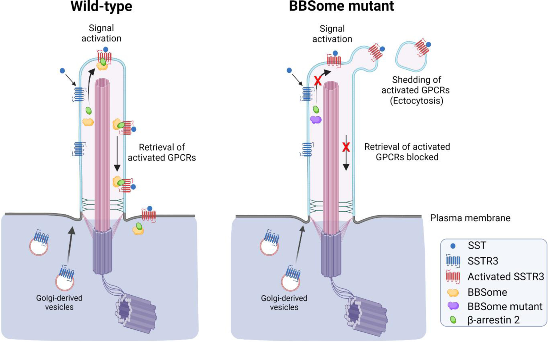
Extracellular vesicles (EVs) provide a mechanism for intercellular communication that transports complex signals in membrane delimited structures between cells, tissues and organisms. Cells secrete EVs of various subtypes defined by the pathway leading to release and by the pathological condition of the cell. Cilia are evolutionarily conserved organelles that can act as sensory structures surveilling the extracellular environment. Here we discuss the secretory functions of cilia and their biological implications. Studies in multiple species - from the nematode Caenorhabditis elegans and the chlorophyte alga Chlamydomonas reinhardtii to mammals - have revealed that cilia shed bioactive EVs (ciliary EVs or ectosomes) by outward budding of the ciliary membrane. The content of ciliary EVs is distinct from that of other vesicles released by cells. Peptides regulate numerous aspects of metazoan physiology and development through evolutionarily conserved mechanisms. Intriguingly, cilia-derived vesicles have recently been found to mediate peptidergic signaling. C. reinhardtii releases the peptide α-amidating enzyme (PAM), bioactive amidated products and components of the peptidergic signaling machinery in ciliary EVs in a developmentally regulated manner. Considering the origin of cilia in early eukaryotes, it is likely that release of peptidergic signals in ciliary EVs represents an alternative and ancient mode of regulated secretion that cells can utilize in the absence of dedicated secretory granules.

Keywords: Amidation; Chlamydomonas; Cilia; Ectosome; Peptidergic Signaling.


Conflict of interest statement

Conflict of Interest Statement

The authors declare no competing interests.

Figures

Figure 1 -
Figure 1 -. Extracellular vesicles of different origin.
The generation of three different types of extracellular vesicle based on their origin is illustrated. a) Ciliary extracellular vesicles. The ciliary membrane is very different from the plasma membrane in terms of both its lipid and protein content. Membrane proteins are transported to cilia via Golgi-derived vesicles or through lateral diffusion from the plasma membrane. Outward budding of the ciliary membrane forms a microdomain that contains membrane and soluble proteins that are present in the ciliary matrix. Fission of the ciliary membrane generates ciliary ectosomes, also known as cilia-derived vesicles, which carry specific protein cargoes that transmit signals and enzymatic activities over long distances. Importantly, ectosome content is controlled by regulated entry of proteins into cilia and by their subsequent sorting to sites of ectosome release. b) Ectosomes/microvesicles. These EVs form by outward budding of the plasma membrane. Receptors, membrane and soluble proteins, lipids and nucleic acids accumulate at the cytosolic face of the outwardly budding plasma membrane; subsequent fission leads to the formation of ectosomes. Ectosomes are larger (100 nm-1 µm in diameter) then exosomes, but surface receptors and membrane proteins (red and blue) share the same topology in both structures. c) Exosomes. Surface receptors and membrane proteins (red and green) internalize through the endosomal pathway. Inward budding of the endosomal membrane leads to the production of various intraluminal vesicles (ILVs), and thus the formation of multivesicular bodies (MVBs); MVBs can be also be formed from Golgi-derived vesicles. Subsequently, fusion of MVBs with the plasma membrane releases these ILVs into the extracellular milieu where they are termed exosomes. Exosomes are small (50–100 nm diameter) membrane limited structures that carry soluble proteins, membrane proteins and surface receptors, lipids and nucleic acid cargoes that are largely different from those of the endocytic system. Illustrations were created with BioRender.com.
Figure 2 -
Figure 2 -. Ectosome formation on cilia
a) Three optical sections of a single C. reinhardtii cell showing the two ~12 µm cilia (one is indicated by an arrow); differential interference contrast microscopy. b) Thin-section electron micrograph through a C. reinhardtii cilium. The transition zone gate and a nascent ectosome budding from the ciliary membrane are indicated. c) Section through a pellet of isolated ectosomes derived from the cilia of mating C. reinhardtii gametes. These structures may be purified in sufficient quantity for detailed biochemical and functional analysis. d) Electron micrograph of the neuroepithelium from the third ventricle of a mouse. A single ectosome budding from the surface of a primary cilium and the basal body that templates cilia formation are indicated. A second orthogonally oriented primary cilium in an adjacent cell is visible in cross section. Bars represent 5 µm (a), 250 nm (b), and 500 nm (c and d).
Figure 3 -
Figure 3 -. Regulated release of PAM and amidated products in ciliary ectosomes.
The secretion of PAM and amidated peptide products in ectosomes released from the cilia of mating gametes but not those of vegetative cells is illustrated. The left panel show the presence of flagellar membrane glycoprotein 1 (FMG1) and PAM in vegetative cell cilia and the release of FMG1, but not PAM, in ciliary ectosomes. The PAM luminal domain is exposed to the extracellular environment while its cytosolic domain is tethered to the axoneme through unknown interactions (green box). The right panel shows the release of PAM, proGATI amidated product and FMG1 in ectosomes from cilia of mating gametes. During mating, PAM and proGATI precursor are trafficked via Golgi-derived vesicles to the ciliary membrane (black arrow). However, whether the C-terminal amidation of proGATI takes place in Golgi-derived vesicles or on the ciliary membrane or even the ectosomal surface is currently unknown. The proGATI-amide product mediates chemotactic responses in both minus and plus gametes, however the signaling pathway has not yet been determined. Illustrations were created with BioRender.com.
Figure 4 -
Figure 4 -. Shedding of GPCRs in ciliary ectosomes.
Illustration of somatostatin receptor (SSTR3) signaling in primary cilia of wild-type and BBSome mutant cells. SSTR3 is a G-protein coupled receptor (GPCR) localized to primary cilia of hippocampal neurons and contains a motif that interacts with components of the BBSome complex [130] (reviewed in [131]). In wild-type cells, upon receptor activation following binding of somatostatin ligand (SST), SSTR3 is retrieved back into the cell body with the assistance of β-arrestin 2 and the BBSome protein complex (left panel). However, when the retrieval machinery is absent or compromised, as occurs in a BBSome mutant that lacks the BBS2 and BBS4 subunits, activated SSTR3 is shed from the ciliary tip by ectocytosis (right panel). A similar release process occurs in cells lacking β-arrestin 2, Arl6 (an Arf-like GTPase) or IFT27, which act as BBSome regulators [14]. For some GPCRs, such as NPY2R, that lack a retrieval motif, cells employ ectocytosis as a primary mechanism to remove activated receptors from cilia, thus reducing receptor signaling and thereby maintaining cellular homeostasis. Illustrations were created with BioRender.com.

References

    1. Wood CR, Huang K, Diener DR, Rosenbaum JL, The cilium secretes bioactive ectosomes, Curr. Biol 23 (2013), 906–911. - PMC - PubMed
    1. Wood CR, Rosenbaum JL, Ciliary ectosomes: transmissions from the cell’s antenna, Trends Cell Biol 25 (2015), 276–285. - PMC - PubMed
    1. Woith E, Fuhrmann G, Melzig MF, Extracellular vesicles-connecting kingdoms, Int. J. Mol. Sci 20 (2019), 5695. - PMC - PubMed
    1. Wang J, Silva M, Haas LA, Morsci NS, Nguyen KCQ, Hall DH, Barr MM, elegans C ciliated sensory neurons release extracellular vesicles that function in animal communication, Curr. Biol 24 (2014), 519–525. - PMC - PubMed
    1. Wang J, Barr MM, Cell-cell communication via ciliary extracellular vesicles: clues from model systems, Essays Biochem 62 (2018), 205–213. - PMC - PubMed

Publication types

LinkOut - more resources