Review
. 2022 May;298(5):101866.
doi: 10.1016/j.jbc.2022.101866. Epub 2022 Mar 26.

Understanding the physical basis of memory: Molecular mechanisms of the engram

Affiliations
Review

Understanding the physical basis of memory: Molecular mechanisms of the engram

Clara Ortega-de San Luis et al. J Biol Chem. 2022 May.

Erratum in

Abstract

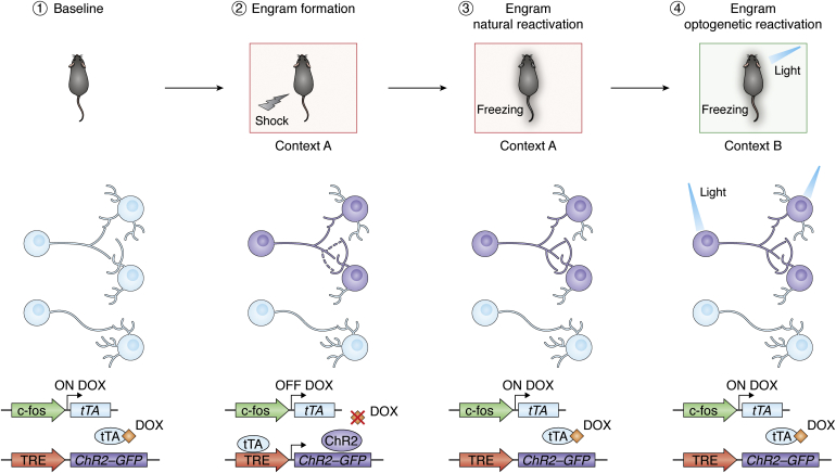
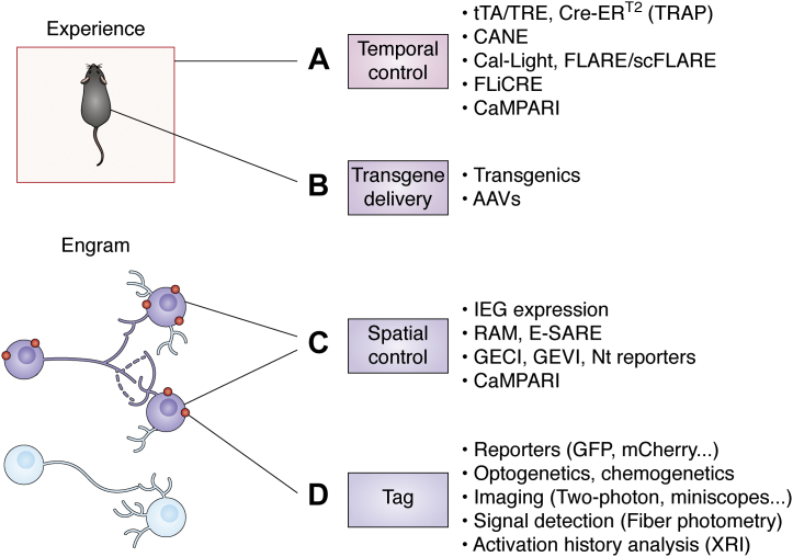
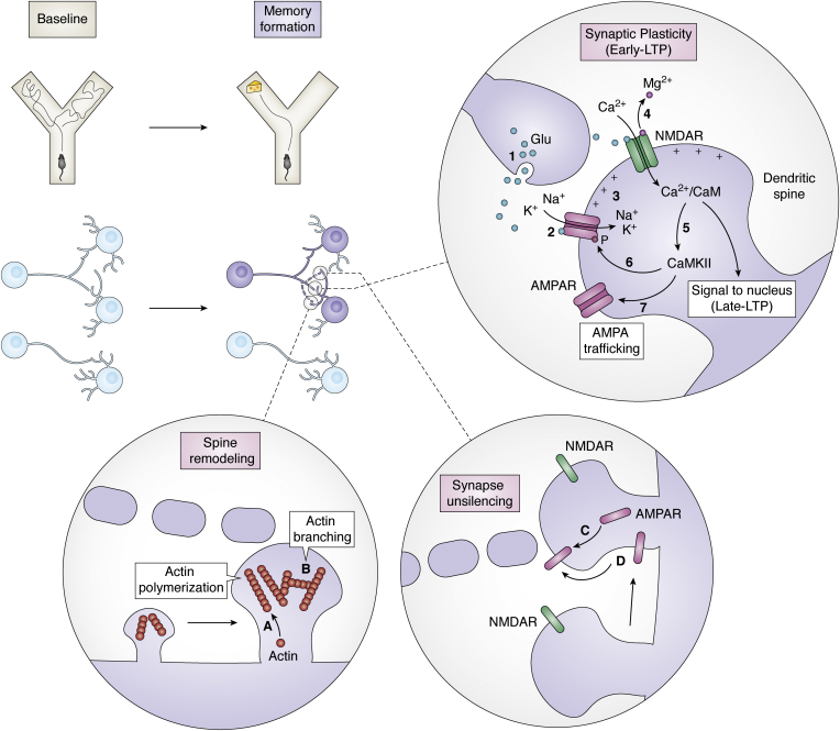
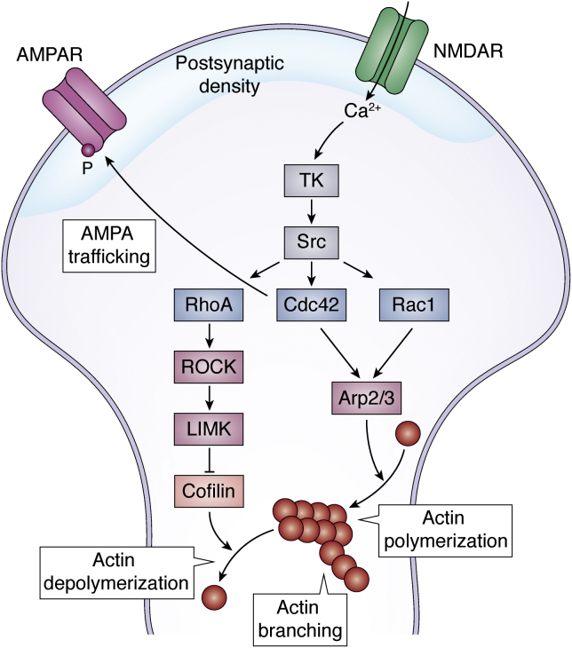
Memory, defined as the storage and use of learned information in the brain, is necessary to modulate behavior and critical for animals to adapt to their environments and survive. Despite being a cornerstone of brain function, questions surrounding the molecular and cellular mechanisms of how information is encoded, stored, and recalled remain largely unanswered. One widely held theory is that an engram is formed by a group of neurons that are active during learning, which undergoes biochemical and physical changes to store information in a stable state, and that are later reactivated during recall of the memory. In the past decade, the development of engram labeling methodologies has proven useful to investigate the biology of memory at the molecular and cellular levels. Engram technology allows the study of individual memories associated with particular experiences and their evolution over time, with enough experimental resolution to discriminate between different memory processes: learning (encoding), consolidation (the passage from short-term to long-term memories), and storage (the maintenance of memory in the brain). Here, we review the current understanding of memory formation at a molecular and cellular level by focusing on insights provided using engram technology.

Keywords: code; engram; memory; molecular mechanism; plasticity; synapse.


Conflict of interest statement

Conflict of interest The authors declare that they have no conflicts of interest with the contents of this article.

Figures

Figure 1
Figure 1
Engram labeling technology. (1) The immediate early gene c-fos promoter drives the expression of tTA. Doxycycline (DOX), delivered through diet, prevents tTA-TRE element binding. (2) An electric shock is delivered when the animal is in a particular context (context A). An engram for that episodic experience is formed (purple cells). In the absence of DOX, engram neurons that are active during the encoding of that fear memory express channelrhodopsin-2 (ChR2) transgene and GFP reporter. (3) Return to context A will reactivate the engram and will induce a freezing response in the animal—behavior associated with fear. (4) The engram cells tagged with ChR2 can be optogenetically reactivated by delivering light into the brain of the animal. This artificial recall of the memory induces freezing behavior in the animal in a neutral context B. TRE, tetracycline response element; tTA, tetracycline transactivator.
Figure 2
Figure 2
The expanded engram toolbox. Engram cell tagging has been achieved with several strategies or tools that can be used individually or combined. A, temporal control allows labeling engrams responsible for encoding a particular experience and can be achieved with different strategies. The tTA/TRE or Cre-ERT2 (targeted recombination in active populations [TRAP]) genetic strategies are temporally controlled by the delivery of doxycycline or tamoxifen, respectively. The capturing activated neuronal ensembles (CANE) technology allows high temporal precision by engineering viruses to specifically infect activated neurons. Other tools (Cal-Light, Fast light–regulated and activity-regulated expression [FLARE] and its improved version single-chain FLARE [scFLARE], and fast light–regulated and calcium-regulated expression [FLiCRE]) rely on the combination of two requirements to achieve temporal control: increase in intracellular Ca2+ and delivery of light. A similar strategy based on the coincidence of activity and light is used by CaMPARI to label only activated cells. B, transgenes can be delivered by generating transgenic mice models or by the use of vectors such as adeno-associated viruses (AAVs). C, only activated cells (engram cells, purple) are tagged; thanks to several spatial control strategies. The expression of immediate early genes (IEGs) such as c-fos can be used to manipulate only activated cells. Engineered artificial promoters have been also used in the robust activity marking (RAM) or the enhanced synaptic activity–regulated element (E-SARE) systems. Intracellular Ca2+ levels are detected by genetically encoded calcium or voltage indicators (GECI and GEVI, respectively) as well as reporters that detect specific neurotransmitter (Nt) release. CaMPARI is a fluorescent indicator that responds to intracellular Ca2+ levels. D, engram cells can be tagged with reporters (e.g., GFP or mCherry fluorescent reporters). They can also be tagged with tools that will allow future manipulation by light (optogenetics) or by drugs (chemogenetics). They can be imaged with several techniques such as two-photon or head-mounted miniature microscopes (miniscopes). Their activity can be monitored by techniques that allow activity readings such as fiber photometry. Activation history over time can be investigated by the use of the expression recording island (XRI) technology. CaMPARI, calcium-modulated photoactivatable ratiometric integrator; GECI, genetically encoded calcium indicator; GEVI, genetically encoded voltage indicator; tTA, tetracycline transactivator.
Figure 3
Figure 3
Molecular mechanisms of learning. When an animal encodes a new memory (such as the encounter of food in a particular arm of a Y-maze, upper panel), a subset of neurons gets activated and an engram is formed (purple neurons, middle panel). Certain synapses in between engram cells (dotted lines) undergo synaptic plasticity changes (early long-term potentiation [LTP]). The (1) neurotransmitter glutamate (Glu) is released from the presynaptic neuron, Glu binds to (2) AMPARs in the postsynaptic membrane, at the level of the dendritic spine, allowing K+ and Na+ to enter the postsynaptic neuron and depolarizing it (3). The positive charges inside the postsynaptic neurons allow the release of the Mg2+ ion from the NMDARs and if the Glu release is sustained enough, it will open the channel (4). Ca2+ enters the postsynaptic neuron, activating the (5) Ca2+/calmodulin-dependent protein kinase II (CaMKII). CaMKII phosphorylates AMPARs (6) to increase their sensitivity to Glu and drives more active channels to the membrane, AMPA trafficking (7). The number and shape of dendritic spines also get modified during learning. Spine remodeling to strengthen the synapse involves cytoskeletal modification such as (A) actin polymerization and (B) actin branching. Engram cells become unsilenced by the trafficking of AMPAR to the synapses by (C) secretion from the intracellular pool and (D) diffusion from other membrane areas. AMPAR, α-amino-3-hydroxy-5-methylisoxazole-4-propionic acid receptor; NMDAR, N-methyl-d-aspartate receptor.
Figure 4
Figure 4
Cytoskeletal modifications after learning. The increase in intracellular Ca2+ provoked by neuronal activation activates tyrosin kinases (TKs) and Src kinases. These activate members of the Rho family small GTPases: RhoA, Cdc42, and Rac1. RhoA activates ROCK and subsequently LIMK kinases, which in turn inhibits effector cofilin and eventually inhibits actin depolymerization. Rac1 and Cdc42 activate Arp2/3, an actin nucleation factor that induces actin polymerization, the elongation of actin filaments, and actin branching, and the formation of new ramifications in the actin filaments. Rho GTPases also anchor, stabilize, phosphorylate, and insert AMPARs to the postsynaptic density area of the membrane. AMPAR, α-amino-3-hydroxy-5-methylisoxazole-4-propionic acid receptor; LIMK, LIM-domain kinases; NMDAR, N-methyl-d-aspartate receptor; Rac1, Ras-related C3 botulinum toxin substrate 1; RhoA, Ras homologous member A; ROCK, Rho-associated coiled-coil kinase.
Figure 5
Figure 5
Molecular mechanisms of consolidation. After the encoding of a memory (the encounter of food in a Y-maze, upper panel), memory undergoes a series of processes that allow its consolidation into long-term memories resistant to the passage of time. Engram synapses (purple, middle panel) undergo synaptic plasticity changes (late phase of long-term potentiation [or late-LTP]). Triggered by the intracellular increase in Ca2+, adenylyl cyclase is activated and the intracellular concentration of cAMP increases. The cAMP increase triggers the activation of protein kinase A (PKA) and mitogen-activated protein kinases (MAPKs). PKA translocates to the nucleus, phosphorylates the transcription factor CREB, and ultimately triggers the transcription of genes containing CREB-responsive element (CRE), such as immediate early genes (c-fos, Arc, or Zif268), kinases such as Ca2+/calmodulin-dependent protein kinase II (CaMKII) and protein kinase Mzeta (PKMζ). In the dendrites, protein synthesis occurs locally by ribosomes translating mRNAs in a localized manner in the dendritic spines.

References

    1. Kandel E.R., Mack S., Jessell T.M., Schwartz J.H., Siegelbaum S.A., Hudspeth A.J. McGraw-Hill Education; New York, NY: 2013. Principles of Neural Science, Fifth Edition.
    1. Tulving E. Episodic memory: From mind to brain. Annu. Rev. Psychol. 2002;53:1–25. - PubMed
    1. Dudai Y., Roediger H.L., Tulving E. In: Science of Memory: Concepts. Roedinger H.L., Dudai Y., Fitzpatrick S.M., editors. Oxford University Press; New York, NY: 2007. Memory concepts; pp. 1–9.
    1. McGaugh J.L. Memory - a century of consolidation. Science. 2000;287:248–251. - PubMed
    1. Schacter D.L., Tulving E. MIT Press; Cambridge, MA: 2020. Memory Systems 1994.

Publication types