. 2022 Mar;10(3):e004026.
doi: 10.1136/jitc-2021-004026.

eEF2K promotes PD-L1 stabilization through inactivating GSK3β in melanoma

Affiliations

eEF2K promotes PD-L1 stabilization through inactivating GSK3β in melanoma

Xisha Chen et al. J Immunother Cancer. 2022 Mar.

Abstract

Background: Immune checkpoint blockade (ICB) targeting programmed death ligand-1 (PD-L1)/programmed cell death protein-1 (PD-1) pathway has become an attractive strategy for cancer treatment; however, unsatisfactory efficacy has limited its clinical benefits. Therefore, a more comprehensive understanding of the regulation of PD-L1 expression is essential for developing more effective cancer immunotherapy. Recent studies have revealed the important roles of eukaryotic elongation factor 2 kinase (eEF2K) in promoting epithelial-mesenchymal transition (EMT), angiogenesis, tumor cell migration and invasion; nevertheless, the exact role of eEF2K in the regulation of tumor immune microenvironment (TIME) remains largely unknown.

Methods: In this study, we used a cohort of 38 patients with melanoma who received anti-PD-1 treatment to explore the association between eEF2K expression and immunotherapy efficacy against melanoma. Immunoprecipitation-mass spectrometry analysis and in vitro assays were used to examine the role and molecular mechanism of eEF2K in regulating PD-L1 expression. We also determined the effects of eEF2K on tumor growth and cytotoxicity of CD8+ T cells in TIME in a mouse melanoma model. We further investigated the efficacy of the eEF2K inhibition in combination with anti-PD-1 treatment in vivo.

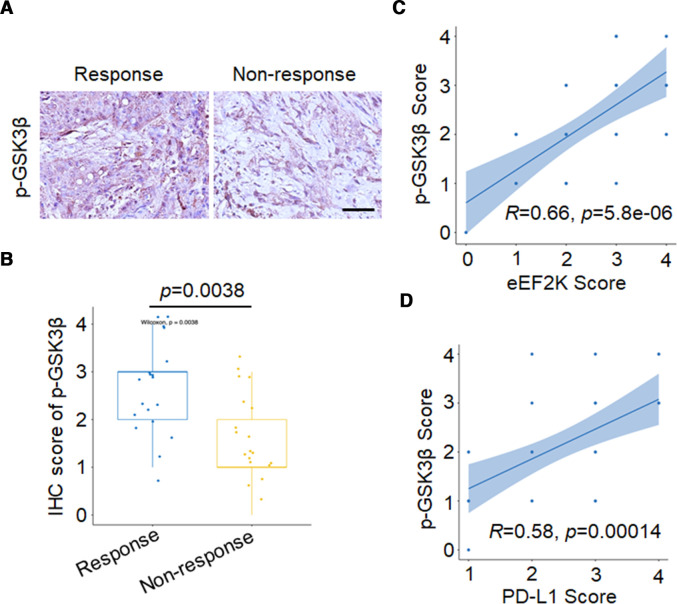
Results: High eEF2K expression is correlated with better therapeutic response and longer survival in patients with melanoma treated with PD-1 monoclonal antibody (mAb). Moreover, eEF2K protein expression is positively correlated with PD-L1 protein expression. Mechanistically, eEF2K directly bound to and inactivated glycogen synthase kinase 3 beta (GSK3β) by phosphorylating it at serine 9 (S9), leading to PD-L1 protein stabilization and upregulation, and subsequently tumor immune evasion. Knockdown of eEF2K decreased PD-L1 expression and enhanced CD8+ T cell activity, thus dramatically attenuating murine B16F10 melanoma growth in vivo. Clinically, p-GSK3β/S9 expression is positively correlated with the expressions of eEF2K and PD-L1, and the response to anti-PD-1 immunotherapy. Furthermore, eEF2K inhibitor, NH125 treatment or eEF2K knockdown enhanced the efficacy of PD-1 mAb therapy in a melanoma mouse model.

Conclusions: Our results suggest that eEF2K may serve as a biomarker for predicting therapeutic response and prognosis in patients receiving anti-PD-1 therapy, reveal a vital role of eEF2K in regulating TIME by controlling PD-L1 expression and provide a potential combination therapeutic strategy of eEF2K inhibition with ICB therapy.

Keywords: biomarkers, tumor; drug therapy, combination; immunotherapy; melanoma; tumor microenvironment.


Conflict of interest statement

Competing interests: None declared.

Figures

Figure 1
Figure 1
High eEF2K expression positively correlates with immunotherapeutic benefits and PD-L1 level in patients with melanoma. (A) Kaplan-Meier overall survival curves in patients with melanoma with or without response to PD-1 mAb therapy. (B) Kaplan-Meier plots of the overall survival rates in PD-1 blockade-treated patients with melanoma with high (IHC overall score is between 3 and 4) or low (IHC overall score is between 0 and 2) expression of eEF2K. (C) IHC staining of 38 human melanoma specimens was performed with the eEF2K antibody. (D) Representative IHC staining images of eEF2K from respond group and non-respond group are shown, scale bar is 50 μm. (E) Schematic diagram for eEF2K as a predictor for the efficacy and prognosis of anti-PD-L1/PD-1 therapy (good prognosis means improvement or full resolution, poor prognosis means no recovery or death due to melanoma). (F, G) The positive correlation between eEF2K and PD-L1 expression in human melanoma specimens. (F) The IHC staining images, and the correlation analyses were performed (G). eEF2K, eukaryotic elongation factor 2 kinase; IHC, immunohistochemical; mAb, monoclonal antibody; PD-1, programmed cell death protein-1; PD-L1, programmed death ligand-1.
Figure 2
Figure 2
eEF2K upregulates PD-L1 expression by inhibiting its proteasome-mediated degradation. Immunoblotting analyses were performed with the indicated antibodies (A–G). (A) Human melanoma cell lines were transfected with a non-targeting siRNA or homo sapiens eEF2K-targeted siRNAs for 72 hours. (B) B16F10 cells were transfected with a non-targeting siRNA or mouse eEF2K-targeted siRNAs for 72 hours. (C) A375, SK-5, SK-28 cells were transfected with a control plasmid or a Flag-eEF2K plasmid for 48 hours. (D) PD-L1-depleted A375 cells with reconstituted expression of HA-PD-L1 were transfected with a control plasmid or a Flag-eEF2K plasmid for 48 hours. (E) A375 cells with or without eEF2K knockdown were treated with CHX (50 µg/mL) for the indicated times. (F) A375 cells with or without eEF2K knockdown were treated with or without MG132 (10 µM) for 4 hours. (G) A375 cells with or without eEF2K knockdown were incubated with recombinant human PD-1 Fc protein and then antihuman Alexa Fluor 488 dye. Immunofluorescence assays were performed to detect PD-1 binding intensity. CHX, cycloheximide; eEF2K, eukaryotic elongation factor 2 kinase; IHC, immunohistochemical; mAb, monoclonal antibody; PD-1, programmed cell death protein-1; PD-L1, programmed death ligand-1; siRNA, small interfering RNA.
Figure 3
Figure 3
Physical interaction of eEF2K with GSK3β. (A) Proteins from the immunoprecipitates assay were separated by polyacrylamide gel eletrophoresis and detected by sliver staining (left). The proteins immunoprecipitated by anti-Flag antibody were further analyzed by mass spectrometry, and several reported eEF2K-interacting proteins like eEF2 and Homer1 were also identified in our Immunoprecipitation-mass spectrometry (IP-MS) analysis (right). Immunoblotting analyses were performed with the indicated antibodies (B, C and E). (B) HEK293T cells were transfected with Flag-eEF2K and HA-GSK3β plasmids, and then subjected to immunoprecipitation with anti-Flag or anti-HA antibodies. The lysates and immunoprecipitates were then blotted. (C) Immunoprecipitation analysis with the indicated antibodies was performed to detect endogenous eEF2K and GSK3β interaction in A375 cells. (D) eEF2K and GSK3β are co-localized in the cytoplasm of A375 cells. The cellular location of eEF2K and GSK3β was examined by immunofluorescence staining. DAPI was used to stain the DNA. Scale bar, 7.5 μm. (E) Purified recombinant GST-eEF2K interacts with GSK3β. GST-eEF2K and GST proteins were pulled down with glutathione beads. GSK3β was detected by WB. DAPI, 4,6-diamidino-2-phenylindole; eEF2K, eukaryotic elongation factor 2 kinase; GSK3β, glycogen synthase kinase 3 beta.
Figure 4
Figure 4
eEF2K phosphorylates GSK3β at serine 9. Immunoblotting analyses were performed with the indicated antibodies. (A) A375 or SK-5 cells were transfected with a non-targeting siRNA or eEF2K-targeted siRNAs for 72 hours. ∗∗P<0.01; ∗∗∗p<0.001. (B) A375 cells with or without GSK3β depletion and reconstituted expression of wild-type (WT) HA-rGSK3β or HA-rGSK3β S9A mutant were transfected with a control plasmid or a Flag-eEF2K plasmid for 48 hours. Immunoprecipitation analysis was performed. (C) Purified GSK3β was incubated with active eEF2K kinase. Western blot analysis was performed with the p-GSK3β/S9 antibody. eEF2K, eukaryotic elongation factor 2 kinase; GSK3β, glycogen synthase kinase 3 beta; siRNA, small interfering RNA.
Figure 5
Figure 5
eEF2K-mediated GSK3β inactivation promotes the stabilization of PD-L1. Immunoblotting analyses were performed with the indicated antibodies. (A) Flag or Flag-eEF2K-expressing A375 or SK-5 cells were transfected with or without HA-rGSK3β S9A for 48 hours. (B) A375 or SK-5 cells with or without eEF2K knockdown were transfected with a non-targeting siRNA or GSK3β-targeted siRNA for 72 hours. ∗P<0.05; ∗∗p<0.01; ∗∗∗p<0.001. eEF2K, eukaryotic elongation factor 2 kinase; GSK3β, glycogen synthase kinase 3 beta; PD-L1, programmed death ligand-1; siRNA, small interfering RNA.
Figure 6
Figure 6
Depletion of eEF2K suppresses B16F10 xenograft tumor growth and promotes T cell activity. Ctrl and two sheEF2K (#1 and #2)-transfected B16F10 cells were injected subcutaneously into 6-week-old male Balb/c nude mice and C57BL/6 mice, the tumor sizes were measured on the days as indicated. (A, B) Subcutaneous tumors from B16F10 xenograft Balb/c nude mice were excised and photographs were taken at the termination of the experiment (A) and tumor weights were measured (B). Tumor inhibition rate (%, TIR), 67.5%. (C, D) Subcutaneous tumors from B16F10 xenograft C57BL/6 mice were excised and photopraphs were taken at the termination of the experiment (C) and xenograft tumor weights were measured (D). TIR, 80.7%. Data represents the mean±SD of tumor weights of each group (n=6). (E) FACS of CD8+ in CD3+ and GZMB+CD8+ in CD8+ TILs from B16F10 xenografts. (F) Representative images of IF staining of eEF2K, p-GSK3β/S9 and PD-L1 of sheEF2K and Ctrl B16F10 xenografts. (G–K) ShNT and sheEF2K-transfected B16F10 cells were injected subcutaneously into 6-week-old male C57BL/6 mice, and received CD8α mAb treatment or IgG isotype control. (G) A schematic view of the treatment plan. (H) Photopraphs of mice tumors of each group at the end of the experiment. (I) Curves of tumor growth. (J) Plots for tumor weight. (K) FACS of CD8+ in CD3+ TILs from B16F10 xenografts. ∗P<0.05; ∗∗p<0.01; ∗∗∗p<0.001. eEF2K, eukaryotic elongation factor 2 kinase; GSK3β, glycogen synthase kinase 3 beta; IF, immunofluorescence; PD-L1, programmed death ligand-1; mAb, monoclonal antibody; TIL, tumor-infiltrating lymphocyte.
Figure 7
Figure 7
High p-GSK3β/S9 expression is positively correlated with response to anti-PD-1 immunotherapy, eEF2K and PD-L1 levels in samples from patients with melanoma. Representative images (A) and quantitative expression (B) of immunohistochemistry staining of p-GSK3β/S9 in patients with melanoma with or without response to PD-1 mAb therapy. The positive correlation between p-GSK3β/S9 and eEF2K (C), PD-L1 (D) in human melanoma specimens. eEF2K, eukaryotic elongation factor 2 kinase; GSK3β, glycogen synthase kinase 3 beta; IHC, immunohistochemical; mAb, monoclonal antibody; PD-1, programmed cell death protein-1; PD-L1, programmed death ligand-1.
Figure 8
Figure 8
eEF2K inhibition synergistically enhanced the therapeutic efficacy of PD-1 blockade in vivo. (A–E) C57BL/6 mice were implanted with B16F10 cells and co-treated with NH125 and PD-1 mAb. (A) A schematic view of the treatment protocol. (B) Photopraphs of B16F10 tumors harvested after euthanizing the mice. n=6 for each group. (C) The tumor growth of B16F10 cells in NH125 and/or anti-PD-1 antibody-treated C57BL/6 mice. (D) Plots for tumor weight. (E) CD8α and granzyme B in mouse tumor tissues of each group were determined by immunofluorescence. (F–I) C57BL/6 mice were implanted with Ctrl or sheEF2K-transfected B16F10 cells, and received PD-1, CD8α mAb treatment or IgG isotype control. (F) A schematic view of the treatment plan. (G) Photopraphs of mice tumors of each group at the end of the experiment. (H) Curves of tumor growth. (I) Plots for tumor weight. ∗∗P<0.01; ∗∗∗p<0.001. (J) A proposed model for eEF2K-induced GSK3β-phosphorylation-dependent PD-L1 stabilization and immunoregulation in melanoma. eEF2K overexpression in cancer cells phosphorylates GSK3β at serine 9 for GSK3β inactivation, leading to PD-L1 stabilization, enhanced PD-1 interaction and subsequent immunosuppressive microenvironment as a consequent. Therapeutic depletion or inhibition of eEF2K maintains GSK3β activity for phosphorylation-dependent proteasome degradation of PD-L1, thereby decreasing the cancer cells PD-L1 expression level and synergistically enhancing the therapeutic efficacy of PD-1 mAb therapy. eEF2K, eukaryotic elongation factor 2 kinase; GSK3β, glycogen synthase kinase 3 beta; mAb, monoclonal antibody; PD-1, programmed cell death protein-1; PD-L1, programmed death ligand-1.

References

    1. Freeman GJ, Long AJ, Iwai Y, et al. . Engagement of the PD-1 immunoinhibitory receptor by a novel B7 family member leads to negative regulation of lymphocyte activation. J Exp Med 2000;192:1027–34. 10.1084/jem.192.7.1027 - DOI - PMC - PubMed
    1. Chen L, Han X. Anti-PD-1/PD-L1 therapy of human cancer: past, present, and future. J Clin Invest 2015;125:3384–91. 10.1172/JCI80011 - DOI - PMC - PubMed
    1. Dong H, Strome SE, Salomao DR, et al. . Tumor-associated B7-H1 promotes T-cell apoptosis: a potential mechanism of immune evasion. Nat Med 2002;8:793–800. 10.1038/nm730 - DOI - PubMed
    1. Topalian SL, Drake CG, Pardoll DM. Immune checkpoint blockade: a common denominator approach to cancer therapy. Cancer Cell 2015;27:450–61. 10.1016/j.ccell.2015.03.001 - DOI - PMC - PubMed
    1. Sharma P, Allison JP. The future of immune checkpoint therapy. Science 2015;348:56–61. 10.1126/science.aaa8172 - DOI - PubMed