Case Reports
. 2022 Mar 28;8(1):36.
doi: 10.1038/s41394-022-00505-x.

Iatrogenic dorsal spinal cord herniation and repair with clip-based expansile duraplasty: a case report

Affiliations
Case Reports

Iatrogenic dorsal spinal cord herniation and repair with clip-based expansile duraplasty: a case report

Axumawi Gebreyohanes et al. Spinal Cord Ser Cases. .

Abstract

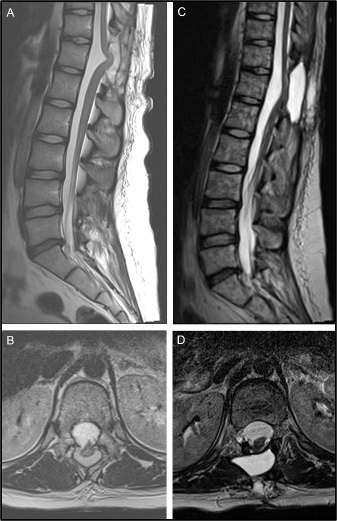
Introduction: Myelopathy arising due to dorsal herniation of the spinal cord is a rare phenomenon, particularly so in the thoracic region. Where cases of thoracic dorsal cord herniation have been reported, the aetiology has typically been non-iatrogenic.

Case presentation: We report the case of a paediatric oncology patient who presented with neurological deterioration secondary to thoracic dorsal spinal cord herniation, manifesting three months after laminectomy for biopsy of a spinal medulloblastoma lesion. We repaired the dural defect using non-penetrating titanium clips to create a secure expansile duraplasty, resulting in radiologically evident reduction of the cord herniation as well as corresponding clinical improvement.

Discussion: Thoracic dorsal spinal cord herniation is an extremely rare occurrence after spinal surgery. Non-penetrating titanium clips can be used to form a secure expansile duraplasty following reduction of the cord herniation. Successful repair of the dural defect re-anteriorises the cord and can confer neurological benefit.


Conflict of interest statement

The authors report no conflicts of interest and have no personal, financial or institutional interest in any drugs, materials or devices mentioned or described in this article.

Figures

Fig. 1
Fig. 1. Magnetic resonance imaging from time of initial clinical presentation.
Multiple spinal and cauda equina lesions are demonstrated on a sagittal whole-neuraxis slice (A): note also the distended bladder. The cord ends at L1. Cranial lesions are seen on parasagittal (B) and axial (C) slices. Selected lesions are highlighted by the white arrows.
Fig. 2
Fig. 2. Pre- and post-operative T2-weighted magnetic resonance imaging.
Sagittal (A) and axial (B) pre-operative images show T12 dorsal dural defect and associated thoracic spinal cord herniation, with a subsequently re-anteriorised cord following dural repair shown in sagittal (C) and axial (D) views.
Fig. 3
Fig. 3. Intraoperative microscopic images depicting reduction of thoracic cord herniation.
The spinal cord was found to be visibly herniated through a large dural defect (A). Arachnoid granulations were released via careful dissection (B), permitting re-anteriorisation of the cord (C). A dural patch was affixed to the surrounding dura using non-penetrating titanium clips (D), with fibrin sealant also used to complete dural repair (E).

References

    1. Heller RS, Hwang SW, Riesenburger RI. Dorsal cervical spinal cord herniation precipitated by kyphosis deformity correction for spinal cord tethering. World Neurosurg. 2017;100:709.e1–709.e4. doi: 10.1016/j.wneu.2017.02.020. - DOI - PubMed
    1. Abd Elwahab SM, O’Sullivan MJ. Spinal cord herniation after resection of cervical spinal neurofibroma with a unique presentation. Spine J. 2015;15:e1–e3. doi: 10.1016/j.spinee.2014.12.007. - DOI - PubMed
    1. Moriyama T, Tachibana T, Maruo K, Inoue S, Okada F, Yoshiya S, et al. Postoperative spinal cord herniation with pseudomeningocele in the cervical spine: a case report. Spine J. 2013;13:e43–e45. doi: 10.1016/j.spinee.2013.05.057. - DOI - PubMed
    1. Zakaria R, Ellenbogen JR, Grewal IS, Buxton N. Posterior spinal cord herniation: a novel occurrence following surgery for an intramedullary cyst at the thoracolumbar junction. Eur Spine J. 2013;22(SUPPL). 10.1007/s00586-012-2515-5 - PMC - PubMed
    1. Le TC, Grunch BH, Karikari IO, Mehta A, Owens T, Gottfried O, et al. Dorsal thoracic spinal cord herniation: report of an unusual case and review of the literature. Spine J. 2012;12:e9–e12. doi: 10.1016/j.spinee.2012.09.039. - DOI - PubMed

Publication types