. 2022 Mar 28;5(1):272.
doi: 10.1038/s42003-022-03186-2.

Molecular basis of specificity and deamidation of eIF4A by Burkholderia Lethal Factor 1

Affiliations

Molecular basis of specificity and deamidation of eIF4A by Burkholderia Lethal Factor 1

George W Mobbs et al. Commun Biol. .

Abstract

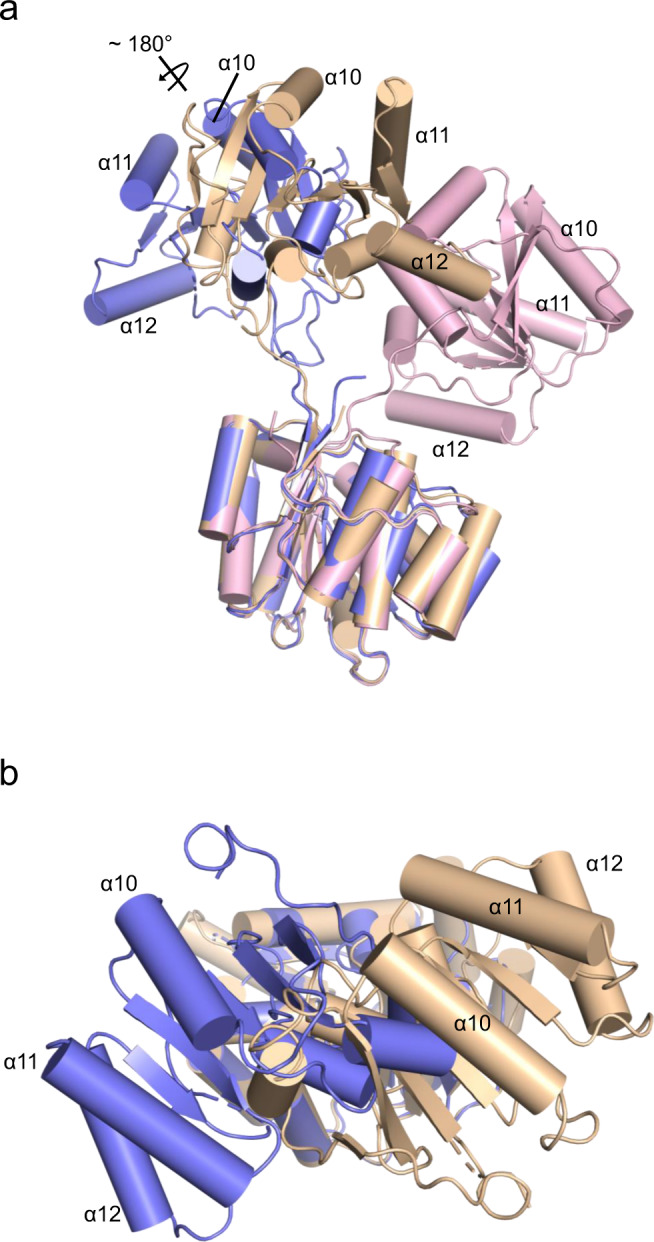
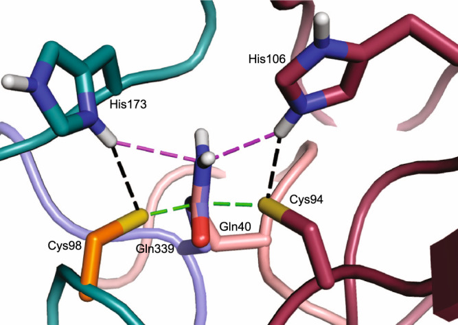
Burkholderia pseudomallei lethal factor 1 (BLF1) exhibits site-specific glutamine deamidase activity against the eukaryotic RNA helicase, eIF4A, thereby blocking mammalian protein synthesis. The structure of a complex between BLF1 C94S and human eIF4A shows that the toxin binds in the cleft between the two RecA-like eIF4A domains forming interactions with residues from both and with the scissile amide of the target glutamine, Gln339, adjacent to the toxin active site. The RecA-like domains adopt a radically twisted orientation compared to other eIF4A structures and the nature and position of conserved residues suggests this may represent a conformation associated with RNA binding. Comparison of the catalytic site of BLF1 with other deamidases and cysteine proteases reveals that they fall into two classes, related by pseudosymmetry, that present either the re or si faces of the target amide/peptide to the nucleophilic sulfur, highlighting constraints in the convergent evolution of their Cys-His active sites.


Conflict of interest statement

The authors declare no competing interests.

Figures

Fig. 1
Fig. 1. BLF1 C94S binds in the cleft between the two domains of eIF4A∆20.
a Stereo pair for cross-eyed viewing of the structure of the BLF1 C94S:eIF4A∆20 complex (PDB:7PQ0 (BLF1; maroon, eIF4A; blue)). b Surface representation of the N- and C-terminal domains of eIF4A. The two patches that interact with BLF1 C94S are shown in gold and orange, respectively with the remainder of the surface colored white. Gln339 of eIF4A is colored red. c Surface representation of the corresponding patches (gold and orange) on BLF1 that interact with eIF4A with the remainder of the surface colored gray. The view is rotated about the horizontal axis by 90° compared to the orientation of eIF4A shown in panel b so that the patches on BLF1 face the reader. d In a second view, modified by a rotation of 90° around the vertical axis, the surface of BLF1 flanking the active site is shown with the Cys/His pair colored green.
Fig. 2
Fig. 2. BLF1 C94S binds to a twisted conformation of the two eIF4A∆20 domains.
Superposition of three different eIF4A structures on the basis of their N-terminal domains reveals the quite different positions of the C-terminal domain. Helices α10, α11, and α12 of eIF4A are labeled to enable the different orientations to be seen. a In the apo yeast eIF4A, (PDB:1FUU; wheat) the two domains adopt an open conformation whilst a complex of yeast eIF4A with eIF4G (PDB:2VSX; pink) reveals a closed conformation for eIF4A. Human eIF4A in complex with BLF1 (PDB:7PQ0; blue) adopts a different open conformation in which the eIF4A domains are twisted compared to apo. The approximate axis of rotation about which the C-terminal domain of eIF4A twists in the complex with BLF1 is indicated. b A view of the superposition between the N-terminal domains of apo yeast eIF4A and Human eIF4A in complex with BLF1 (background), shown in wheat and blue respectively, highlighting the approximate 180° rotation relating their C-terminal domains in the foreground.
Fig. 3
Fig. 3. Active site of hybrid WT BLF1:eIF4A∆20 model.
In the hybrid WT BLF1: eIF4A model, the eIF4a substrate Gln339 carboxamide forms hydrogen bonds to the main chain nitrogen of BLF1 Ser92 and Cys94 (black dashes) and lies close to the main chain carbonyl of Tyr90. His106 Nε2 donates a hydrogen bond to the sulfur of Cys94 (black dashes) and lies 3.5 Å from the side chain amide nitrogen of Gln339 with the Cys94 sulfur 2.1 Å above the amide carbon (magenta dashes). The diagram was prepared using PyMOL (sulfur: yellow, oxygen: red, nitrogen: dark blue, carbon: (BLF1; maroon, eIF4A; blue).
Fig. 4
Fig. 4. The relative positions of the substrate and catalytic cysteines in WT BLF1:eIF4A∆20 model compared to other structures.
a Stereo view of the active site of WT BLF1:eIF4A∆20 complex (BLF1; maroon, eIF4A; blue) superimposed with that of the TEV C151A protease with its substrate (PDB:1LVB; yellow), and with the WT TEV protease (PDB:1LVM; green). The target carboxamide group of the side chain of Gln339 of eIF4A (blue) superposes well with the scissile peptide of the TEV substrate (yellow). The superposition shows that the nucleophilic cysteine of TEV and BLF1 approach the re face of their respective substrates. b Cys151 of TEV showing it adopts the gauche+ conformation. c Stereo view of the local superposition between the WT BLF1:eIF4A∆20 complex (BLF1; maroon, eIF4A; blue) and the Cif C117A:NEDD8 complex (PDB:4F8C (Cif; teal, Nedd8 substrate; pink)), and AvrPphB (PDB:1UKF; orange), maximizing the overlap of their Cys/His pairs. The superposition shows that the nucleophilic cysteine of Cif approaches the si face of Gln40 Nedd8 substrate, compared to the re face seen in the BLF1:eIF4A∆20 complex. d Cys98 of the Cif-like protease AvrPphB showing it adopts a gauche conformation. e Stereo view of the WT BLF1:eIF4A∆20 complex (BLF1; maroon, eIF4A; blue) superimposed with the papain-ZFLG inhibitor complex (PDB:1KHP; white), and with the papain-like protease, Amb an 11 (PDB:5EF4; purple) revealing the common position of the oxyanion. f Cys155 of the papain-like protease, Amb an 11 showing it adopts a gauche conformation.
Fig. 5
Fig. 5. Superposition of the catalytic histidine from the three families.
Local superposition of the catalytic histidine and cysteine residues from BLF1 (Cys94/His106; maroon), TEV (Cys151/His46; yellow), and CHYP Cif/ AvrPphB (Cys98/ His173; white). In both BLF1 and TEV, the imidazole rings occupy the same approximate position and form a hydrogen bond with their respective cysteine residues using the same, Nε2, Nitrogen, despite differences in the position of the main chain. In contrast, in Cif, the main chain approaches from a third location, to leave the imidazole ring of His173 occupying a similar position but utilizing the nitrogen Nδ1 to generate a hydrogen bond with its catalytic cysteine.
Fig. 6
Fig. 6. Comparisons of the active site architectures between deamidases attacking the re and si face of the target amide.
A superposition between complexes of the WT BLF1:eIF4A∆20 hybrid model (BLF1; maroon, eIF4A; blue) and Cif C117A:NEDD8 complex (PDB:4F8C (Cif; teal, NEDD8 substrate; pink))/ Cys98 of AvrPphB (PDB:1UKF; orange). The superposition is based on the glutamine side chain substrates to show the catalytically active Cys-His pairs are positioned on opposite faces of the amide substrate in a pseudo-mirror plane arrangement. Hydrogen bonds between the histidine imidazolium ring and the cysteine thiolate are shown in black dashed lines, with the distances between the thiolate and glutamine carboxamide carbon highlighted in green, and the imidazolate Nε2 hydrogen and the glutamine carboxamide nitrogen in magenta.

References

    1. Ray K, Marteyn B, Sansonetti PJ, Tang CM. Life on the inside: the intracellular lifestyle of cytosolic bacteria. Nat. Rev. Microbiol. 2009;7:333–340. - PubMed
    1. Wiersinga WJ, van der Poll T, White NJ, Day NP, Peacock SJ. Melioidosis: insights into the pathogenicity of Burkholderia pseudomallei. Nat. Rev. Microbiol. 2006;4:272–282. - PubMed
    1. Wiersinga WJ, et al. Melioidosis. Nat. Rev. Dis. Prim. 2018;4:17107. - PMC - PubMed
    1. Nandi T, et al. A genomic survey of positive selection in Burkholderia pseudomallei provides insights into the evolution of accidental virulence. PLoS Pathog. 2010;6:1000845. - PMC - PubMed
    1. Dance DAB. Ecology of Burkholderia pseudomallei and the interactions between environmental Burkholderia spp. and human–animal hosts. Acta Trop. 2000;74:159–168. - PubMed

Publication types