Clinical Trial
. 2022 Apr;28(4):780-788.
doi: 10.1038/s41591-022-01737-y. Epub 2022 Mar 28.

In vivo topical gene therapy for recessive dystrophic epidermolysis bullosa: a phase 1 and 2 trial

Affiliations
Clinical Trial

In vivo topical gene therapy for recessive dystrophic epidermolysis bullosa: a phase 1 and 2 trial

Irina Gurevich et al. Nat Med. 2022 Apr.

Abstract

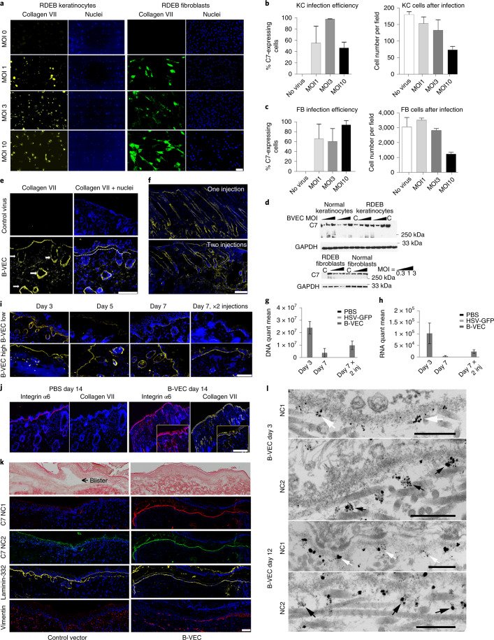
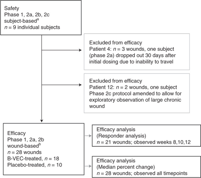
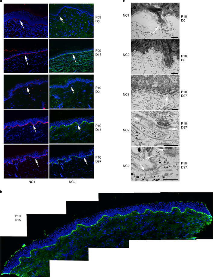
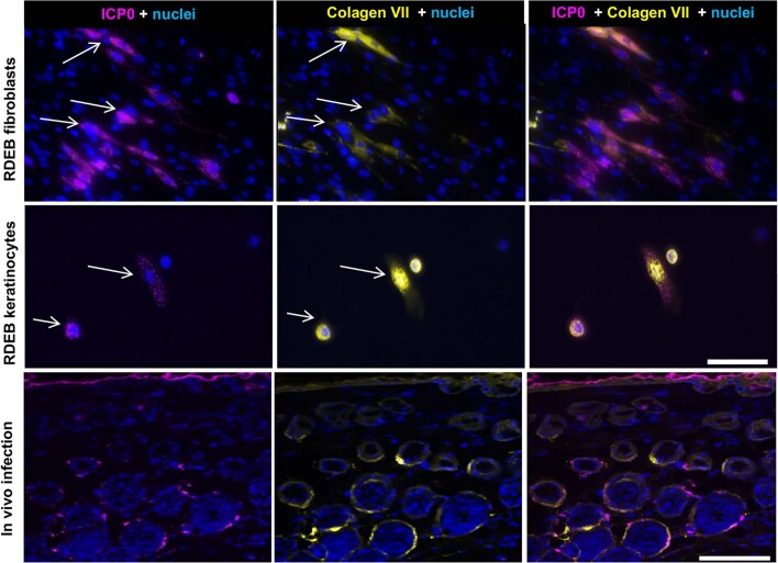
Recessive dystrophic epidermolysis bullosa (RDEB) is a lifelong genodermatosis associated with blistering, wounding, and scarring caused by mutations in COL7A1, the gene encoding the anchoring fibril component, collagen VII (C7). Here, we evaluated beremagene geperpavec (B-VEC), an engineered, non-replicating COL7A1 containing herpes simplex virus type 1 (HSV-1) vector, to treat RDEB skin. B-VEC restored C7 expression in RDEB keratinocytes, fibroblasts, RDEB mice and human RDEB xenografts. Subsequently, a randomized, placebo-controlled, phase 1 and 2 clinical trial (NCT03536143) evaluated matched wounds from nine RDEB patients receiving topical B-VEC or placebo repeatedly over 12 weeks. No grade 2 or above B-VEC-related adverse events or vector shedding or tissue-bound skin immunoreactants were noted. HSV-1 and C7 antibodies sometimes presented at baseline or increased after B-VEC treatment without an apparent impact on safety or efficacy. Primary and secondary objectives of C7 expression, anchoring fibril assembly, wound surface area reduction, duration of wound closure, and time to wound closure following B-VEC treatment were met. A patient-reported pain-severity secondary outcome was not assessed given the small proportion of wounds treated. A global assessment secondary endpoint was not pursued due to redundancy with regard to other endpoints. These studies show that B-VEC is an easily administered, safely tolerated, topical molecular corrective therapy promoting wound healing in patients with RDEB.


Conflict of interest statement

M.P.M. received funding from Krystal Biotech to conduct this study through a sponsored research award administered through the Stanford University Office of Research Management. M.P.M. is also an investigator for the following companies that are studying molecular corrective therapies for recessive dystrophic epidermolysis bullosa: Castle Creek Pharmaceuticals, Abeona Therapeutics, WINGS therapeutics and Phoenix Tissue Repair. A.P.S. owns stock in Krystal Biotech. P.A., P.P.Z. and S.O., as well as H.L., N.R., N.S., M.O. and S.M.K. are employees of Krystal Biotech. All other authors have no competing interests.

Figures

Fig. 1
Fig. 1. Collagen VII (C7) expression in primary RDEB patient cells, RDEB mice, and human RDEB xenografts on immunodeficient mice following B-VEC therapy.
a, RDEB keratinocytes and fibroblasts were infected in vitro with B-VEC vector at various ratios of cell to viral particle (MOI). Cells were fixed 48 h after infection and analyzed by indirect immunofluorescence microscopy (IDIF) to validate C7 expression in keratinocytes (kc; yellow) and fibroblasts (fb; green). Scale bar, 100 µm. b,c, Infection efficiency and live cell number in cultures after infection (n = 3 for each condition). d, Keratinocytes and fibroblasts were collected after various MOI (n = 3 for each condition) and analyzed by Western blot. C, control vector. e, Mice were injected intradermally at four separate sites on the back (one vehicle control site and three B-VEC treatment sites, one injection per site). Some mice received a second injection on day 3 at the same four sites. At day 3 after B-VEC treatment (dose of 4.6 x 107 p.f.u. per 50 µl per injection), RDEB mice injected intradermally with B-VEC vector had linear C7 basement membrane zone (BMZ) expression (yellow, dotted line) including in hair follicle basement membranes (arrows). Scale bar, 100 µm. f, At day 7, C7 expression (yellow) was analyzed using IDIF after one or two injections in RDEB mouse skin. Scale bar, 200 µm. g,h, COL7A1 DNA (g) and C7 transcript expression levels (h) were analyzed using qPCR or RT–qPCR, after one or two injections (dose of 4.6 x 107 p.f.u. per 50 µl per injection) in RDEB mouse skin, on day 3 and 7 after single or repeated injections (n = 9 injections, triplicate for each condition, per timepoint). i, Demonstration of C7 expression dose dependency in RDEB mouse skin for low-dose injections (top row) and high-dose injections (bottom row) at days 3, 5, and 7 after injection. The far right panels show C7 expression at high and low B-VEC doses 7 days after both injections. j, Heterozygous RDEB mice were treated with daily topical B-VEC or vehicle (PBS) applications on wounded skin for 5 days. Fourteen days after the first application, linear C7 expression using human-specific antibody is shown in yellow at the epidermal–dermal junction and hair follicle epithelial–dermal junctions, and co-localization with the BMZ marker α6 integrin staining is shown in red. The inserts in the right panels demonstrate higher magnification of the sample region. Four areas on the back were treated per mouse (1 control and 3 B-VEC) and four mice in total were tested. Scale bar, 100 µm. k, Xenografts comprising human RDEB fibroblasts and keratinocytes were treated by topical B-VEC and imaged 5 days later. The grafts were analyzed by light (first row) and immunofluorescence (rows 2–5) microscopy. Light microscopy demonstrated areas of dermal–epidermal blistering (left panel, arrow), which was not seen after B-VEC treatment (right panel). In the treated region, expression of the NC1 (red) and NC2 (green) domains of C7 co-localized with the control BMZ marker laminin-332 (yellow). Row 5 demonstrates the persistence and location of human fibroblasts in the dermis of the graft. Scale bar, 100 µm. Representative of eight grafts treated with B-VEC (mice) and 2 with placebo. l, Human RDEB skin xenografts were treated with topical B-VEC and analyzed after 3 and 12 days using immunoelectron microscopy with C7 NC1-directed antibodies (NP185), followed by gold nanoparticles (first, third rows; white arrows), and NC2-directed primary antibodies (LH24), followed by gold nanoparticles (second, fourth rows; black arrows). Error bars on all panels represent standard error of the mean of all replicates. Representative of eight grafts treated with B-VEC and two grafts treated with placebo. Scale bars, 300 nm. Data plots, including error bars and P values, were generated using GraphPad Prism v8.3.0.
Fig. 2
Fig. 2. Trial profile.
The safety population was evaluated considering an individual systemic patient response being the independent event (n = 9 patients). The efficacy assessments and analyses were conducted considering an individual wound response as the independent event (n = 28 wounds treated with B-VEC or placebo in the efficacy evaluable population: 18 B-VEC-treated wounds, 10 placebo-treated wounds). Responder analysis included patient wounds observed at weeks 8, 10 and 12 (21 wounds). aNine individuals of whom three participated in both 2a and 2b. One of nine individuals dropped out 30 days after initial dosing due to inability to travel. bIntra-patient wound randomization: phase 1, 1:1; phase 2a, 2b, 2:1.
Fig. 3
Fig. 3. Assessment of RDEB patient skin wound healing following topical B-VEC or placebo.
a,b Photos of B-VEC treated wounds (a) or placebo-treated wounds (b) on the indicated patients at baseline (before therapy) and at approximately 3 months after treatment. c, Mean percent change from baseline in the area of all target wounds, in patients 1–11 over the 12 week treatment period. KB103, B-VEC. P value was determined using the Wilcoxon rank-sum test. d, number of days from the start of therapy; p, patient number; w, wound number.
Fig. 4
Fig. 4. C7 expression and AF formation after B-VEC topical therapy.
a, Indirect immunofluorescence analysis of C7 NC1 and NC2 expression in topical B-VEC-treated patient skin. In a subset of seven patients (Supplementary Tables 3 and 4), biopsies from intact B-VEC-treated skin were evaluated for C7 NC1 and NC2 expression. Representative images are shown from patients (P) 9 and 10 (collected on the indicated days (D)). These were analyzed using dual label immunofluorescence for expression of the C7 NC1 and NC2 domains using anti-NC1 antisera (red) and an anti-NC2 monoclonal antibody, LH24 (green), and counterstained with nuclear stain (blue). The arrows indicate the dermal–epidermal junction. b, Extended examination of C7 NC2 expression across an entire tissue section of topical B-VEC-treated patient skin. Multiple sections of a skin biopsy taken from a healed wound area 15 days after treatment with topical B-VEC were analyzed using immunofluorescence and tiled together to show the results across the entire tissue section. c, Immunoelectron microscopy of C7 NC1 and NC2 expression and AFs in B-VEC-treated patient skin. Representative images are shown from patient 10 (collected at the indicated times) and were analyzed with immunoelectron microscopy using antibodies to the C7 NC1 domain (NP185) and C7 NC2 domain (LH24). (C7 NC1 and NC2 expression was assessed in patients whose specimens were amenable to immunoelectron microscopy analysis (n = 3; Supplementary Table 4.) The arrow in the upper panel and center panel shows positive immuno-gold staining for the NC1 domain in the lamina densa region. Arrows in the lower two panels show the presence of mature banded AFs associated with immuno-gold staining for the NC2 domain approximately 300 nm from the lamina densa. Scale bars, 500 nm.
Extended Data Fig. 1
Extended Data Fig. 1. Co-expression of C7 with B-VEC viral markers.
RDEB fibroblasts and keratinocytes were infected in vitro with B-VEC vector (top 2 rows). Representative of 3 replicates for each condition. RDEB mice were injected intradermally with B-VEC vector (bottom row). Representative of 3 mice; 3 treated back areas per mouse. Co-expression of C7 (yellow) and ICP0 (pink) is shown in RDEB fibroblast culture (top panel), keratinocyte culture (middle panel), and in mouse skin (bottom panel). Scale bar is 100 μm. B-VEC, beremagene geperpavec; RDEB, recessive dystrophic epidermolysis bullosa.
Extended Data Fig. 2
Extended Data Fig. 2. In vivo co-expression of B-VEC and keratinocyte, fibroblast-specific markers.
Three days following B-VEC injection, RDEB mouse skin was biopsied and analyzed by dual label immunofluorescence microscopy using antibodies to the fibroblast marker vimentin (red, arrow upper panel), or antibodies to keratin 14 (yellow, arrow lower panel) combined with antibodies to ICP0. Scale bar is 50 μm. Representative of 3 mice; 3 treated back areas per mouse. B-VEC, beremagene geperpavec; RDEB, recessive dystrophic epidermolysis bullosa.
Extended Data Fig. 3
Extended Data Fig. 3. Overview of Phase 1 and 2 study design.
aThree (3) patients were enrolled into both Phase 2a and Phase 2b. These patients’ Phase 2b wounds were treatment-naïve except for chronic (5 year) dorsal foot wound on Pt 3. A wash-out period of 3 months passed between treatments in Phase 2a and 2b. Patients were enrolled in Phase 2a as Pts 3, 5, and 6, and in Phase 2b as Pts 11, 8, and 7, respectively. 1Phase 1, 2a, 2b, 2c; all interventions all wounds. 2Phase 1, 2a, 2b; randomized wounds (Pt 12, 2c considered separately for efficacy). 3Patients were enrolled in Phase 2a as Pts 3, 5, and 6 and in Phase 2b as Pts 11, 8, and 7, respectively. Pt, Patient.
Extended Data Fig. 4
Extended Data Fig. 4. Patient antibody analysis.
A. Quantification of collagen VII antibodies in patient sera. Patient sera was collected at baseline (day 0) or at indicated times following initiation of B-VEC treatment and analyzed for presence of collagen VII IgG antibodies using ELISA. A level greater than or equal to 20 reference units (RU) per mL is considered to be positive. B. Quantification of HSV-1 antibodies in patient sera. Patient sera was collected at baseline (day 0) or at indicated times following initiation of B-VEC treatment and analyzed for the presence of antibodies against HSV using a qualified Plaque Reduction Neutralization Test (PRNT), which utilizes B-VEC. *Patients 3, 5, and 6 subsequently re-enrolled as Patients 11, 8, and 7, respectively. B-VEC, beremagene geperpavec; ELISA, enzyme-linked immunosorbent assay; HSV-1, herpes simplex virus type 1; IgG, immunoglobulin G; RDEB, recessive dystrophic epidermolysis bullosa.
Extended Data Fig. 5
Extended Data Fig. 5. B-VEC treatment of large chronic wound.
Results of Patient 12 treatment after two cycles of topical B-VEC therapy. Left panel shows presence of chronic wound 1 year prior to treatment. Center panel shows baseline appearance. Right panel shows the wound following B-VEC treatment. B-VEC, beremagene geperpavec.
Extended Data Fig. 6
Extended Data Fig. 6. Patient 12, % change from baseline over time.
The percent change in wound surface area for both B-VEC and placebo wounds was calculated over the course of treatment of Patient 12 and was plotted as a bar graph (end of cycle, % change area of wound from baseline). B-VEC, beremagene geperpavec.

Comment in

References

    1. Fine J-D. Epidemiology of inherited epidermolysis bullosa based on incidence and prevalence estimates from the National Epidermolysis Bullosa Registry. JAMA Dermatol. 2016;152:1231–1238. doi: 10.1001/jamadermatol.2016.2473. - DOI - PubMed
    1. Rousselle P, et al. Laminin 5 binds the NC-1 domain of type VII collagen. J. Cell Biol. 1997;138:719–728. doi: 10.1083/jcb.138.3.719. - DOI - PMC - PubMed
    1. Chen M, et al. Interactions of the amino-terminal noncollagenous (NC1) domain of type VII collagen with extracellular matrix components. A potential role in epidermal–dermal adherence in human skin. J. Biol. Chem. 1997;272:14516–14522. doi: 10.1074/jbc.272.23.14516. - DOI - PubMed
    1. Bruckner-Tuderman L, et al. Immunohistochemical and mutation analyses demonstrate that procollagen VII is processed to collagen VII through removal of the NC-2 domain. J. Cell Biol. 1995;131:551–559. doi: 10.1083/jcb.131.2.551. - DOI - PMC - PubMed
    1. Marinkovich, M. P. Inherited epidermolysis bullosa. In Fitzpatrick’s Dermatology 9th edn (eds Kang, S. et al.) Ch. 60 (McGraw Hill Medical, 2019).

Publication types

Associated data