

Clinical impact of assessing thrombus age using magnetic resonance venography prior to catheter-directed thrombolysis
- PMID: 35347362
- PMCID: PMC9213279
- DOI: 10.1007/s00330-022-08599-5
Clinical impact of assessing thrombus age using magnetic resonance venography prior to catheter-directed thrombolysis
Abstract
Objectives: Magnetic resonance venography (MRV) is underutilized in the evaluation of thrombus properties prior to endovascular treatment but may improve procedural outcomes. We therefore investigated the clinical impact of using a dedicated MRV scoring system to assess thrombus characteristics prior to endovascular intervention for iliofemoral deep vein thrombosis (DVT).
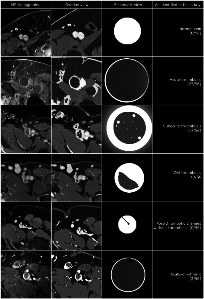
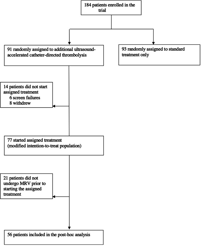
Methods: This is a post hoc analysis of data from the CAVA trial ( Clinicaltrials.gov :NCT00970619). MRV studies of patients receiving ultrasound-accelerated catheter-directed thrombolysis (CDT) for iliofemoral DVT were reviewed. Thrombus age-related imaging characteristics were scored and translated into an overall score (acute, subacute, or old). MRV scores were compared to patient-reported complaints. MRV-scored groups were compared for CDT duration and success rate.
Results: Fifty-six patients (29 men; age 50.8 ± 16.4 years) were included. Using MRV, 27 thrombi were classified acute, 17 subacute, and 12 old. Based on patient-reported complaints, 11 (91.7%) of these old thrombi would have been categorized acute or subacute, and one (3.7%) of the acute thrombi as old. Average duration of CDT to > 90% restored patency differed significantly between groups (p < 0.0001): average duration was 23 h for acute thromboses (range: 19-25), 43 h for subacute (range: 41-62), and 85 h for old thromboses (range: 74-96). CDT was almost eleven times more successful in thromboses characterized as acute and subacute compared to old thromboses (OR: 10.7; 95% CI 2.1-55.5).
Conclusion: A dedicated MRV scoring system can safely discriminate between acute, subacute, and old thromboses. MRV-based selection is predictive of procedural duration and success rate and can help avoid unnecessary complications.
Key points: • Thrombus age, characterized by MRV as acute, subacute, and old, can predict CDT duration and probability of success. • Accurate pre-interventional MRV-based thrombus aging has the potential to facilitate identification of eligible patients and may thus prevent CDT-related complications.
Keywords: Magnetic resonance venography; Thrombolysis; Thrombosis; Thrombus.
© 2022. The Author(s).
Conflict of interest statement
The authors declare no competing interests.
Figures

References
-
- Notten P, Ten Cate-Hoek AJ, Arnoldussen CWKP, et al. Ultrasound-accelerated catheter-directed thrombolysis versus anticoagulation for the prevention of post-thrombotic syndrome (CAVA): a single-blind, multicentre, randomised trial. Lancet Haematol. 2020;7(1):e40–e49. doi: 10.1016/S2352-3026(19)30209-1. - DOI - PubMed
MeSH terms
Associated data
Grants and funding
LinkOut - more resources
Full Text Sources
Medical
