. 2022 Apr 5;11(7):e023021.
doi: 10.1161/JAHA.121.023021. Epub 2022 Mar 29.

Platelet-Derived Growth Factor Receptor Type α Activation Drives Pulmonary Vascular Remodeling Via Progenitor Cell Proliferation and Induces Pulmonary Hypertension

Affiliations

Platelet-Derived Growth Factor Receptor Type α Activation Drives Pulmonary Vascular Remodeling Via Progenitor Cell Proliferation and Induces Pulmonary Hypertension

Julien Solinc et al. J Am Heart Assoc. .

Abstract

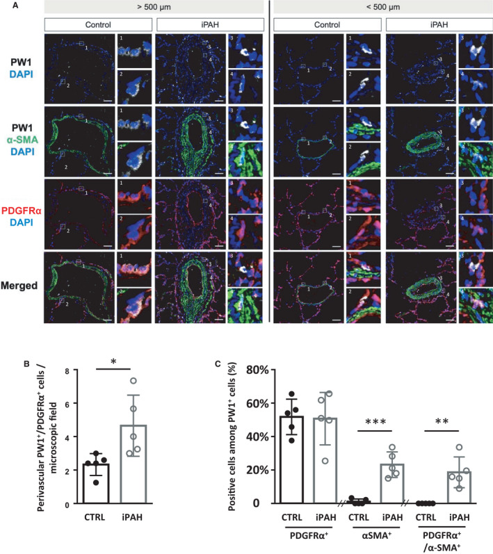
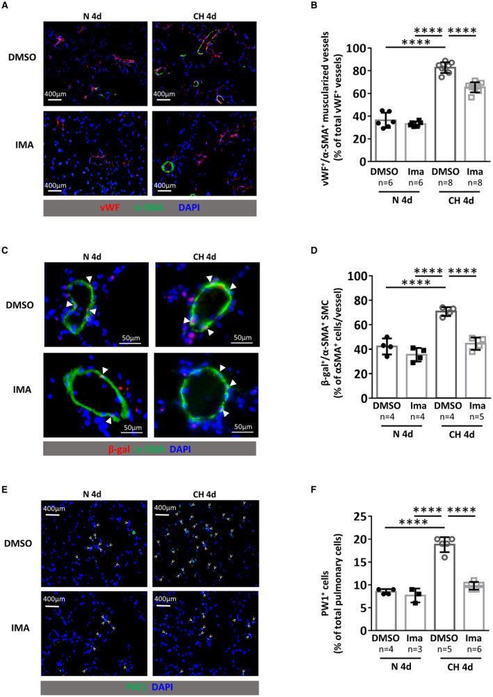
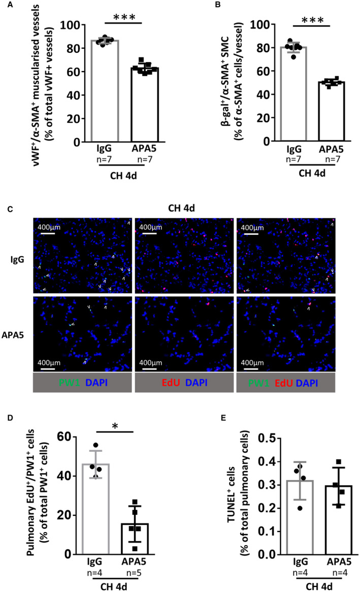
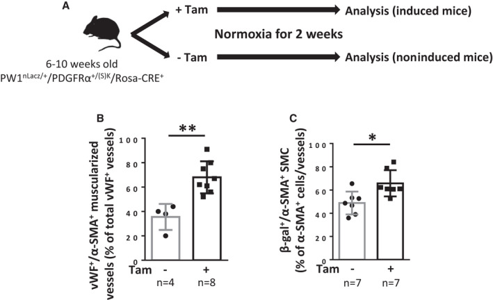
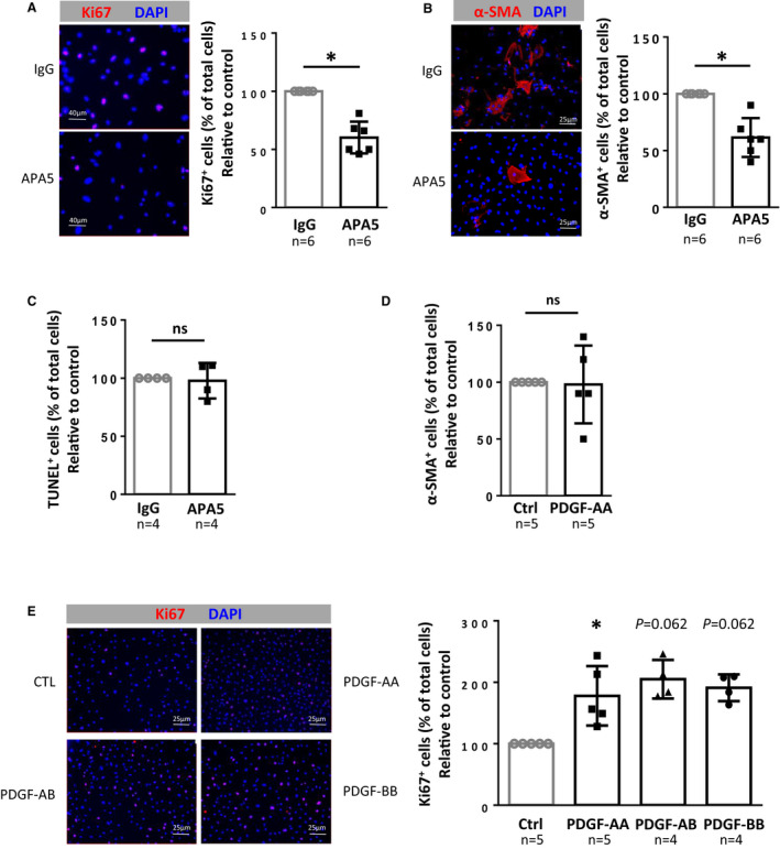
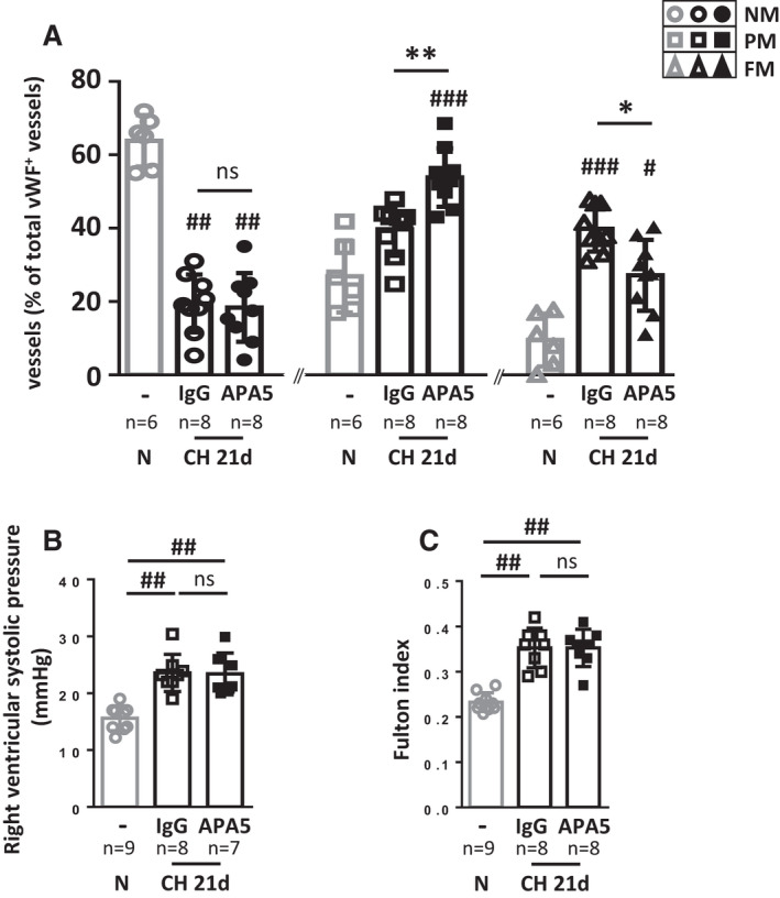
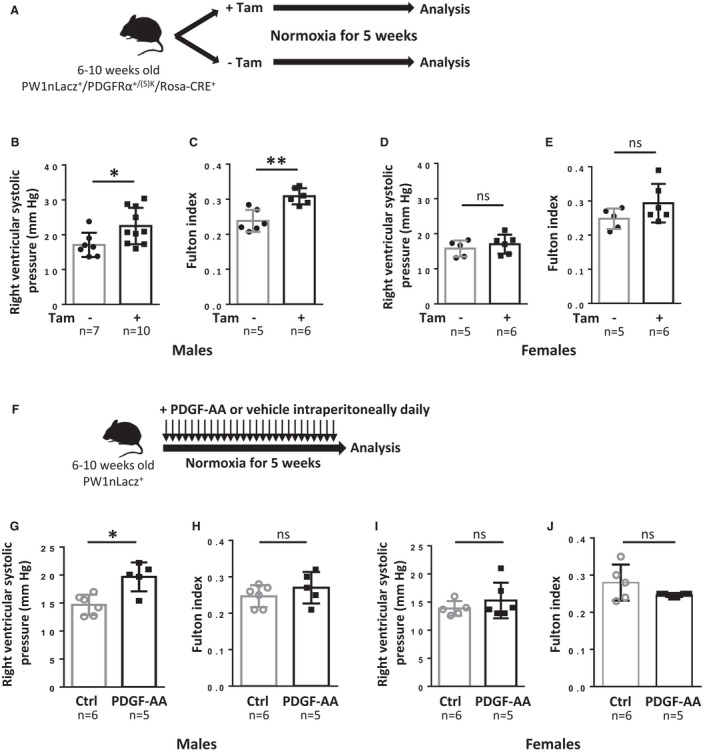
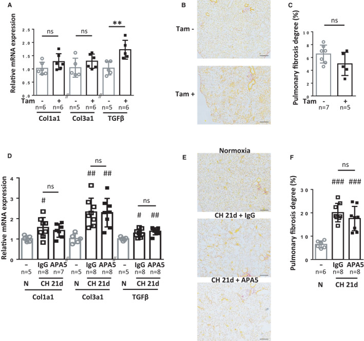
Background Platelet-derived growth factor is a major regulator of the vascular remodeling associated with pulmonary arterial hypertension. We previously showed that protein widely 1 (PW1+) vascular progenitor cells participate in early vessel neomuscularization during experimental pulmonary hypertension (PH) and we addressed the role of the platelet-derived growth factor receptor type α (PDGFRα) pathway in progenitor cell-dependent vascular remodeling and in PH development. Methods and Results Remodeled pulmonary arteries from patients with idiopathic pulmonary arterial hypertension showed an increased number of perivascular and vascular PW1+ cells expressing PDGFRα. PW1nLacZ reporter mice were used to follow the fate of pulmonary PW1+ progenitor cells in a model of chronic hypoxia-induced PH development. Under chronic hypoxia, PDGFRα inhibition prevented the increase in PW1+ progenitor cell proliferation and differentiation into vascular smooth muscle cells and reduced pulmonary vessel neomuscularization, but did not prevent an increased right ventricular systolic pressure or the development of right ventricular hypertrophy. Conversely, constitutive PDGFRα activation led to neomuscularization via PW1+ progenitor cell differentiation into new smooth muscle cells and to PH development in male mice without fibrosis. In vitro, PW1+ progenitor cell proliferation, but not differentiation, was dependent on PDGFRα activity. Conclusions These results demonstrate a major role of PDGFRα signaling in progenitor cell-dependent lung vessel neomuscularization and vascular remodeling contributing to PH development, including in idiopathic pulmonary arterial hypertension patients. Our findings suggest that PDGFRα blockers may offer a therapeutic add-on strategy to combine with current pulmonary arterial hypertension treatments to reduce vascular remodeling. Furthermore, our study highlights constitutive PDGFRα activation as a novel experimental PH model.

Keywords: fibrosis; platelet‐derived growth factor receptor alpha; pulmonary hypertension; stem cells; vascular remodeling.


Figures

Figure 1
Figure 1. Perivascular PW1+/PDGFRα+ cells are increased in the perivascular zone and within the vascular wall of remodeled arteries in patients with iPAH.
Lung sections from control (CTRL) or patients with idiopathic pulmonary arterial hypertension (iPAH) were labeled for PW1 (white), PDGFRα (red), and α‐SMA (green). A, Representative confocal images of pulmonary vessels (>500 µm diameter, left panels; <500 µm diameter, right panels) from control patients (n=5) and patients with iPAH (n=5). Panels 1, 2, and 3 show details of perivascular PW1+/PDFGFRα+ cells. Panel 4 shows details of PW1+/PDFGFRα+/α‐SMA+ cells found within arteries in patients with iPAH. B, quantification of perivascular PW1+/PDGFRα+ cells. C, quantification of PDGFRα+ or α‐SMA+ or PDGFRα+/α‐SMA+ cells among perivascular and vascular PW1+. Scale bar, 50 µm. Bars represent means and whiskers represent SD. *P<0.05, **P<0.01, ***P<0.001; ns indicates not significant (2‐tailed Mann‐Whitney); PDGFRα indicates platelet‐derived growth factor receptor type α; PW1, protein widely 1; and SMA, smooth muscle actin.
Figure 2
Figure 2. Imatinib treatment prevents early chronic hypoxia (CH)‐induced neomuscularization and PW1+ progenitor cell recruitment and differentiation in SMCs.
PW1nLacz mice were maintained under normoxia (N 4d) or chronic hypoxia (CH 4d) for 4 days and treated daily with DMSO or imatinib (IMA). A, Representative images of von Willebrand factor (vWF, red) and α‐smooth muscle actin (α‐SMA, green) staining in lungs. B, Quantification of muscularized vessels (fully+partially) (n=6–8 mice/group). C Representative images of pulmonary muscularized vessel stained with α‐SMA (green) and β‐galactosidase (red) in lungs (double positive are marked by arrowheads). D, Quantification of lung β‐galactosidase (β‐gal)‐expressing SMC in fully‐muscularized vessels (n=4–5 mice/group). E, Representative images of lung parenchyma stained for PW1 (PW1 in green, positive cells are marked by arrowheads). F, Quantification of PW1+ cells in lung parenchyma (n=3–6 mice/group). Bars represent means and whiskers represent SD. ****P<0.0001, 2‐way ANOVA and Tukey. PW1 indicates protein widely 1; and SMCs, smooth muscle cells.
Figure 3
Figure 3. PDGFRα inhibition with the specific blocking antibody APA5 prevents early chronic hypoxia (CH)‐induced neomuscularization and PW1+ progenitor cell proliferation and differentiation in SMCs.
PW1nLacz mice were maintained under CH for 4 days and treated with control antibody (IgG) or PDGFRα blocking antibody (APA5). A, Quantification of pulmonary muscularized vessels (fully+partially). For each animal (n=7 mice/group), at least 100 vWF+ vessels (<100 µm) were analyzed for muscularization (α‐SMA+). B, Quantification of lung β‐galactosidase (β‐gal)‐expressing PW1‐derived SMCs in fully‐muscularized vessels (<100 µm) (n=7 mice/group). C, Representative images of lung parenchyma from CH mice treated with control IgG or APA5 and stained for PW1 (green) and EdU (red), double‐positive cells are marked by arrowheads. D, Quantification of EdU+/PW1+ cells in lung parenchyma (n=4–5 mice/group). E, Quantification of pulmonary apoptotic cells. The percentage of TUNEL+ cells in lung parenchyma was determined by immunofluorescence (n=4 mice/group). Bars represent means and whiskers represent SD. *P<0.05, **P<0.01, ***P<0.001. 2‐tailed Mann‐Whitney. Scale bar 400 µm. EdU indicates ethynyl‐deoxyuridine; PDGFRα, platelet‐derived growth factor receptor type α; PW1, protein widely 1; SMA, smooth muscle actin; SMC, smooth muscle cell; TUNEL, terminal deoxynucleotidyl transferase dUTP nick end labeling; and vWF, von Willebrand factor.
Figure 4
Figure 4. Constitutive PDGFRα activation leads to pulmonary vessel neomuscularization because of increased numbers of PW1+ progenitor‐derived SMCs.
A, Timeline of tamoxifen (Tam) induction and analysis of PW1nLacz/+/PDGFRα+/(S)K/Rosa‐CRE+ mice (males + females). B and C, Analysis of mice lungs 2 weeks after tamoxifen induction. B, Quantification of muscularized vessels in lungs from control or tamoxifen‐treated mice. Pulmonary vessel muscularization was determined by immunofluorescence using anti–α‐smooth muscle actin (α‐SMA) and anti‐von Willebrand factor (vWF) antibodies (n=4–8). For each animal, ≈100 vWF+ vessels (<100 µm) were analyzed for muscularization (α‐SMA+). C, Quantification of lung β‐galactosidase (β‐gal)‐expressing SMC in fully‐muscularized pulmonary vessels. The percentage of lung β‐gal+/α‐SMA+ cells among α‐SMA+ cells in fully‐muscularized vessels (<100 µm) was determined by immunofluorescence (n=7 for each condition). Bars represent means and whiskers represent SD. *P<0.05, **P<0.01 (2‐tailed Mann‐Whitney). PDGFRα indicates platelet‐derived growth factor receptor type α; PW1, protein widely 1; SMA, smooth muscle actin; and SMC, smooth muscle cell.
Figure 5
Figure 5. PDGFRα regulates PW1+ progenitor cell proliferation in vitro.
A, Effect of PDGFRα inhibition on PW1+ progenitor cell proliferation. Pulmonary PW1+ progenitor cells were sorted by FACS, incubated for 5 days with IgG control or APA5 and proliferative cells were then labeled with anti‐Ki67 antibody (red). (Left) Representative immunofluorescence images and (right) quantification of PW1+ progenitor cell proliferation as a ratio of Ki67+ cells over total cells reported to control (n=6). B, Effect of PDGFRα inhibition on PW1+ progenitor cell differentiation into SMC. FACS‐sorted pulmonary PW1+ progenitor cells were incubated for 5 days with IgG control or APA5 and then labeled with anti‐α‐SMA antibody (red). (Left) Representative immunofluorescence images and (right) quantification of PW1+ progenitor cell differentiation as a ratio of α‐SMA+ cells over total cells reported to control (n=6). C, Effect of PDGFRα inhibition on PW1+ progenitor cell apoptosis. FACS‐sorted pulmonary PW1+ progenitor cells were incubated for 5 days with IgG control or APA5 and then labeled following the TUNEL method. The ratio of TUNEL+ cells over total cells was determined and reported to control (n=4). D, Effect of PDGFRα activation on PW1+ progenitor cell differentiation into SMCs. FACS‐sorted pulmonary PW1+ progenitor cells were incubated for 5 days with IgG control or PDGF‐AA, ‐AB, or ‐BB (20 ng/mL) and labeled with anti–α‐SMA antibody (red). The ratio of α‐SMA+ cells over total cells was reported to control (n=5). E, Effect of PDGFRα activation on PW1+ progenitor cell proliferation. FACS‐sorted pulmonary PW1+ progenitor cells were incubated for 5 days with either vehicle, PDGF‐AA, PDGF‐AB, or PDGF‐BB (20 ng/mL) and then labeled with anti‐Ki67 antibody (red). (Left) Representative immunofluorescence images and (right) quantification of PW1+ progenitor cell proliferation as a ratio of Ki67+ cells over total cells reported to control (n=4–5). Bars represent means and whiskers represent SD. *P<0.05, ns indicates not significant (1‐tailed Wilcoxon signed‐ranked test). FACS indicates fluorescence‐activated cell sorting; PDGF, platelet‐derived growth factor; PDGFRα, platelet‐derived growth factor receptor type α; PW1, protein widely 1; SMA, smooth muscle actin; and SMC, smooth muscle cell; and TUNEL, terminal deoxynucleotidyl transferase dUTP nick end labeling; and vWF, von Willebrand factor.
Figure 6
Figure 6. PDGFRα inhibition with APA5 limits vascular neomuscularization but does not prevent chronic hypoxia (CH)‐induced PH.
PW1nLacz mice were maintained untreated under normoxia (N) or under CH for 21 days and treated with control IgG or PDGFRα blocking antibody APA5. A, Pulmonary vessel muscularization was determined by immunofluorescence in lungs of control untreated normoxic mice (n=6) or of IgG‐ and APA5‐treated mice after 21 days of chronic hypoxia (CH 21d, n=9) using anti–α‐smooth muscle actin (α‐SMA) and anti–von Willebrand factor (vWF) antibodies. For each animal, >100 vWF+ vessels (<100 μm) were analyzed for muscularization (α‐SMA+). Vessels were identified as nonmuscularized (NM), partially muscularized (PM) or fully muscularized (FM). B, Right ventricular systolic pressure (RVSP, mm Hg) was measured in untreated normoxic mice (n=9) and in IgG‐ and APA5‐treated mice after 21 days of CH (CH 21d, n=7–8). C, Right ventricle hypertrophy was measured as the Fulton index in normoxic untreated mice (n=9) and in IgG‐ and APA5‐treated mice in CH (CH 21d, n=8). Bars represent means and whiskers represent SD. *P<0.05, **P<0.01, ns indicates not significant for APA5 vs IgG within CH 21d group; # P<0.05, ## P<0.01 vs normoxic; ns indicates not significant (Kruskal‐Wallis and Dunn). PDGFRα, platelet‐derived growth factor receptor type α; PH, pulmonary hypertension; PW1, protein widely 1; and SMA, smooth muscle actin.
Figure 7
Figure 7. Constitutive PDGFRα activation leads to pulmonary hypertension.
A, Timeline of tamoxifen (Tam) induction and analysis of PW1nLacz/PDGFRα+/(S)K/Rosa‐CRE+ mice. B to E, Analysis of mice 5 weeks after tamoxifen induction. B and D, Right ventricular systolic pressure (RVSP) was measured in control or tamoxifen‐treated (B), males (n=7–10) and (D), females (n=5–6). C and E, Right ventricular hypertrophy was determined using the Fulton index (right ventricular weight to left ventricular+septum weight ratio) in control or tamoxifen‐treated (C), males (n=5–6) and (E), females (n=5–6). F, Timeline of PDGF‐AA treatment and analysis of PW1nLacz+ mice. G through J, Analysis of mice after 5 weeks of PDGF‐AA treatment. G and I, Right RVSP was measured in control or PDGF‐AA‐treated (G), males (n=5–6) and (I), females (n=5–6). H and J, Right ventricular hypertrophy was determined using the Fulton index in control or PDGF‐AA–treated (H), males (n=5–6) and (J), females (n=5–6). Bars represent means and whiskers represent SD. *P<0.05, **P<0.01; ns indicates not significant (2‐tailed Mann‐Whitney); PDGF, platelet‐derived growth factor; PDGFRα, platelet‐derived growth factor receptor type α; and PW1, protein widely 1.
Figure 8
Figure 8. Lung fibrosis is not induced by PDGFRα activation and is not reduced by PDGFRα inhibition.
A through C, Fibrosis evaluation in noninduced (n=6–7) or tamoxifen‐induced PW1nLacz/+/PDGFRα+/(S)K/Rosa‐CRE+ mice 5 weeks after tamoxifen induction (n=5–6). D to F, Fibrosis evaluation in untreated mice under normoxia (n=5) or IgG and APA5‐treated mice after 21 days of CH (n=7–8). A and D, RT‐qPCR measurements of mRNA expression for collagen 1a1, collagen 3a1, and transforming growth factor‐β in lungs. B and E, Representative images of picrosirius red staining for collagen (red) in pulmonary parenchyma. Larger images are displayed in Figures S10 and S11. C and F, Quantification of the picrosirius red‐stained area as a measure of fibrosis using Histolab analysis. Fibrosis area (% of total area) was determined as the mean fibrotic area of four large lung sections for each animal. Bars represent means and whiskers represent SD. *P<0.05, **P<0.01 Tam+ vs Tam‐ group, 2‐tailed Mann‐Whitney; # P<0.05, ## P<0.01 vs normoxic, Kruskal‐Wallis and Dunn. CH indicates chronic hypoxia; ns, not significant for APA5 vs IgG within CH 21d group in D and F (scale bar 200 µm); PDGFRα, platelet‐derived growth factor receptor type α; PW1, protein widely 1; and RT‐qPCR, reverse transcriptase quantitative polymerase chain reaction.

Similar articles

Cited by

References

    1. Ricard N, Tu LY, Le Hiress M, Huertas A, Phan C, Thuillet R, Sattler C, Fadel E, Seferian A, Montani D, et al. Increased pericyte coverage mediated by endothelial‐derived fibroblast growth factor‐2 and interleukin‐6 is a source of smooth muscle‐like cells in pulmonary hypertension. Circulation. 2014;129:1586–1597. doi: 10.1161/CIRCULATIONAHA.113.007469 - DOI - PubMed
    1. Sheikh AQ, Misra A, Rosas IO, Adams RH, Greif DM. Smooth muscle cell progenitors are primed to muscularize in pulmonary hypertension. Sci Transl Med. 2015;7:308ra159. doi: 10.1126/scitranslmed.aaa9712 - DOI - PMC - PubMed
    1. Dierick F, Héry T, Hoareau‐Coudert B, Mougenot N, Monceau V, Claude C, Crisan M, Besson V, Dorfmüller P, Marodon G, et al. Resident PW1+ progenitor cells participate in vascular remodeling during pulmonary arterial hypertension. Circ Res. 2016;118:822–833. doi: 10.1161/circresaha.115.307035 - DOI - PubMed
    1. Bordenave J, Tu LY, Berrebeh N, Thuillet R, Cumont A, Le Vely B, Fadel E, Nadaud S, Savale L, Humbert M, et al. Lineage tracing reveals the dynamic contribution of pericytes to the blood vessel remodeling in pulmonary hypertension. Arterioscler Thromb Vasc Biol. 2020;40:766–782. doi: 10.1161/ATVBAHA.119.313715 - DOI - PubMed
    1. Perros F, Montani D, Dorfmüller P, Durand‐Gasselin I, Tcherakian C, Le Pavec J, Mazmanian M, Fadel E, Mussot S, Mercier O, et al. Platelet‐derived growth factor expression and function in idiopathic pulmonary arterial hypertension. Am J Respir Crit Care Med. 2008;178:81–88. doi: 10.1164/rccm.200707-1037OC - DOI - PubMed

Publication types

Substances