. 2022 Jun 3;145(5):1610-1623.
doi: 10.1093/brain/awac114.

Human lesions and animal studies link the claustrum to perception, salience, sleep and pain

Affiliations

Human lesions and animal studies link the claustrum to perception, salience, sleep and pain

Huriye Atilgan et al. Brain. .

Abstract

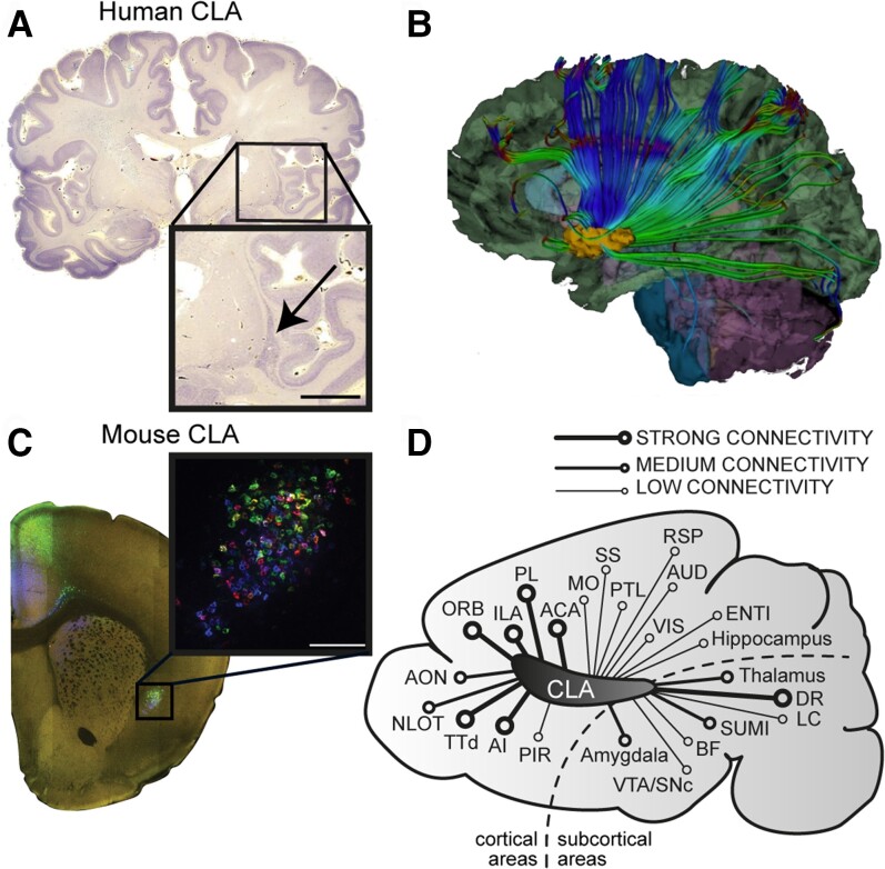
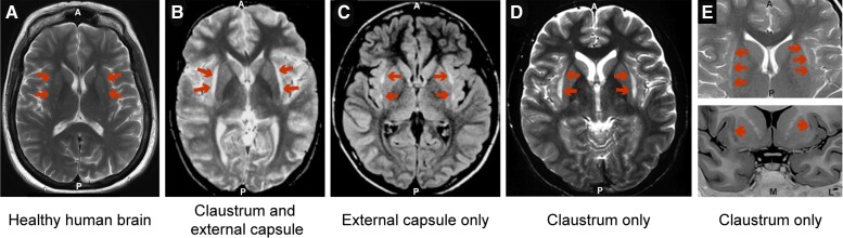
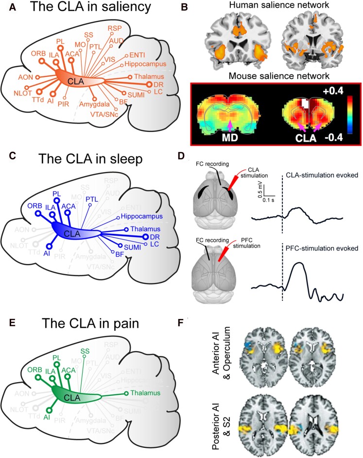
The claustrum is the most densely interconnected region in the human brain. Despite the accumulating data from clinical and experimental studies, the functional role of the claustrum remains unknown. Here, we systematically review claustrum lesion studies and discuss their functional implications. Claustral lesions are associated with an array of signs and symptoms, including changes in cognitive, perceptual and motor abilities; electrical activity; mental state; and sleep. The wide range of symptoms observed following claustral lesions do not provide compelling evidence to support prominent current theories of claustrum function such as multisensory integration or salience computation. Conversely, the lesions studies support the hypothesis that the claustrum regulates cortical excitability. We argue that the claustrum is connected to, or part of, multiple brain networks that perform both fundamental and higher cognitive functions. As a multifunctional node in numerous networks, this may explain the manifold effects of claustrum damage on brain and behaviour.

Keywords: claustrum; lesion; pain; perception‌; salience; sleep.


Figures

Figure 1
Figure 1
The location and connectivity of the claustrum. (A) Nissl stained human coronal brain section with inset showing the location of the claustrum. (B) White matter tractography image showing outgoing connections from the claustrum reprinted from Torgerson et al. with permission from John Wiley and Sons ©2015. (C) Mouse coronal brain section with inset showing the claustrum labelled by multi-colour retrograde tracers. (D) Schematic illustration of a sagittal mouse brain section showing the relative connectivity strength between the claustrum and other brain regions. Connectivity strength was assessed based on the data provided in Zingg et al. and Wang et al. Most but not all connections are reciprocal (see reviews Mathur and Jackson et al.). ACA = anterior cingulate area; AI = anterior insular cortex; Aud = auditory cortex; AON = anterior olfactory nucleus; BF = basal forebrain; CLA = claustrum; DR = dorsal raphe; ENTI = entorhinal cortex; ILA = infralimbic area; LC = locus coeruleus; MO = motor cortex; LOT = the nucleus of the lateral olfactory tract; ORB = orbital area; PL = prelimbic area; PTL = parietal cortex; PIR = piriform area; RSP = retrosplenial cortex; SUMI = supramammillary; SS = somatosensory cortex; TTd = dorsal taenia tecta; VTA/SNc = ventral tegmental area/substantia nigra; VIS = visual cortex.
Figure 2
Figure 2
Similar lesions have been described as affecting either the claustrum, the external capsule, or both. (A) Representative T2-weighted image of a healthy human brain. (B) T2-weighted image showing a lesion reported as affecting the claustrum and external capsule. Reprinted from Sperner et al. with permission from Springer Nature ©1996. (C) Fluid-attenuated inversion recovery (FLAIR) image showing a lesion reported as affecting the external capsule. Reprinted from Mumoli et al. with permission from Springer Nature ©2014. (D) T2-weighted image showing a lesion reported as affecting the claustrum. Reprinted from Ishii et al. with permission from American Society of Neuroradiology ©2011. (E) T2-weighted image (top) and T1-weighted image (bottom) showing a lesion reported as affecting the claustrum. Reprinted from Silva et al. with permission from Springer Nature ©2018.
Figure 3
Figure 3
The claustrum is connected with or part of multiple brain networks. (A) Schematic illustration of a sagittal mouse brain section highlighting the connections between the claustrum (CLA) and regions important in salience. (B) Representative functional MRI images showing the human and rodent salience networks adapted from Seeley et al. and Smith et al., respectively (© Society for Neuroscience). (C) Schematic illustration of a sagittal mouse brain section highlighting the connections between the claustrum and regions important in sleep. (D) Claustrum and prefrontal cortex (PFC) stimulation both evoke slow-wave like activity. Reprinted from Narikiyo et al. and Vyazovskiy et al., respectively, with permission from Springer Nature ©2020. (E) Schematic illustration of a sagittal mouse brain section highlighting the connections between the claustrum and regions important in pain processing. (F) Brain activity to physical pain using multi-study data and machine learning classification tools. Images kindly provided by Tor Wager. FC = frontal cortex; MD = mediodorsal thalamus.

References

    1. Atlan G, Terem A, Peretz-Rivlin N, Groysman M, Citri A. Mapping synaptic cortico-claustral connectivity in the mouse. J Comp Neurol. 2017;525(6):1381–1402. - PubMed
    1. Atlan G, Terem A, Peretz-Rivlin N, et al. . The claustrum supports resilience to distraction. Curr Biol. 2018;28(17):2752–2762.e7. - PMC - PubMed
    1. Carman JB, Cowan WM, Powell TP. The cortical projection upon the claustrum. J Neurol Neurosurg Psychiatry. 1964;27(1):46–51. - PMC - PubMed
    1. Goll Y, Atlan G, Citri A. Attention: the claustrum. Trends Neurosci. 2015;38(8):486–495. - PubMed
    1. Hoover WB, Vertes RP. Anatomical analysis of afferent projections to the medial prefrontal cortex in the rat. Brain Struct Funct. 2007;212(2):149–179. - PubMed

Publication types