. 2023 Mar 1;146(3):991-1005.
doi: 10.1093/brain/awac113.

The bidirectional lung brain-axis of amyloid-β pathology: ozone dysregulates the peri-plaque microenvironment

Affiliations

The bidirectional lung brain-axis of amyloid-β pathology: ozone dysregulates the peri-plaque microenvironment

Hendrik J Greve et al. Brain. .

Abstract

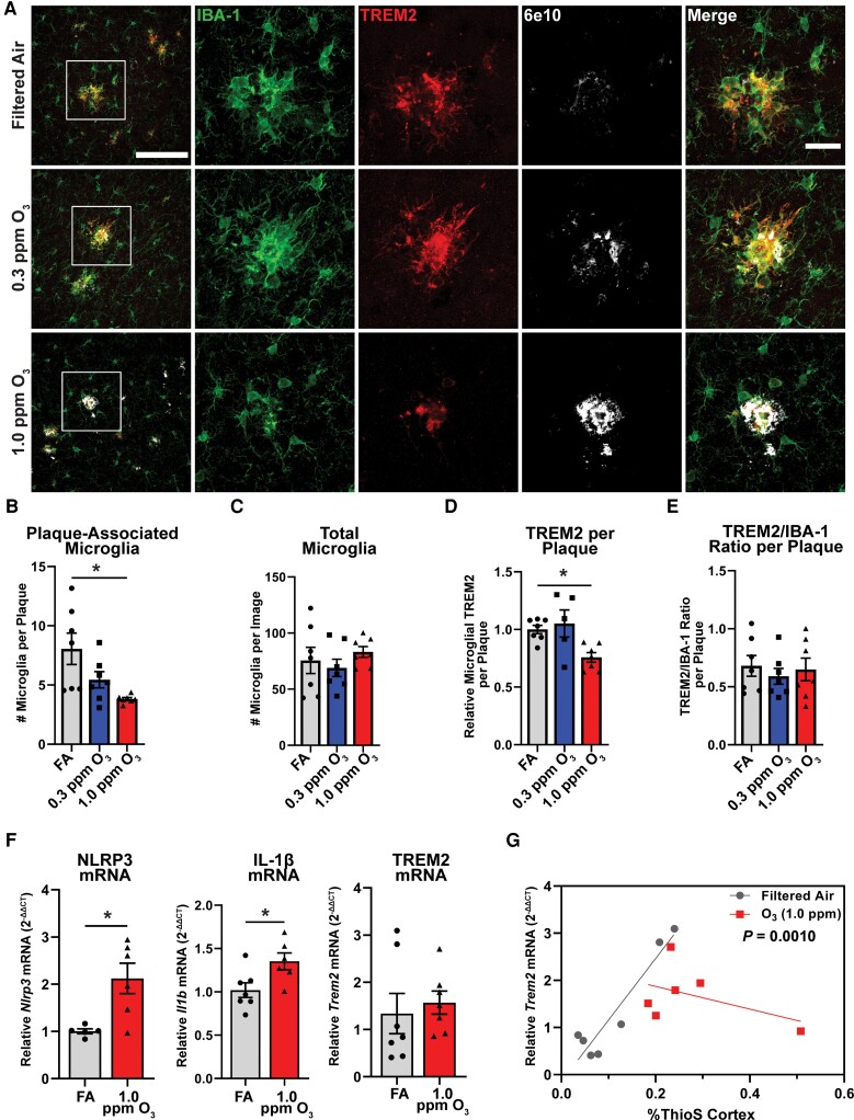
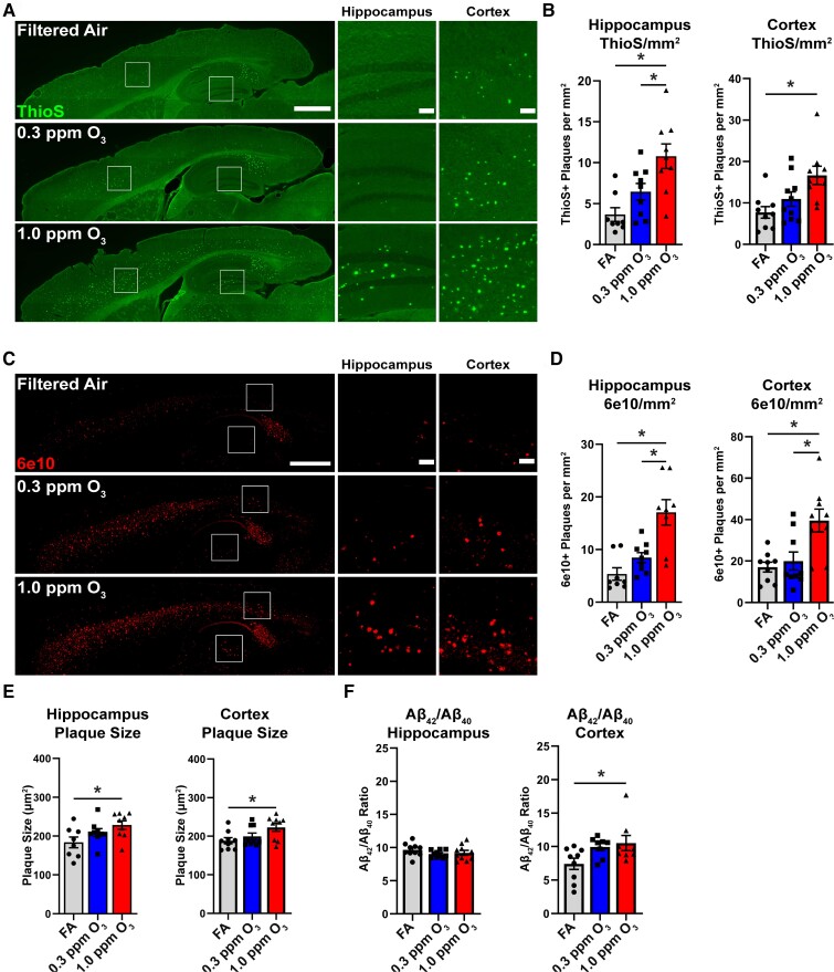
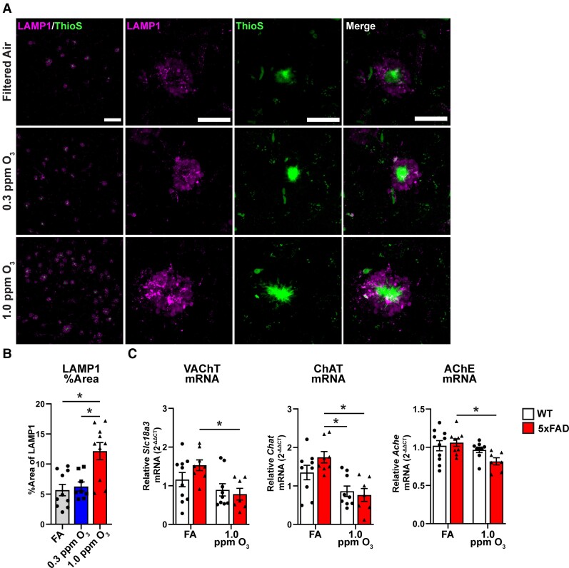
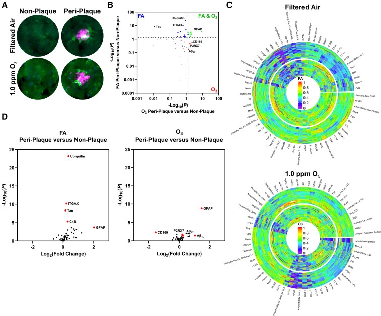
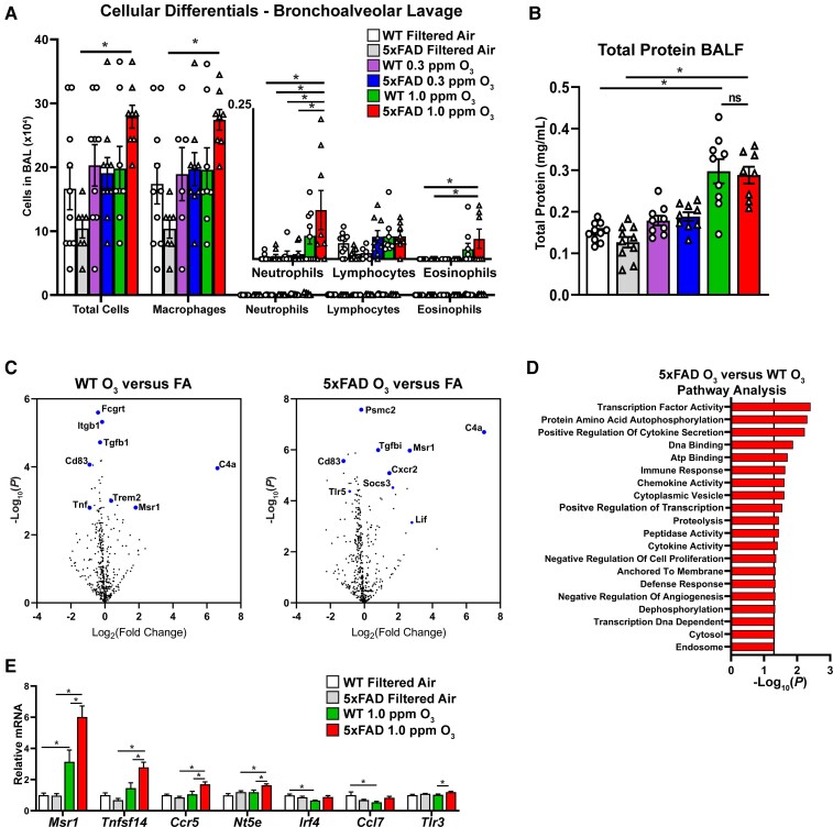
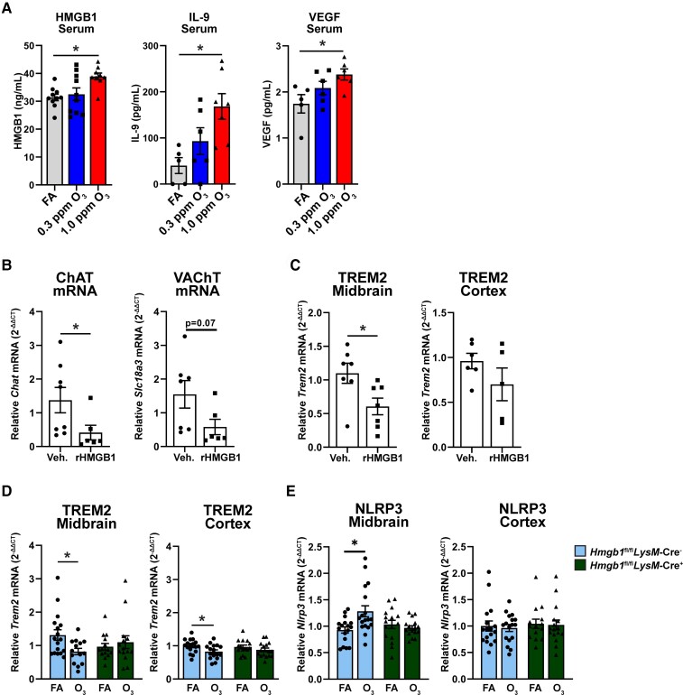
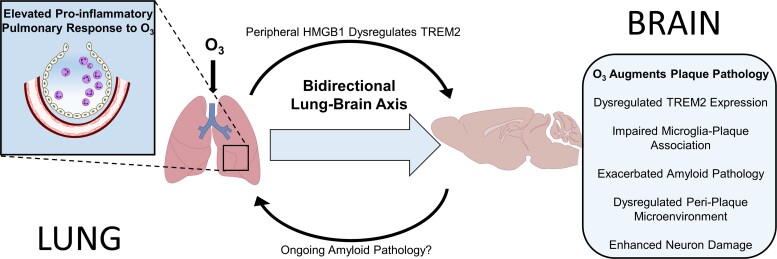
The mechanisms underlying how urban air pollution affects Alzheimer's disease (AD) are largely unknown. Ozone (O3) is a reactive gas component of air pollution linked to increased AD risk, but is confined to the respiratory tract after inhalation, implicating the peripheral immune response to air pollution in AD neuropathology. Here, we demonstrate that O3 exposure impaired the ability of microglia, the brain's parenchymal immune cells, to associate with and form a protective barrier around Aβ plaques, leading to augmented dystrophic neurites and increased Aβ plaque load. Spatial proteomic profiling analysis of peri-plaque proteins revealed a microenvironment-specific signature of dysregulated disease-associated microglia protein expression and increased pathogenic molecule levels with O3 exposure. Unexpectedly, 5xFAD mice exhibited an augmented pulmonary cell and humoral immune response to O3, supporting that ongoing neuropathology may regulate the peripheral O3 response. Circulating HMGB1 was one factor upregulated in only 5xFAD mice, and peripheral HMGB1 was separately shown to regulate brain Trem2 mRNA expression. These findings demonstrate a bidirectional lung-brain axis regulating the central and peripheral AD immune response and highlight this interaction as a potential novel therapeutic target in AD.

Keywords: HMGB1; TREM2; air pollution; amyloid plaque; lung-brain axis; microglia.


Conflict of interest statement

The authors report no competing interests.

Figures

Figure 1
Figure 1
Ozone impairs microglial-plaque association and TREM2 on plaque-associated microglia. 5xFAD mice (10–11 weeks old) were exposed to O3 (0.3 or 1.0 ppm) or FA by inhalation for 13 weeks. (A) Representative confocal images of TREM2 (red), IBA1 (green) and 6e10 (white) in the cortex of 5xFAD mice exposed to O3 or FA control. Scale bars = 100 μm; Inset = 25 μm. (B and C) Number of (B) plaque-associated microglia and (C) total microglia per image. (D and E) Quantification of (D) microglial TREM2 relative to plaque area and (E) the ratio microglial TREM2 to IBA-1. (F) RT-qPCR of the pro-inflammatory genes Nlrp3 and Il1b as well as Trem2 in the cortex of O3 exposed mice. (G) Correlation of %ThioS in the cortex and Trem2 mRNA. Data are represented as mean ± SEM, n = 5–7 mice/exposure group. *P < 0.05; one-way ANOVA, Bonferroni post hoc or Welch’s t-test.
Figure 2
Figure 2
Ozone exposure exacerbates amyloid pathology in 5xFAD mice. 5xFAD mice (10–11 weeks old) were exposed to O3 (0.3 or 1.0 ppm) or FA by inhalation for 13 weeks. (A) Representative images of ThioS+ plaques in the cortex and hippocampus. Scale bars = 1000 μm; Inset = 100 μm. (B) Quantification of the number of ThioS+ plaques per mm2 in the hippocampus and cortex. (C) Representative images of 6e10+ Aβ in the cortex and hippocampus. Scale bars = 1000 μm; Inset = 100 μm. (D) Quantification of the number of 6e10+ plaques per mm2 in the hippocampus and cortex. (E) Quantification of the average size of 6e10+ plaques in the hippocampus and cortex. (F) The ratio of DEA soluble Aβ42 to Aβ40 in the hippocampus and cortex. Data are represented as mean ± SEM, n = 8–10 mice/exposure group. *P < 0.05, one-way ANOVA, Bonferroni post hoc.
Figure 3
Figure 3
Ozone exposure exacerbates neuritic dystrophy and dysregulates acetylcholinergic gene expression. 5xFAD mice (10–11 weeks old) were exposed to O3 (0.3 or 1.0 ppm) or FA by inhalation for 13 weeks. (A) Representative confocal images of ThioS (green) and LAMP1 (magenta) taken at ×20 (top) or ×63 (bottom) in the M1/M2 cortex of 5xFAD mice exposed to O3. Scale bars: top = 100 µm; bottom = 25 µm. (B) Quantification of LAMP1 per cent area. (C) mRNA expression of Slc18a3 (VAChT), Chat and Ache in the cortex of O3 exposed 5xFAD and WT mice. n = 8–10 mice/exposure group. *P < 0.05; one-way ANOVA, Bonferroni post hoc or Welch’s t-test.
Figure 4
Figure 4
O3 alters the peri-plaque microenvironment and dysregulates key DAM proteins. 5xFAD mice (10–11 weeks old) were exposed to O3 (0.3 or 1.0 ppm) or FA by inhalation for 13 weeks. (A) Representative images from NanoString GeoMX DSP platform of IBA-1 (green) and Aβ (magenta) in the cortex of 5xFAD mice. (B) Plot comparing changes from peri-plaque to non-plaque areas in FA and O3. (C) Radial heat map of protein changes from non-plaque (outer circle) to peri-plaque (inner circle) regions in O3 exposed 5xFAD mice. (D) Volcano plots comparing peri-plaque versus non-plaque areas for FA and O3. n = 4 mice/group (34–36 peri-plaque and 36 plaque-distant ROIs per mouse).
Figure 5
Figure 5
Ongoing AD pathology augments pulmonary and peripheral immune responses to O3. 5xFAD mice (10–11 weeks old) were exposed to O3 (0.3 or 1.0 ppm) or FA by inhalation for 13 weeks. (A) Cellular differentials from bronchoalveolar lavage (BAL) from O3-exposed 5xFAD and control littermates. (B) Total protein in BAL fluid. (C) Volcano plots from NanoString analysis of BAL cells from O3 WT versus FA and O3 5xFAD versus FA. Highlighted genes represent a subset of significantly changed genes after FDR correction (Benjamani-Hochberg, P < 0.01). (D) Undirected pathway analysis of 5xFAD O3 versus WT O3 showing the top 20 significantly changed pathways (P < 0.05). (E) Analysis of gene changes between 5xFAD and WT mice exposed to O3.
Figure 6
Figure 6
Peripheral HMGB1 regulates O3-induced CNS changes. 5xFAD mice (10–11 weeks old) were exposed to O3 (0.3 or 1.0 ppm) or FA by inhalation for 13 weeks. (A) Serum analysis of HMGB1, IL-9, and VEGF in 5xFAD mice exposed to O3. C57Bl/6 mice were injected with rHMGB1 (32.5 μg) or vehicle (Veh.) by tail and samples were collected 3 h after injection (B) mRNA analysis of acetylcholinergic genes in mice injected with rHMGB1. (C) mRNA analysis of Trem2 in midbrain and cortex of mice injected with rHMGB1. Hmgb1fl/flLysM-cre and Hmgb1fl/flLysM-cre+were exposed to O3 (1.0 ppm) or FA for 3 days, 4 h/day. (D and E) mRNA expression of (D) Trem2 and (E) Nlrp3 in cortex and midbrain of Hmgb1fl/flLysM-cre and Hmgb1fl/flLysM-cre+exposed to O3. n = 5–10 mice/exposure group for 13 wk O3 exposure, n = 6–7 mice/treatment group for rHMGB1, n = 16–18 mice/exposure group for short term O3. *P < 0.05; Welch’s t-test, or one- or two-way ANOVA, Bonferroni post hoc.
Figure 7
Figure 7
O3 regulates the amyloid plaque micro-environment through the bidirectional lung-brain axis. Exposure to O3, a reactive air pollutant unable to translocate to the brain to exert CNS effects, caused: reduced microglial-plaque association, impaired CNS Trem2 upregulation in response to plaques, exacerbated amyloid and neuronal pathology, and dysregulated the peri-plaque microenvironment in 5xFAD mice, effects commonly associated with loss of TREM2 function. 5xFAD mice also exhibited enhanced circulating factors (HMGB1), elevated pulmonary immune cell trafficking, and an augmented pulmonary immune cell pro-inflammatory transcriptome in response to O3, when compared to control mice, supporting a role for ongoing Aβ neuropathology in the pulmonary response to air pollution, revealing a bi-directional lung-brain axis governing central and peripheral immune responses. Mechanistically, iv injection of recombinant HMGB1 in control mice impaired brain Trem2 expression and genetic ablation of HMGB1 in peripheral myeloid cells abolished O3-induced reduction of TREM2, revealing a role for peripheral HMGB1 in the regulation of CNS Trem2 expression. These findings implicate urban air pollution, the periphery, and the lung-brain axis in amyloid pathology and highlight the potential role of TREM2 and HMGB1 in this process, identifying novel potential therapeutic targets for the regulation of CNS Trem2 expression and Alzheimer’s disease.

References

    1. Nichols E, Steinmetz JD, Vollset SE, et al. Estimation of the global prevalence of dementia in 2019 and forecasted prevalence in 2050: an analysis for the Global Burden of Disease Study 2019. Lancet Public Health. 2022;7(2):e105–e125. - PMC - PubMed
    1. Long JM, Holtzman DM. Alzheimer disease: An update on pathobiology and treatment strategies. Cell. 2019;179(2):312–339. - PMC - PubMed
    1. Jayaraj RL, Rodriguez EA, Wang Y, Block ML. Outdoor ambient air pollution and neurodegenerative diseases: The neuroinflammation hypothesis. Curr Environ Health Rep. 2017;4(2):166–179. - PubMed
    1. Croze ML, Zimmer L. Ozone atmospheric pollution and Alzheimer’s disease: From epidemiological facts to molecular mechanisms. J Alzheimer's Dis. 2018;62:503–522. - PubMed
    1. Kilian J, Kitazawa M. The emerging risk of exposure to air pollution on cognitive decline and Alzheimer's disease – Evidence from epidemiological and animal studies. Biomed J. 2018;41(3):141–162. - PMC - PubMed