. 2022 Jun;35(3):573-589.
doi: 10.1007/s10534-022-00389-2. Epub 2022 Mar 29.

Crystal structure and metal binding properties of the periplasmic iron component EfeM from Pseudomonas syringae EfeUOB/M iron-transport system

Affiliations

Crystal structure and metal binding properties of the periplasmic iron component EfeM from Pseudomonas syringae EfeUOB/M iron-transport system

Mohan B Rajasekaran et al. Biometals. 2022 Jun.

Abstract

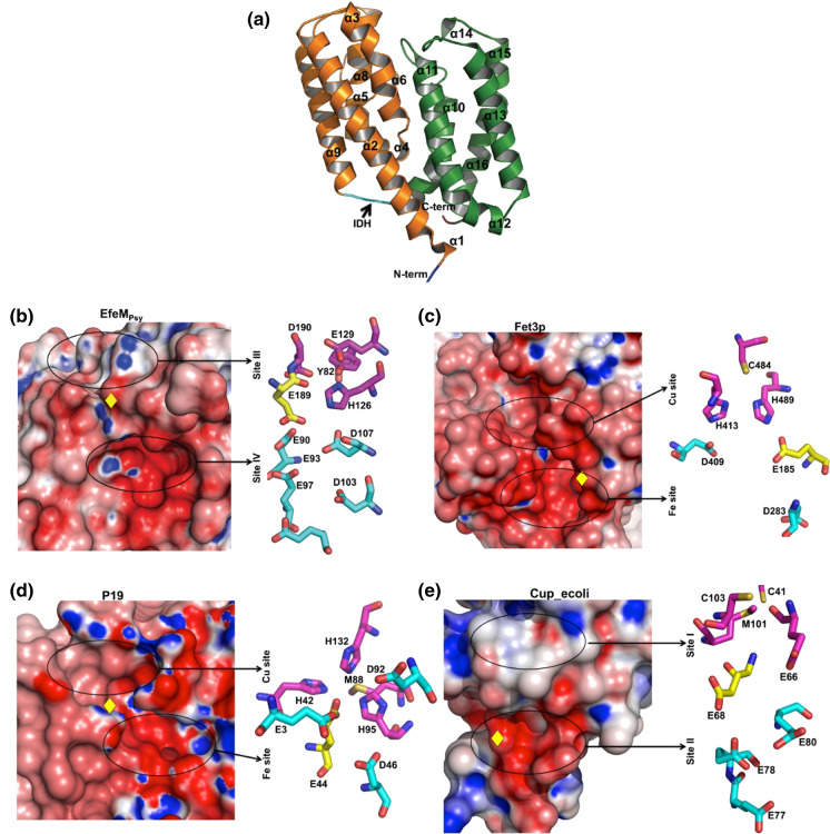
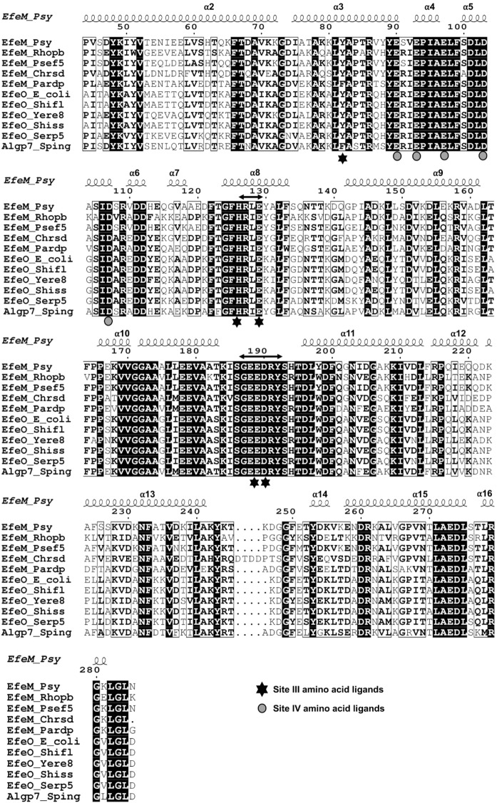
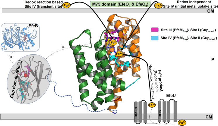
EfeUOB/M has been characterised in Pseudomonas syringae pathovar. syringae as a novel type of ferrous-iron transporter, consisting of an inner-membrane protein (EfeUPsy) and three periplasmic proteins (EfeOPsy, EfeMPsy and EfeBPsy). The role of an iron permease and peroxidase function has been identified for the EfeU and EfeB proteins, respectively, but the role of EfeO/M remains unclear. EfeMPsy is an 'M75-only' EfeO-like protein with a C-terminal peptidase-M75 domain (EfeOII/EfeM family). Herein, we report the 1.6 Å resolution crystal structure of EfeMPsy, the first structural report for an EfeM component of P. syringae pv. syringae. The structure possesses the bi-lobate architecture found in other bacterial periplasmic substrate/solute binding proteins. Metal binding studies, using SRCD and ICP-OES, reveal a preference of EfeMPsy for copper, iron and zinc. This work provides detailed knowledge of the structural scaffold, the metal site geometry, and the divalent metal binding potential of EfeM. This work provides crucial underpinning for a more detailed understanding of the role of EfeM/EfeO proteins and the peptidase-M75 domains in EfeUOB/M iron uptake systems in bacteria.

Keywords: Acidic patch; EfeM; EfeUOB iron-transport system; Metal-binding; Peptidase-M75 domain; X-ray crystallography.


Conflict of interest statement

All authors have no conflict of interest.

Figures

Fig. 1
Fig. 1
Crystal structure of EfeMPsy from P. syringae pv. syringae (PDB code: 7Q1G) and its comparison with periplasmic iron transport components from representative partially related EFeUOB systems. a Overall structure of EfeMPsy showing two helix bundle domains ‘B1’ and ‘B2’ coloured light orange and green, respectively, with inter-domain hinge (IDH) loop in cyan; b Electrostatic surface representation highlighting metal-binding pockets and amino acids lining Site III and Site IV of EfeMPsy. As in EfeMPsy, the other related proteins from various EfeUOB-like systems, namely; c Fet3p; Fet3p from S. cerevisiae (PDB code: 1ZPU); d P19 protein from C. jejuni (PDB code: 3LZQ); e Homology model for cupredoxin domain; Cup_ecoli (Cup-I family) of EfeO from E. coli (Rajasekaran et al. 2010b), all showing a two metal site network consisting of a copper site/Site I, coupled to an acidic patch with Glu/Asp ligands coordinating Mn2+ (as an iron analog) or Fe2+/Fe3+. The residues lining Site I/Site III/Cu and Site IV/Fe site are highlighted in magenta and in cyan (ball and sticks), respectively. The approximate location for the Asp residue situated at the interface between these two sites is highlighted as a yellow diamond in the electrostatic surface representations. The black ovals show the location of these metal sites. For b, c, d and e, the first part of the figure represents the electrostatic potentials for the respective protein, followed by representation of the metal site amino acid ligands (as ball and sticks)
Fig. 2
Fig. 2
Multiple sequence alignment (MSA) for peptidase-M75 domain region of EfeMPsy with selected homologues belonging to the EfeO (Cup domain with peptidase-M75) and EfeM (peptidase-M75 only) families. The search for representative homologues was performed using the gene name for EfeMPsy (‘Psyr_3370’) as the search probe in the STRING database (Szklarczyk et al. 2019). The protein sequences, for both families, were found by this search and the MSA was calculated using the CLUSTAL OMEGA server (Sievers et al. 2011). Visualisation of the alignment was carried out using the ESPript server (Robert and Gouet 2014). For Site III, all five residues (Tyr82, His126, Glu129, Glu189, Asp190) are completely conserved whereas, similar residues Tyr/Phe and Asp/Glu align for positions 82 and 190, respectively. In the case of Site IV, all five residues (Glu90, Glu93, Glu97, Asp103 and Asp107) are completely conserved throughout the alignment. The ‘HXXE’ and ‘GEEDRY’ motifs are highlighted by double headed arrows. In addition, Algp7, a prominent member from Algp7-imelysin like family is also included in the alignment for comparing the metal site conservation between EfeO/EfeM and Algp7-imelysin like family. The UNIPROTKB database accession numbers for the representative entry sequences are listed out in the Supplementary Table S1
Fig. 3
Fig. 3
Characterisation of the EfeMPsy metal binding Site IV and its interaction with select metals by GRID-FLAP. a GRID molecular interaction field (shown as gold contour) in the region of our proposed metal binding site, Site IV for EfeMPsy, highlights the key Glu/Asp amino acids lining the binding pocket. Glu90 also is identified as a potential ligand by GRID-FLAP, however, it is not displayed here in this orientation, which displays the best possible close-up view of the MIF contours b Representative GRID molecular interaction fields, shown as three-dimensional contours (solid grey regions), shows specific favourable regions of EfeMPsy for interaction with Fe3+ with the highest MIF energy of − 81.58 kcal/mol suggesting Fe3+ as the most favoured metal followed almost equally by the other select metals Fe2+, Cu2+, Zn2+ with MIF energies of − 54.26, − 54.17 and − 53.43 kcal/mol, respectively. The three-dimensional constellation of amino acid residues permits favourable six coordinate geometry for metal binding
Fig. 4
Fig. 4
Far–UV (200–260 nm) SRCD spectra of apo EfeMPsy and its titration with select metals. The SRCD spectrum of EfeMPsy (180 μl of pure protein, ~ 10 μM, ~ 0.3 mg/ml) with metals was measured in 0.5 mm path-length Suprasil quartz cells (Hellma®) in the far-UV region (180–260 nm). Each spectrum is the average of four or eight scans expressed in molar circular dichroism (Δε) using average amino acid residue molecular weight of 113. For the purpose of clarity, the titration spectral data between 200 and 260 nm are presented. All the experiments were performed at 25 °C. a Left panel shows SRCD spectra of EfeMPsy with Zn2+ additions with molar stoichiometry starting from 0.25 (2.5 μM) to 4 (40 μM); b Right panel shows SRCD spectra of EfeMPsy with Cu2+ additions with molar stoichiometry starting from 0.4 (4 μM) to 5 (50 μM)
Fig. 5
Fig. 5
Hypothetical functional schema for the engagement of metal binding sites of M75 domain in EfeUOB/M iron transport systems. The four main components of EfeUOB/M system are EfeU, EfeO, EfeM and EfeB. EfeU is represented schematically on the cytoplasmic membrane (putative seven transmembrane helices are highlighted as grey cylinders with loop connections). Cartoon representation (blue colour) of periplasmic peroxidase, EfeB, our previously determined crystal structure with haem molecule represented as red sticks (PDB code: 2Y4F). The final protein EfeO/EfeM is represented by focusing the M75 domain of EfeMPsy crystal structure (PDB code: 7Q1G). Our homology model for the cupredoxin domain of E. coli (28) is shown in grey cartoon with potential Cu2+ shown as red spheres. As part of hypothetical schema, M75 domain with its putative metal sites could assist in multiple roles according to the iron availability. The putative role of Site III initially for the uptake coupled with oxidation of ferrous iron or involved in direct uptake of ferric iron is a great possibility. Similarly, the involvement of M75 domain in translocating the Fe3+ product (generated either through EfeB mediated oxidation or by initial uptake of ferric iron from environment) to the cytosol through the EfeU component cannot be ruled out. In this case, proposed Site IV could play a possible role as transient Fe3+ holding site or initial Fe3+ uptake site. The selection of a suitable iron uptake pathway, comprising the best combinations of metal sites among these alternative routes, might be decided based upon the availability of iron in environment, its oxidation state and the need for iron acquisition. The subcellular locations are represented by following keywords: CM, cytoplasmic membrane; P, intermembrane space/periplasm; and OM, outer membrane. The putative ~ 20 amino acid linker between Cup and M75 domain in the case of EfeOecoli is shown as blue dash lines

References

    1. Adams PD, et al. PHENIX: a comprehensive Python-based system for macromolecular structure solution. Acta Crystallogr D. 2010;66:213–221. doi: 10.1107/S0907444909052925. - DOI - PMC - PubMed
    1. Andrews SC. Iron storage in bacteria. Adv Microb Physiol. 1998;40:281–351. doi: 10.1016/S0065-2911(08)60134-4. - DOI - PubMed
    1. Andrews SC, Robinson AK, Rodriguez-Quinones F. Bacterial iron homeostasis. FEMS Microbiol Rev. 2003;27:215–237. doi: 10.1016/S0168-6445(03)00055-X. - DOI - PubMed
    1. Askwith C, Kaplan J. An oxidase-permease-based iron transport system in Schizosaccharomyces pombe and its expression in Saccharomyces cerevisiae. J Biol Chem. 1997;272:401–405. doi: 10.1074/jbc.272.1.401. - DOI - PubMed
    1. Baichoo N, Wang T, Ye R, Helmann JD. Global analysis of the Bacillus subtilis Fur regulon and the iron starvation stimulon. Mol Microbiol. 2002;45:1613–1629. doi: 10.1046/j.1365-2958.2002.03113.x. - DOI - PubMed

Publication types

MeSH terms

LinkOut - more resources